Collie Eye Anomaly in Australian Kelpie dogs in Poland - BMC ...
←
→
Page content transcription
If your browser does not render page correctly, please read the page content below
Kucharczyk et al. BMC Veterinary Research              (2019) 15:392
https://doi.org/10.1186/s12917-019-2143-y

 CASE REPORT                                                                                                                                        Open Access

Collie Eye Anomaly in Australian Kelpie
dogs in Poland
Natalia Kucharczyk1, Anna Cislo-Pakuluk1 and Peter Bedford2*

  Abstract
  Background: To report the occurrence of choroidal hypoplasia in the Australian Kelpie breed in Poland, the affected
  dogs testing positive for the Collie Eye Anomaly NHEJ1 gene mutation.
  Case presentations: Choroidal hypoplasia (CH) was initially diagnosed in a young female Australian Kelpie presented
  for routine ophthalmological examination prior to breeding. Indirect ophthalmoscopy revealed tigroid fundi bilaterally
  with areas of abnormally arranged choroidal vasculature temporal to the optic disc. These lesions had the appearance
  of the choroidal hypoplasia diagnostic for Collie Eye Anomaly, a genetically determined disease seen most commonly
  in Collie types.
  The DNA based test for the NHEJ1 gene mutation that is confirmatory for Collie Eye Anomaly proved the dog to be
  homozygous for this mutation. Twenty one other related dogs were subsequently examined genetically, the dam
  proving to be affected and eight others were shown to be carriers.
  Conclusions: This report demonstrates that Collie Eye Anomaly is present in a Polish bred Australian Kelpie line and as
  such breeders in this country and those importing dogs or semen internationally should be aware of other possible
  cases.
  Keywords: Australian Kelpie, Choroidal hypoplasia, Collie Eye Anomaly, NHEJ1 gene

Background                                                                               and intraocular haemorrhage are also described, but
Collie Eye Anomaly (CEA) is a congenital canine pleo-                                    although potentially blinding, these features are of low in-
morphic ocular disease characterized by two main lesions,                                cidence [1–5]. Thus CEA affected dogs can vary from
choroidal hypoplasia/chorioretinal dysplasia (CH/CRD)                                    mildly to moderately affected without vision impairment
and papillary/peripapillary colobomata. CH/CRD, referred                                 or possibly present with partial or total blindness.
to as CH in this paper, is characterized by the focal                                      A number of studies concerning the genetic background
absence of pigmented choroidal tissue and tapetum tem-                                   of the disease have been completed [6–12], one initial
poral to the optic disc and the presence of choroidal blood                              suggestion being that CEA might be inherited as a com-
vessels abnormal in both appearance and arrangement. If                                  plex trait involving multiple genetic factors [5]. Subse-
the lesion also involves part of the non-tapetal fundus the                              quently it has been shown to be an autosomal recessive
overlying retinal pigment epithelium lacks pigment. CH is                                trait in Collie types and Lowe et al. localized a 3.9 - cM
always bilaterally present, but to varying degrees between                               locus associated with CH on chromosome 37 [9]. Fine -
affected dogs and even within the same individual. How-                                  mapping techniques have been used to identify a 7.8 kb
ever, no matter how extensive, CH appears to be of no                                    deletion in intron 4 of the NHEJ1 gene(non-homologous
clinical significance in terms of an effect on sight. Colobo-                            end-joining factor 1), the CEA locus [10]. CH has been
matous defects can vary considerably in size, the larger                                 shown to be due to the same NHEJ1 deletion in several
lesions affecting vision and potentially being involved in                               other breeds and a confirmatory genetic test is now com-
post-natal retinal detachment. In addition to these two                                  mercially available.
features both congenital and post-natal retinal detachment                                 CEA was first described as an hereditary disorder in
                                                                                         the Border Collie, Rough and Smooth Collies and the
* Correspondence: profg1@btinternet.com                                                  Shetland Sheepdog [1–5, 13, 14]. It has also been vari-
2
 Royal Veterinary College, London, UK
Full list of author information is available at the end of the article                   ously reported in several other breeds including the

                                           © The Author(s). 2019 Open Access This article is distributed under the terms of the Creative Commons Attribution 4.0
                                           International License (http://creativecommons.org/licenses/by/4.0/), which permits unrestricted use, distribution, and
                                           reproduction in any medium, provided you give appropriate credit to the original author(s) and the source, provide a link to
                                           the Creative Commons license, and indicate if changes were made. The Creative Commons Public Domain Dedication waiver
                                           (http://creativecommons.org/publicdomain/zero/1.0/) applies to the data made available in this article, unless otherwise stated.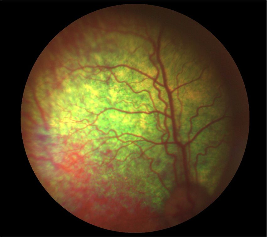
Kucharczyk et al. BMC Veterinary Research (2019) 15:392 Page 2 of 4
Australian Shepherd, the Boykin Spaniel, the Lanca-
shire Heeler, the Longhaired Whippet, the Nova Scotia
Duck Tolling Retriever, the Hokkaido dog and the
Silken Windhound [15–20]. A phenotypically identical
lesion has been seen in other non-collie breeds includ-
ing the German Shepherd Dog, Miniature and Toy
Poodles, the Beagle and a mixed-breed dog [17, 18, 20].
The causal genetic defect has not been determined for
all of these breeds.
This is the first published report of CEA in the Austra-
lian Kelpie breed. The initial ophthalmoscopic finding was
subsequently confirmed using the specific DNA based
test. Based on analysis of the probands’ pedigree the dog’s
dam proved to be homozygous for the mutation and one
other affected dog plus several carriers were found.
Case presentation
The initial finding was in a two-and-a-half year old
female Australian Kelpie which was being considered for
breeding purposes. There were no reported visual defi-
cits and performance in a maze test, the menace
response, the pupillary light reflexes and the dazzle re-
flex were all considered to be normal. Biomicroscopic
examination (SL-17 Portable Slit Lamp, Kowa) of the an-
terior segment revealed minor bilateral stromal iridal at-
rophy. Mydriasis was effected using tropicamide
(Tropicamidum WZF 1%, Polfa Warszawa) and fundus
examination completed using direct and indirect oph-
thalmoscopy (Keeler Standard Direct and Keeler Vant-
age Plus Indirect ophthalmoscopes). Fundus imaging
was obtained using a 30D condensing lens (Volk 2) and
photographs were taken using the ClearView Fundus
Camera (Optibrand). Both fundi had a tigroid appear-
ance in the non-tapetal areas, but an irregular arrange-
ment of the choroidal vasculature was observed
bilaterally in the temporal region of the tapetal fundus.
The lesions were easily identifiable with the choroidal
blood vessels being fewer in number and thicker than
normal. The degree of CH allowed the white appearance
of the sclera to be visible between the abnormal blood
vessels (Fig. 1a-c). Colobomatous lesions were not
present and no other ocular abnormalities were found.
Based on the presence of CH a tentative diagnosis of
CEA was suggested, this diagnosis being subsequently
confirmed using a real time PCR DNA test for the CEA
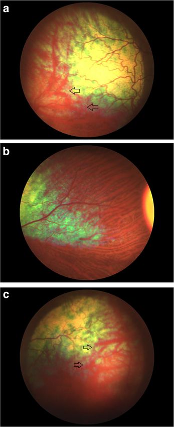
NHEJ1 7.8 kb deletion gene mutation (Laboklin GmbH Fig. 1 Fundus photographs of the affected female to demonstrate
the abnormal arrangement of choroidal vessels in the temporal
& Co.KG, 8304).
tapetal fundus. a Left eye, temporal area, the arrowsdemonstrating
Using an analysis of the probands’ pedigree twenty other the large area of choroidal hypoplasia. b Right eye, medial area,
dogs related to the affected female were subsequently demonstrating a normal appearance. c Left eye, temporal area,
screened using the DNA test for the CEA mutation. Five demonstrating choroidal hypoplasia
of these dogs were also examined clinically and CH lesions
were found in one, the dog’s dam. Here the small size of
lesions rendered a positive ophthalmoscopic diagnosis dif-
ficult (Fig. 2), demonstrating the value of litter screeningKucharczyk et al. BMC Veterinary Research (2019) 15:392 Page 3 of 4
Fig. 2 The temporal area of the right fundus of the affected dam demonstrating a small area of choroidal hypoplasia
at six to 8 weeks of age to rule out a putative “go normal” the more traditional “collie” breeds were established.
diagnosis. However DNA testing confirmed the CEA diag- This background indicated that CEA could be present in
nosis for this dog whilst eight others were shown to be the Kelpie and was the reason for this small study.CH le-
carriers for the disease (Fig. 3). Colobomatous lesions and sions and the association with the CEA related NHEJ1
other possible features of CEA were not found in any of mutation could be reported,but colobomatous defects
these dogs. and the other features of CEA were not seen in either of
Permission has been obtained from all the owners and the affected dogs. Further screening is required to evalu-
breeders whose dogs were involved in this study to record ate the prevalence of the NEHJ1 gene mutation in this
and publish both the clinical findings and the DNA results. breed and breeders should be aware that in addition to
the use of the DNA test and the ophthalmoscopic exam-
Discussion and conclusions ination of the breeding stock, litter screening at six to 8
This is the first report of CEA in the Australian Kelpie weeks of age will identify both CH and possible colobo-
breed, diagnosed on the basis of bilateral CH seen matous lesions. The existence of a “go normal” change
ophthalmoscopically and confirmed by genetic testing. in which CH lesions can be masked by pigmentation be-
The breed originated from British dogs imported into yond 12 weeks of age means that in adult dogs ophthal-
Australia for stock work in the early nineteenth century. moscopic examination needs to be supported by DNA
They were simply described as collies or colleys, before testing to effect efficient disease control.
Fig. 3 Pedigree analysis of the 21 related Australian Kelpie dogs examined on the survey. The subject of this case report is the affected dog on
the second line and asterisks mark the six dogs clinically examinedKucharczyk et al. BMC Veterinary Research (2019) 15:392 Page 4 of 4
Abbreviations cosegregates with Collie eye anomaly across multiple dog breeds. Genome
CEA: Collie Eye Anomaly; CH: Choroidal hypoplasia, focal absence of Res. 2007;17(11):1562–71.
choroidal tissue temporal to the optic disc; CRD: Chorioretinal dysplasia; 12. Chang H-S, Mizukami K, Yabuki A, Hossain MA, Rahman MM, Uddin MM,
DNA: Deoxyribonucleic acid; NHEJ1: Non-homologous end-joining factor 1 et al. A novel rapid genotyping technique for Collie eye anomaly: SYBR
Green-based real-time polymerase chain reaction method applicable to
Acknowledgements blood and saliva specimens on Flinders Technology Associates filter paper. J
The authors acknowledge the held of the several breeders who volunteered Vet Diagn Invest. 2010;22(5):708–15.
their dogs for the study. 13. Barnet KC, Stades FC. Collie Eye Anomaly in the Shetland Sheepdog in the
Netherlands. J Small Anim Pract. 1979;20(6):321–9.
Authors’ contributions 14. Bedford PGC. Collie Eye Anomaly in the Border Collie. Vet Rec. 1982;111(2):34–5.
All three authors were equally involved in the clinical assessment of the 15. Rubin L, Nelson B, Sharp C. Collie eye anomaly in Australian Shepherd dogs.
dogs (NK, AC-P and PB), the manuscript preparation (NK, AC-P and PB) and Prog Vet Comp Ophthalmol. 1991;1:105–8.
all have read and approved the final manuscript (NK AC-P and PB). 16. Bedford PG. Collie eye anomaly in the Lancashire heeler. Vet Rec. 1998;
143(13):354–6.
17. Rampazzo A, D’Angelo A, Capucchio MT, Sereno S, Peruccio C. Collie eye
Funding anomaly in a mixed-breed dog. Vet Ophthalmol. 2005;8(5):357–60.
There were no funding issues as all costs were met by the breeders. 18. Mizukami K, Chang H-S, Ota M, Yabuki A, Hossain MA, Rahman MM, et al.
Collie eye anomaly in Hokkaido dogs: case study. Vet Ophthalmol. 2012;
Availability of data and materials 15(2):128–32.
The datasets used during the current study are available from the 19. Brown EA, Thomasy SM, Murphy CJ, Bannasch DL. Genetic analysis of optic
corresponding author on reasonable request. nerve head coloboma in the Nova Scotia Duck Tolling Retriever identifies
discordance with the NHEJ1 intronic deletion (collie eye anomaly mutation).
Ethics approval and consent to participate Vet Ophthalmol. 2018;21(2):144–50.
There were no associated ethical issues in this clinical study and all the dog 20. Priester WA. Congenital ocular defects in cattle, horses, cats and dogs. J Am
breeders involved gave written consent for their participation, seeing the Vet Med Assoc. 1972;160(11):1504–11.
study as vital to future breeding programmes and the wellbeing of the
breed.
Publisher’s Note
Springer Nature remains neutral with regard to jurisdictional claims in
Consent for publication published maps and institutional affiliations.
The authors would like to thank the breeders of all the dogs included in this
clinical study for their participation and their permission to publish the
results. Written consent in both Polish and English to publish all the personal
and clinical details together with the identifying images and the pedigree
analysis has been obtained and the statements are available for scrutiny.
These records are held by both the authors and the breeders.
Competing interests
The authors declare that they have no competing interests.
Author details
1
Viva Veterinary Clinic, Wroclaw, Poland. 2Royal Veterinary College, London,
UK.
Received: 23 April 2019 Accepted: 14 October 2019
References
1. Donovan EF, Wyman M. Ocular fundus anomaly in the collie. J Am Vet Med
Assoc. 1965;147(12):1465–9.
2. Roberts SR. The collie eye anomaly. J Am Vet Med Assoc. 1969;155(6):859–65.
3. Wyman M, Donovan EF. Eye anomaly of the collie. J Am Vet Med Assoc.
1969;155(6):866–70.
4. Donovan RH, Freeman HM, Schepens CL. Anomaly of the collie eye. J Am
Vet Med Assoc. 1969;155(6):872–7.
5. Bedford PGC. Collie eye anomaly in the United Kingdom. Vet Rec. 1982;
111(12):263–7.
6. Yakely WL, Wyman M, Donovan EF, Fechheimer NS. Genetic transmission of an
ocular fundus anomaly in Collies. J Am Vet Med Assoc. 1968;152(5):457–61.0.
7. Yakely WL. Collie eye anomaly: decreased prevalence through selective
breeding. J Am Vet Med Assoc. 1972;161(10):1103–7.
8. Wallin-Håkanson B, Wallin-Håkanson N, Hedhammar A. Influence of selective
breeding on the prevalence of chorioretinal dysplasia and coloboma in the
rough collie in Sweden. J Small Anim Pract. 2000;41(2):56–9.
9. Wallin-Håkanson B, Wallin-Håkanson N, Hedhammar A. Collie eye anomaly
in the rough collie in Sweden: genetic transmission and influence on
offspring vitality. J Small Anim Pract. 2000;41(6):254–8.
10. Lowe JK, Kukekova AV, Kirkness EF, Langlois MC, Aguirre GD, Acland GM,
et al. Linkage mapping of the primary disease locus for collie eye anomaly.
Genomics. 2003 Jul;82(1):86–95.
11. Parker HG, Kukekova AV, Akey DT, Goldstein O, Kirkness EF, Baysac KC, et al.
Breed relationships facilitate fine-mapping studies: a 7.8-kb deletionYou can also read