Case Report: Anti-MOG Antibody Seroconversion Accompanied by Dimethyl Fumarate Treatment - Frontiers
←
→
Page content transcription
If your browser does not render page correctly, please read the page content below
CASE REPORT
                                                                                                                                         published: 15 February 2021
                                                                                                                                   doi: 10.3389/fimmu.2021.625465

                                            Case Report: Anti-MOG Antibody
                                            Seroconversion Accompanied by
                                            Dimethyl Fumarate Treatment
                                            Keita Takahashi 1 , Hideyuki Takeuchi 1*, Ryoko Fukai 1 , Haruko Nakamura 1 ,
                                            Keisuke Morihara 1 , Yuichi Higashiyama 1 , Toshiyuki Takahashi 2 , Hiroshi Doi 1 and
                                            Fumiaki Tanaka 1*
                                            1
                                                Department of Neurology and Stroke Medicine, Yokohama City University Graduate School of Medicine, Yokohama, Japan,
                                            2
                                                Department of Neurology, Tohoku University Graduate School of Medicine, Sendai, Japan

                                            Here we report three cases of anti-myelin oligodendrocyte glycoprotein (MOG)
                                            antibody–associated disease (MOGAD) mimicking multiple sclerosis in which
                                            seropositivity for anti-MOG antibodies occurred during disease-modifying drug dimethyl
                                            fumarate (DMF) treatment. These patients developed relapses with anti-MOG antibody
                         Edited by:         seroconversion after switching from fingolimod or steroid pulse therapy to DMF, which
              Pamela Ann McCombe,
                    The University of       was associated with peripheral lymphocyte recovery. MOGAD is considered a humoral
                Queensland, Australia       immune disease, and DMF reportedly enhances Th2-skewed humoral immune activity.
                        Reviewed by:        Therefore, we suggest that DMF, but not fingolimod, may exacerbate humoral immune
                    Giacomo Casella,
                                            imbalance and enhance autoantibody production, leading to aggravation of MOGAD.
         Thomas Jefferson University,
                         United States      Keywords: anti-myelin oligodendrocyte glycoprotein antibody-associated disease, dimethyl fumarate, fingolimod,
                       Alice Mariottini,    multiple sclerosis, seroconversion
           University of Florence, Italy

                 *Correspondence:
                   Hideyuki Takeuchi        INTRODUCTION
            htake@yokohama-cu.ac.jp
                     Fumiaki Tanaka         Anti-myelin oligodendrocyte glycoprotein (MOG) antibody–associated disease (MOGAD), which
          ftanaka@yokohama-cu.ac.jp
                                            makes up ∼40% of anti-aquaporin 4 (AQP4) antibody–negative neuromyelitis optica spectrum
                                            disorders, is often difficult to distinguish from multiple sclerosis (MS) (1). Whether disease-
                   Specialty section:
                                            modifying drugs (DMDs) widely used in MS are also effective in MOGAD remains unclear. Here
         This article was submitted to
                Multiple Sclerosis and
                                            we report three cases of MOGAD treated as atypical MS due to negativity for specific antibodies
                    Neuroimmunology,        in which seroconversion for anti-MOG antibodies occurred when DMD was switched to dimethyl
               a section of the journal     fumarate (DMF).
              Frontiers in Immunology

       Received: 03 November 2020           CASE REPORT
        Accepted: 27 January 2021
       Published: 15 February 2021          The first patient was a 27-year-old woman who experienced two attacks of optic neuritis and
                            Citation:       transverse myelitis with short cervical and thoracic cord lesions in 4 years (Figure 1A). She
    Takahashi K, Takeuchi H, Fukai R,       experienced severe attacks of left retrobulbar neuritis and transverse myelitis at the age of
             Nakamura H, Morihara K,        24. Magnetic resonance imaging (MRI) showed T2-hyperintense lesions in the left optic nerve
    Higashiyama Y, Takahashi T, Doi H       (Figure 1B) and left dorsal horn and fasciculus at the C3 level (Figures 1C,D). Optical coherence
   and Tanaka F (2021) Case Report:
                                            tomography (OCT) detected a significant decrease in retinal nerve fiber layer (RNFL) thickness in
  Anti-MOG Antibody Seroconversion
  Accompanied by Dimethyl Fumarate
                                            the left eye (Figure 1E). Cerebrospinal fluid (CSF) analysis revealed a slight elevation in protein
                           Treatment.       levels and increases in myelin basic protein (MBP) levels and IgG index, but not in cell count
          Front. Immunol. 12:625465.        or oligoclonal bands (Table 1; Patient 1, pre). Neither anti-AQP4 nor anti-MOG antibodies were
    doi: 10.3389/fimmu.2021.625465          detected in serum samples using an internationally standardized cell-based assay (CBA) (2–4). She

Frontiers in Immunology | www.frontiersin.org                                      1                                      February 2021 | Volume 12 | Article 625465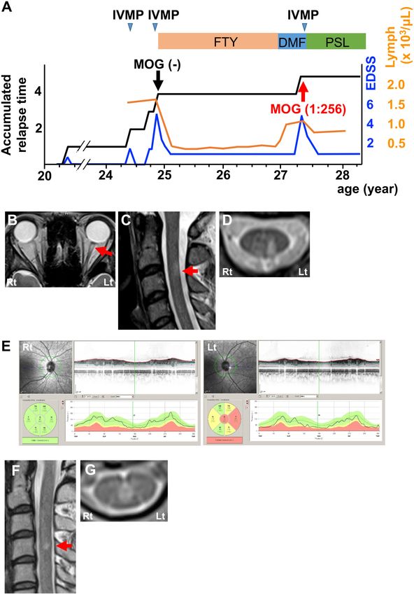
Takahashi et al.                                                                                                                           MOG Seroconversion With DMF

 FIGURE 1 | Clinical features of Patient 1. (A) Graph of the time course for relapses and treatments. (B) Axial T2-weighted orbit MRI showing left optic nerve lesion (red
 arrow). (C) Sagittal T2-weighted cervical MRI showing a short cervical lesion at the C3 level (red arrow). (D) Axial T2-weighted cervical MRI showing a lesion in the left
 dorsal horn and fasciculus at the C3 level. (E) OCT showing significant RNFL thinning in the left eye. Green, normal RNFL thickness (5th−95th percentile); yellow,
 mild-to-moderate RNFL thinning (1st−5th percentile); red, severe RNFL thinning (
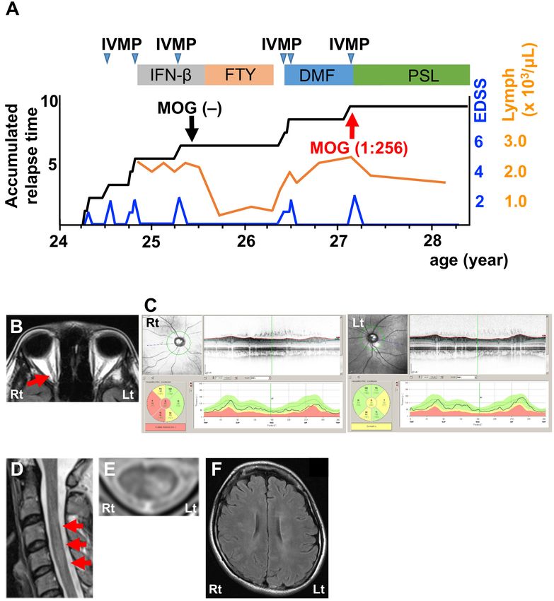
Takahashi et al. MOG Seroconversion With DMF
CBA using the same methodology (3) revealed seroconversion the first CBA was truly seronegative for anti-MOG antibodies.
for anti-MOG antibodies (titer, 1:256; cut-off, ≥1:128). We Intravenous methylprednisolone pulse therapy (1 g/day for 3
reconfirmed that the frozen serum sample from the time of consecutive days; IVMP) markedly decreased symptoms. She
has had no relapses with subsequent oral prednisolone therapy
(20 mg/day) for over 1 year (Figure 1A).
The second patient was a 27-year-old woman who had
TABLE 1 | CSF findings of three patients.
several attacks of optic neuritis and transverse myelitis involving
Patient 1 Patient 2 Patient 3 short lesions in the cervical cord in a year (Figure 2A). She
pre/post pre/post pre/post experienced a severe attack of optic neuritis around the age of
25, as shown on fluid-attenuated inversion recovery (FLAIR)
Cell count (cells/µl) 2/2 20/10 2/1
MRI (Figure 2B). OCT revealed severe RNFL thinning in the
Protein (mg/dl) 44/86 51/31 31/31 right eye and mild RNFL thinning in the left eye (Figure 2C).
Myelin basic protein (pg/ml) 86.5/103.1 ND/ND 1729.8/ND CSF analysis detected pleocytosis, elevations in protein levels
IgG index 0.91/0.64 2.06/1.36 0.36/0.41 and IgG index, and the presence of oligoclonal bands, but not
Oligoclonal band –/– +/+ –/– MBP (Table 1; Patient 2, pre). A CBA did not detect anti-AQP4
Pre, CSF collection before seroconversion for anti-MOG antibodies; post, CSF collection or anti-MOG antibodies in the serum. She had been treated
after seroconversion for anti-MOG antibodies; ND, not detected. with interferon-β followed by fingolimod for the diagnosis of
FIGURE 2 | Clinical features of Patient 2. (A) Graph of the time course for relapses and treatments. (B) Axial FLAIR orbit MRI showing right optic nerve lesions (red
arrow). (C) OCT showing severe RNFL thinning in the right eye and mild RNFL thinning in the left eye. Green, normal RNFL thickness (5th−95th percentile); yellow,
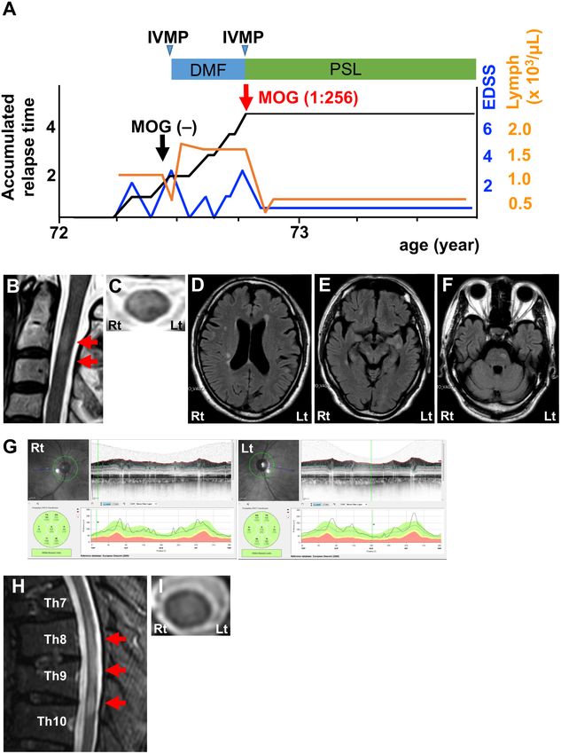
mild-to-moderate RNFL thinning (1st−5th percentile); red, severe RNFL thinning (Takahashi et al. MOG Seroconversion With DMF FIGURE 3 | Clinical features of Patient 3. (A) Graph of the time course for relapses and treatments. (B) Sagittal T2-weighted cervical MRI showing short lesions at the C2–3 level (red arrows). (C) Axial T2-weighted cervical MRI showing a ventral-dominant lesion at the C2 level. (D–F) Axial FLAIR brain MRI showing lesions in the periventricular area, subcortical area, and brain stem. (G) OCT showing normal RNFL thickness. Green, normal RNFL thickness (5th−95th percentile). (H) Sagittal T2-weighted thoracic MRI showing lesions at the Th8–10 levels (red arrows). (I) Axial T2-weighted cervical MRI showing lesion at the Th8 level around the central canal. DMF, dimethyl fumarate; IVMP, intravenous methylprednisolone pulse therapy; PSL, prednisolone; EDSS, Expanded Disability Status Scale score; Lymph, peripheral lymphocyte count. atypical MS for 1 year without any relapses, but DMD was (Table 1; Patient 2, post). This patient also had seroconversion for changed to DMF because of epidural hematoma as an unexpected anti-MOG antibodies (titer, 1:256) using the same methodology. adverse side effect of fingolimod (5). After 1 year, she had a Serum from the time of the first CBA was reconfirmed to be truly relapse of transverse myelitis with a short cervical cord lesion seronegative. IVMP substantially decreased her symptoms. Oral (Figures 2D,E) and asymptomatic MS-like supratentorial lesions prednisolone (20 mg/day) completely suppressed the relapse for (Figure 2F) following lymphocyte recovery (Figure 2A). CSF more than a year (Figure 2A). analysis revealed a profile that was similar to the profile before The third patient was a 73-year-old man with a severe attack DMF treatment, except for normal protein levels at this time of transverse myelitis in the previous year (Figure 3A). FLAIR Frontiers in Immunology | www.frontiersin.org 4 February 2021 | Volume 12 | Article 625465
Takahashi et al.                                                                                                MOG Seroconversion With DMF

MRI showed short lesions in the cervical cord (Figures 3B,C)            that these patients truly had MOGAD. Another possibility is that
as well as asymptomatic lesions in the periventricular area,            DMF treatment enhanced anti-MOG autoantibody production
subcortical area, and brain stem (Figures 3D–F). OCT indicated          whereas fingolimod treatment effectively suppressed it. The
no signs of optic neuritis (Figure 3G). CSF analysis revealed a         main therapeutic effect of fingolimod is immunosuppression
prominent increase in MBP levels, but was otherwise normal              due to the retention of auto-reactive lymphocytes within
(Table 1; Patient 3, pre). Neither anti-AQP4 nor anti-MOG               secondary lymphoid organs, whereas the net activity of DMF
antibodies were detected in the serum using the same CBA                is mainly a pro-tolerogenic lymphocyte shift (9). As humoral
described for the other 2 patients. IVMP effectively resolved           immunosuppression, fingolimod is likely efficacious in MOGAD
his symptoms. He was subsequently treated with DMF for the              whereas DMF is not efficacious. A previous report documented
diagnosis of atypical MS. Lymphocyte counts became higher               a similar seroconversion for anti-MOG antibodies during
than pre-treatment levels and reached a plateau (Figure 3A).            interferon-β treatment, which modulates immune balance
Three months later, he developed a severe relapse of transverse         from Th1/Th17-mediated cellular immunity to Th2-mediated
myelitis. MRI revealed a FLAIR-hyperintense lesion around the           humoral immunity and may promote autoantibody synthesis
central canal extending from the Th8 level to the Th10 level            (10). Like interferon-β, DMF also enhances Th2-skewed
(Figures 3H,I). CSF analysis showed a normal profile, including         humoral immune activity (11). Another report demonstrated
normal MBP levels (Table 1; Patient 3, post). A CBA for anti-           that DMF induces severe relapses of neuromyelitis optica
MOG antibodies turned positive at that time (titer, 1:256). We          spectrum disorders, indicating that DMF has a detrimental
also reconfirmed seronegativity for anti-MOG antibodies in a            effect in humoral immunity-mediated diseases (12). In fact, our
frozen serum sample from the time of the first CBA. IVMP                cases revealed that fingolimod effectively suppresses peripheral
was effective in decreasing his symptoms again. To date, he has         lymphocyte count and switching from fingolimod to DMF or
been in remission with oral prednisolone therapy (20 mg/day) for        initiating DMF leads to a recovery of peripheral lymphocyte
more than a year (Figure 3A).                                           count accompanied by seroconversion for anti-MOG antibodies
                                                                        (Figures 1A, 2A, 3A). These findings suggest that DMF might
                                                                        exacerbate Th2-prone humoral autoimmunity. In addition, two
DISCUSSION                                                              of three patients (Patients 1 and 2) had similar or higher
                                                                        peripheral lymphocyte counts at the time of the first CBA than
Although therapy for MOGAD is yet to be established,                    at the time of the second CBA, suggesting that the Th2-skewed
according to consensus among international experts (6, 7),              peripheral lymphocyte subpopulation shift associated with DMF
immunosuppressive therapy (e.g., corticosteroids, azathioprine,         (11) may affect MOGAD development rather than the higher
tacrolimus, mycophenolate mofetil, and methotrexate) is                 absolute number of peripheral lymphocytes.
the mainstay for treatment to prevent relapse. In particular,               In conclusion, we presented three cases of MOGAD with
B cell depleting therapies such as rituximab have shown                 seroconversion for anti-MOG antibodies accompanied by
good therapeutic responses, but relapse occurs immediately              recurrence of clinical disability after treatment with DMF. Thus,
after B cell recovery. These data strongly suggest that                 DMF should be avoided in MOGAD, whereas fingolimod may be
suppression of humoral immunity is likely the key therapeutic           efficacious because it suppresses humoral immunity.
strategy for MOGAD. DMDs for MS such as interferon-
β, glatiramer acetate, and natalizumab might not be
efficacious, but the effectiveness of fingolimod remains                PRACTICAL IMPLICATIONS
uncertain (6, 7). Although a recent report mentioned
that DMF is ineffective but not harmful in a patient                    Dimethyl fumarate may exacerbate anti-myelin oligodendrocyte
with MOGAD (8), the effectiveness of DMF has yet to                     glycoprotein antibody–associated disease whereas fingolimod
be determined.                                                          may be effective against this disease.
    Initially, our patients were diagnosed with atypical MS
because they were seronegative for specific antibodies and
had good response to IVMP and fingolimod. However,                      DATA AVAILABILITY STATEMENT
seroconversion for anti-MOG antibodies occurred when DMD
therapy was switched to DMF. We propose two possible                    The original contributions presented in the study are included
mechanisms to explain this seroconversion. One possibility              in the article/supplementary material, further inquiries can be
is that the first CBA results in all patients were false-               directed to the corresponding authors.
negative due to fluctuations in autoantibody titers, and the
timing of seroconversion and switching to DMF was just a
coincidence because the titers from the second CBA in all               ETHICS STATEMENT
patients were relatively low (1:256). Despite low titers of anti-
MOG antibodies, all patients have been in remission with                Written informed consent was obtained from the patients for the
oral prednisolone therapy for at least 1 year after their last          publication of this case series in accordance with the Declaration
relapse. This good response to corticosteroid therapy suggests          of Helsinki.

Frontiers in Immunology | www.frontiersin.org                       5                                 February 2021 | Volume 12 | Article 625465
Takahashi et al. MOG Seroconversion With DMF
AUTHOR CONTRIBUTIONS FUNDING
KT, HT, RF, HN, KM, YH, and HD examined and treated the This study was supported partly by Grants-in-Aid for Scientific
patient. TT assessed anti-MOG antibody titers. KT and HT Research from the Ministry of Education, Culture, Sports,
analyzed data. HT and FT designed and supervised this study. Science and Technology of Japan, grants from the Ministry of
KT, HT, and FT wrote the manuscript. All authors contributed to Health, Labor and Welfare of Japan, and a grant for Strategic
the article and approved the submitted version. Research Promotion from Yokohama City University.
REFERENCES glycoprotein antibody disease. Cureus. (2019) 11:e6040. doi: 10.7759/
cureus.6040
1. Narayan R, Simpson A, Fritsche K, Salama S, Pardo S, Mealy M, 9. Rommer PS, Milo R, Han MH, Satyanarayan S, Sellner J, Hauer L, et al.
et al. MOG antibody disease: a review of MOG antibody seropositive Immunological aspects of approved MS therapeutics. Front Immunol. (2019)
neuromyelitis optica spectrum disorder. Mult Scler Relat Disord. (2018) 10:1564. doi: 10.3389/fimmu.2019.01564
25:66–72. doi: 10.1016/j.msard.2018.07.025 10. Pawlitzki M, Campe C, Rolfes L, Heinze HJ, Leypoldt F, Wandinger
2. Takahashi T, Fujihara K, Nakashima I, Misu T, Miyazawa I, Nakamura M, et al. KP, et al. Transient MOG antibody seroconversion associated
Anti-aquaporin-4 antibody is involved in the pathogenesis of NMO: a study with immunomodulating therapy. Mult Scler Relat Disord. (2020)
on antibody titre. Brain. (2007) 130:1235–43. doi: 10.1093/brain/awm062 37:101420. doi: 10.1016/j.msard.2019.101420
3. Sato DK, Callegaro D, Lana-Peixoto MA, Waters PJ, de Haidar Jorge 11. Kira JI, Unexpected exacerbations following initiation of disease-modifying
FM, Takahashi T, et al. Distinction between MOG antibody-positive and drugs in neuromyelitis optica spectrum disorder: Which factor is responsible,
AQP4 antibody-positive NMO spectrum disorders. Neurology. (2014) 82:474– anti-aquaporin 4 antibodies, B cells, Th1 cells, Th2 cells, Th17 cells, or others?
81. doi: 10.1212/WNL.0000000000000101 Mult Scler. (2017) 23:1300–2. doi: 10.1177/1352458517703803
4. Waters P, Woodhall M, O’Connor KC, Reindl M, Lang B, Sato DK, et 12. Yamout BI, Beaini S, Zeineddine MM, Akkawi N. Catastrophic
al. MOG cell-based assay detects non-MS patients with inflammatory relapses following initiation of dimethyl fumarate in two patients
neurologic disease. Neurol Neuroimmunol Neuroinflamm. (2015) with neuromyelitis optica spectrum disorder. Mult Scler. (2017)
2:e89. doi: 10.1212/NXI.0000000000000089 23:1297–300. doi: 10.1177/1352458517694086
5. Fukai R, Takahashi K, Abe H, Higashiyama Y, Doi H, Takeuchi H, et al.
Non-traumatic acute epidural hematoma in multiple sclerosis treated with Conflict of Interest: The authors declare that the research was conducted in the
fingolimod. Front Neurol. (2019) 10:763. doi: 10.3389/fneur.2019.00763 absence of any commercial or financial relationships that could be construed as a
6. Fujihara K, Sato DK, Nakashima I, Takahashi T, Kaneko K, Ogawa potential conflict of interest.
R, et al. Myelin oligodendrocyte glycoprotein immunoglobulin G-
associated disease: an overview. Clin Exp Neuroimmunol. (2018) Copyright © 2021 Takahashi, Takeuchi, Fukai, Nakamura, Morihara, Higashiyama,
9:48–55. doi: 10.1111/cen3.12434 Takahashi, Doi and Tanaka. This is an open-access article distributed under the
7. Reindl M, Waters P. Myelin oligodendrocyte glycoprotein terms of the Creative Commons Attribution License (CC BY). The use, distribution
antibodies in neurological disease. Nat Rev Neurol. (2019) or reproduction in other forums is permitted, provided the original author(s) and
15:89–102. doi: 10.1038/s41582-018-0112-x the copyright owner(s) are credited and that the original publication in this journal
8. Warabi Y, Takahashi T, Isozaki E. Dimethyl fumarate was ineffective is cited, in accordance with accepted academic practice. No use, distribution or
but not harmful for a patient with myelin oligodendrocyte reproduction is permitted which does not comply with these terms.
Frontiers in Immunology | www.frontiersin.org 6 February 2021 | Volume 12 | Article 625465You can also read