High-dose glucocorticoid treatment of near-fatal bocavirus lung infection results in rapid recovery
←
→
Page content transcription
If your browser does not render page correctly, please read the page content below

ORIGINAL RESEARCH LETTER

                                  High-dose glucocorticoid treatment of
                                  near-fatal bocavirus lung infection
                                  results in rapid recovery
                                  To the Editor:
                                  Human bocavirus (HBoV), which belongs to Parvoviridae, is a well-defined pathogen of respiratory
                                  infections, particularly in young children [1]. In contrast, the frequency of HBoV infections in adults with
                                  respiratory symptoms is virtually unknown and its causative role in respiratory failure is debated [1, 2].
                                  Furthermore, the observation that dexamethasone is beneficial in COVID-19 patients with respiratory
                                  failure requiring respiratory support has gained great interest [3]. However, whether glucocorticoid
                                  treatment is useful in other severe viral respiratory diseases is a matter of controversy [4].
                                  Here, we report on a 58-year-old, obese (body mass index 35 kg·m−2, Eastern Cooperative Oncology
                                  Group performance status 1), Caucasian male who presented at Charité University Hospital (Berlin,
                                  Germany) on 25 February 2020 with a 6-day history of fever and shortness of breath. 3 months earlier, he
                                  had received BEAM (carmustine, etoposide, cytarabine and melphalan) high-dose chemotherapy and
                                  autologous stem cell transplantation (autoSCT) for relapsed Hodgkin’s lymphoma, resulting in complete
                                  remission. Relevant medical history included psoriasis, diabetes mellitus type 2, permanent atrial
                                  fibrillation and COPD of Global Initiative for Chronic Obstructive Lung Disease stage II, with no relevant
                                  previously documented pulmonary structural alterations. Concomitant medication on admission consisted
                                  of acyclovir, pantoprazole, digitoxin, bisoprolol, sitagliptin, metformin, insulin and acitretin, all not
                                  considered as causative for the clinical symptoms. On admission, peripheral oxygen saturation
                                  demonstrated hypoxaemia (SpO2 88%), C-reactive protein (CRP) level was 50.9 mg·L−1 (normal range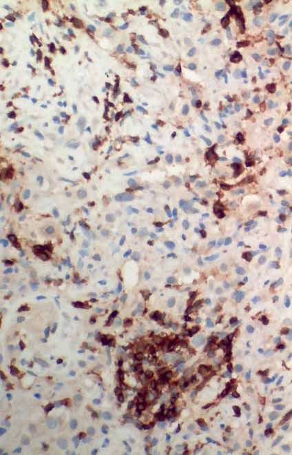
ORIGINAL RESEARCH LETTER | M. OBENAUS ET AL.

 a)                                           b)                                            c)                          d)

 e)                                                f)                                                     g)

FIGURE 1 Radiomorphological and pathological–anatomical findings of a patient with near-fatal human bocavirus (HBoV) infection. a) Computed
tomography (CT) image after a 7-day course of antibiotic therapy shows bilateral pulmonary infiltrates with interstitial pattern and ground-glass
opacities (3 March 2020). b) HBoV-specific fluorescence in situ hybridisation in lung biopsy. Head and tail HBoV genome regions were detected
with specific fluorescence probes and tissue was counterstained with DAPI. Yellow and orange dots (arrows) indicate codetection of the head and
tail regions of HBoV-DNA. c) Lung biopsy showing fibroblast proliferation (arrows) (haematoxylin and eosin staining). d) Strong infiltration with
T-lymphocytes (CD3 staining in brown) in the lung tissue reflecting excessive immune response. e) Chest radiography (anteroposterior view) at
initiation of glucocorticoid treatment (day 1) showing multifocal opacities in both lungs with foci of consolidation in the upper-left lobe (5 March
2020), and f) at day 4, demonstrating resolution of the multifocal opacities with demarked consolidation in the upper left lobe (8 March 2020).
g) CT image 2 months after treatment demonstrates complete resolution of infiltrates with remaining pre-existing small bullae (26 May 2020).

                                    copies (3.2×10⁸ cp·mL−1 in BAL), HBoV detection by fluorescence in situ hybridisation (figure 1b) and
                                    pronounced T-lymphocytic infiltrates (figure 1c and d) in the lung tissue confirmed this scenario. Given
                                    the lack of HBoV-specific treatments, we aimed to mitigate the immune reaction and subsequent
                                    pulmonary damage by pulsed high-dose glucocorticoid therapy (5 March (day 1), 500 mg prednisolone;
                                    days 2 and 3, 1000 mg methylprednisolone) followed by rapid reduction to 20 mg·day−1 prednisolone and
                                    tapering. Respiratory parameters and radiological findings improved rapidly (figure 1e and f ), allowing
                                    termination of mechanical ventilation on day 4 (8 March) of intensive care unit treatment and discharge
                                    on 17 March. In parallel to the clinical improvement, CRP values rapidly decreased and remained normal.
                                    A CT scan 2 months after treatment confirmed complete resolution of all infiltrates (figure 1g).
                                    Given the virtually unknown frequency of HBoV in adult patients with respiratory symptoms [1], we
                                    retrospectively analysed a total of 5328 consecutive adult respiratory samples, mainly consisting of
                                    tracheobronchial secretions and BALs, and which were all examined by PCR-based respiratory panel
                                    analyses, for HBoV. We identified 17 HBoV-positive patients, most of them heavily immunocompromised.
                                    Given the reconstitution of white blood cells (4.45 nL−1, normal range 3.90–10.50 nL−1) with normal
                                    neutrophils (3.03 nL−1, normal range 1.50–7.70 nL−1) and slightly reduced lymphocytes (0.95 nL−1,
                                    normal range 1.10–4.50 nL−1), even though platelets (30 nL−1, normal range 150–370 nL−1) and
                                    haemoglobin (8.9 mg·dL−1, normal range 13.5–17.0 mg·dL−1) were reduced on admission, as well as
                                    almost normal immunoglobulin levels (IgG 6.61 g·L−1, normal range 7.00–16.00 g·L−1; IgA 1.72 g·L−1,
                                    normal range 0.7–4.00 g·L−1; and IgM 0.30 g·L−1, normal range 0.40–2.30 g·L−1) at the time of
                                    HBoV-induced respiratory failure, we judged our patient to be largely immunocompetent. Thus, even
                                    though rare, HBoV must be considered a life-threatening pathogen in adults with atypical pneumonia and
                                    should be included in the respective diagnostic work-up, with HBoV-DNA quantification if positive.

https://doi.org/10.1183/23120541.00060-2021                                                                                                       2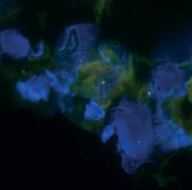
ORIGINAL RESEARCH LETTER | M. OBENAUS ET AL.

                                  Only recently, it has been demonstrated that dexamethasone is beneficial in COVID-19 patients with
                                  respiratory failure requiring respiratory support [3]; however, the benefit of glucocorticoid treatment in
                                  other severe viral respiratory diseases is unclear [4]. In contrast to the dexamethasone scheme used in the
                                  RECOVERY trial in COVID-19 patients (6 mg once daily for up to 10 days), we applied a pulsed
                                  high-dose glucocorticoid therapy scheme deduced from treatment of autoimmune diseases. Notably, the
                                  application of such pulsed “very high-dose” glucocorticoid therapy is supposed to extend the
                                  glucocorticoid mode of action compared to lower doses [5, 6]. We assume that this short-term high-dose
                                  glucocorticoid therapy effectively dampened the detrimental virus-induced immune response, most likely
                                  by glucocorticoid-induced cell death of immune effector cells including infiltrating T-lymphocytes, and
                                  anti-inflammatory effects by modulation of monocyte and macrophage function [7, 8]. Furthermore,
                                  glucocorticoid therapy can inhibit fibroblast proliferation and support the clearance of
                                  inflammation-induced tissue damage [7]. We are aware of glucocorticoid therapy-induced side-effects; that
                                  corticosteroid treatment of ARDS or SARS patients, or patients suffering from viral pneumonia, has
                                  generated conflicting results [4, 5, 9–11]; and that prolonged intake of corticosteroids predisposes to
                                  invasive aspergillosis in the context of severe influenza infection [12]. However, short pulsed high-dose
                                  glucocorticoid therapy has been used for decades for the treatment of various severe autoimmune diseases,
                                  with limited and well-known side-effects [13]. Therefore, we decided to use such a “very high-dose”
                                  short-term scheme and not a lower-dose, longer-lasting scheme for our patient. We believe that in selected
                                  patients, in which the virus-induced immune response is profound and likely to be more harmful to lung
                                  tissue than damage by the virus itself, and in which fatal lung damage has not yet passed the point of no
                                  return, glucocorticoid therapy may preserve lung tissue and function. Notably, most recently, it has been
                                  shown that severe COVID-19 patients benefit from pulsed methylprednisolone administration if given
                                  during the early phase of pulmonary infection [14]. Thus, high-dose glucocorticoid therapy may be
                                  beneficial in patients suffering from severe pulmonary disease caused by HBoV, as demonstrated in our
                                  case, or other viruses.

                                  Matthias Obenaus 1, Oliver Schildgen2, Dirk Schürmann3, Ann-Christin von Brünneck4,
                                  Martin Janz1,5, Ulrich Keller1,5, Bernhard Gebauer6, Johannes Schilling7, Stefan Schwartz 1,
                                  Benedikt Weissbrich8, Thomas Schneider9, Jörg Hofmann10 and Stephan Mathas1,5
                                  1
                                    Hematology, Oncology and Tumor Immunology, Charité – Universitätsmedizin Berlin, corporate member
                                  of Freie Universität Berlin and Humboldt-Universität zu Berlin, Berlin, Germany. 2Institute of Pathology,
                                  Kliniken der Stadt Köln gGmbH, Kliniken der Privaten Universität Witten/Herdecke mit Sitz in Köln,
                                  Cologne, Germany. 3Infectious Diseases and Respiratory Medicine, Charité – Universitätsmedizin Berlin,
                                  Berlin, Germany. 4Pathology, Charité – Universitätsmedizin Berlin, Berlin, Germany. 5Experimental and
                                  Clinical Research Center, a cooperation between the Charité and the Max-Delbrück-Center for Molecular
                                  Medicine, Berlin, Germany. 6Radiology, Charité – Universitätsmedizin Berlin, Berlin, Germany.
                                  7
                                    Nephrology and Medical Intensive Care, Charité – Universitätsmedizin Berlin, Berlin, Germany. 8Institute
                                  for Virology and Immunbiology, University Würzburg, Würzburg, Germany. 9Gastroenterology, Infectious
                                  Diseases and Rheumatology, Charité – Universitätsmedizin Berlin, Berlin, Germany. 10Institute of
                                  Virology, Charité – Universitätsmedizin Berlin, Berlin, Germany.

                                  Correspondence: Stephan Mathas, Hematology, Oncology and Tumor Immunology and Max-Delbrück-
                                  Center for Molecular Medicine, Charité – Universitätsmedizin Berlin, Hindenburgdamm 30, D-12200
                                  Berlin, Germany. E-mail: stephan.mathas@charite.de

                                  Received: 12 Feb 2021 | Accepted: 27 Feb 2021

                                  Conflict of interest: M. Obenaus reports personal fees from Novartis during the conduct of this study. O. Schildgen has
                                  nothing to disclose. D. Schürmann has nothing to disclose. A-C. von Brünneck has nothing to disclose. M. Janz has
                                  nothing to disclose. U. Keller reports personal fees from Takeda, Hexal, Abbvie, Pentizapharm and AstraZeneca, and
                                  personal fees and other support from Roche, Janssen-Cilag, Pfizer and BMS, outside the submitted work. B. Gebauer
                                  reports personal fees from Parexek/CALYX, ICON, Bayer, Siemens, Roche, Merck, IPSEN, Pfizer, Elsai, MSD,
                                  Pharmcept, Guerbet, Ewimed and Sirtex Medicalm outside the submitted work. J. Schilling has nothing to disclose.
                                  S. Schwartz reports personal fees from Pfizer, BTG Intl Inc., MSD and Novartis, personal fees and nonfinancial support
                                  from Gilead Sciences, AMGEN and Jazz Pharmaceuticals, and nonfinancial support from Basilea, outside the submitted
                                  work. B. Weissbrich has nothing to disclose. T. Schneider has nothing to disclose. J. Hofmann has nothing to disclose.
                                  S. Mathas has nothing to disclose.

                                  References
                                  1    Qiu J, Söderlund-Venermo M, Young NS. Human parvoviruses. Clin Microbiol Rev 2017; 30: 43–113.
                                  2    Dieninghoff D, Karagiannidis C, Straßmann S, et al. Fatal HBoV-1 infection in adult female cystic fibrosis patient.
                                       Hum Pathol Case Rep 2017; 7: 51–52.

https://doi.org/10.1183/23120541.00060-2021                                                                                                             3
ORIGINAL RESEARCH LETTER | M. OBENAUS ET AL.
3 RECOVERY Collaborative Group. Dexamethasone in hospitalised patients with Covid-19. N Engl J Med 2021; 384:
693–704.
4 Ruuskanen O, Lahti E, Jennings LC, et al. Viral pneumonia. Lancet 2011; 377: 1264–1275.
5 Stahn C, Buttgereit F. Genomic and nongenomic effects of glucocorticoids. Nat Clin Pract Rheumatol 2008; 4:
525–533.
6 Buttgereit F, da Silva JAP, Boers M, et al. Standardized nomenclature for glucocorticoid dosages and
glucocorticoid treatment regimens: Current questions and tentative answers in rheumatology. Ann Rheum Dis
2002; 61: 718–722.
7 Cain DW, Cidlowski JA. Immune regulation by glucocorticoids. Nat Rev Immunol 2017; 17: 233–247.
8 Ehrchen JM, Roth J, Barczyk-Kahlert K. More than suppression: glucocorticoid action on monocytes and
macrophages. Front Immunol 2019; 10: 2028.
9 Steinberg KP, Hudson LD, Goodman RB, et al. Efficacy and safety of corticosteroids for persistent acute
respiratory distress syndrome. N Engl J Med 2006; 354: 1671–1684.
10 Villar J, Ferrando C, Martínez D, et al. Dexamethasone treatment for the acute respiratory distress syndrome: a
multicentre, randomised controlled trial. Lancet Respir Med 2020; 8: 267–276.
11 Sung JJY, Wu A, Joynt GM, et al. Severe acute respiratory syndrome: report of treatment and outcome after a
major outbreak. Thorax 2004; 59: 414–420.
12 Schauwvlieghe AFAD, Rijnders BJA, Philips N, et al. Invasive aspergillosis in patients admitted to the intensive
care unit with severe influenza: a retrospective cohort study. Lancet Respir Med 2018; 6: 782–792.
13 Weusten BLAM, Jacobs JWG, Bijlsma JWJ. Corticosteroid pulse therapy in active rheumatoid arthritis. Semin
Arthritis Rheum 1993; 23: 183–192.
14 Edalatifard M, Akhtari M, Salehi M, et al. Intravenous methylprednisolone pulse as a treatment for hospitalised
severe COVID-19 patients: Results from a randomised controlled clinical trial. Eur Respir J 2020; 56: 2002808.
https://doi.org/10.1183/23120541.00060-2021 4You can also read