Ultrasonic cavitation erosion mechanism of free-floating Al3Zr intermetallics
←
→
Page content transcription
If your browser does not render page correctly, please read the page content below
CAV2021
                                     11th International Symposium on Cavitation
                                           May 10-13, 2021, Daejon, Korea

       Ultrasonic cavitation erosion mechanism of free-floating Al3Zr intermetallics

Abhinav Priyadarshi 1*, Shazamin Bin Shahrani 1, Tungky Subroto 2, Koulis Pericleous 3 , Dmitry Eskin 2, 4 ,
                              John Durodola 1 , and Iakovos Tzanakis 1, 5
 1   Faculty of Technology, Design and Environment, Oxford Brookes University, Oxford OX33 1HX, United Kingdom
       2 Brunel Centre for Advance Solidification Technology (BCAST), Brunel University London, Uxbridge UB8 3PH,

                                                      United Kingdom
     3 Computational Science and Engineering Group (CSEG), Department of Mathematics, University of Greenwich,

                                             London SE10 9LS, United Kingdom
                                     4 Tomsk State University, Tomsk 634050, Russia

                   5 Department of Materials, University of Oxford, Oxford OX1 3PH, United Kingdom

 Abstract: In this paper, we investigate the cavitation-induced erosion and breakdown mechanism of free-
 floating Al3Zr crystals exposed to ultrasonic vibrations in water at different exposure times using in-situ
 high-speed imaging technique and scanning electron microscopy (SEM). The post-mortem
 microstructural examination of the damaged crystals shows that the micron-sized hierarchical crack
 network structure is initially formed in the outer layer of the crystals. Subsequently, the cracked surface
 undergoes delamination with subsequent layer-by-layer breakdown into micro-fragments in the range of
 5-50 μm. This process is accelerated every time the fragment is dragged into the cavitation zone by the
 recirculating acoustic flow conditions.

 Keywords: Ultrasonic melt treatment, cavitation erosion, primary intermetallic crystal, fragmentation,
 high-speed imaging

1. Introduction
     Grain refinement during solidification of Al-alloy melts can be achieved either using chemical
inoculation by adding refiners or through dynamic nucleation and multiplication of grains when subjected
to an external processing field [1]. The inoculation approach is usually influenced by the formation of a
constitutional supercooling zone and the selection of a nucleant [2]. On the other hand, dynamic nucleation
and multiplication of solid phases primarily depends on the type of external field applied, i.e. mechanical,
electromagnetic or ultrasonic vibration [3–5]. As the chemical inoculation brings along additional solutes
and/or impurities, the application of external fields is a more appropriate choice for high-purity Al cast
alloys.
     Structure refinement using ultrasonic cavitation melt treatment (UST) has gained popularity in casting
technology owing to its simple, effective and eco-friendly response during solidification process [6]. The
mechanism of grain refinement by ultrasonic processing operate through either cavitation-induced
dendritic/crystal fragmentation [3] or cavitation-assisted heterogeneous nucleation [7] through wetting and
activation of inclusions [8]. Fragmentation of primary intermetallic crystals has been identified as the most
promising method of producing microstructural refinement in Al-based alloys during controlled UST [6,9].
However, understanding the interaction dynamics of cavitation bubbles, ultrasound induced acoustic flow
and solidifying intermetallic phases especially at the liquid–solid (L-S) interface in real alloy melts becomes
complex, not least owing to real-time observational difficulties caused by the opaqueness of molten metals.
Lately, studies related to observations of such highly dynamic phenomena have used water [6,10] and other
transparent organic melts [11] to replicate the liquid metal behaviour and monitor the dynamic interaction
of bubbles with solid phases.

* Corresponding Author: Abhinav Priyadarshi, abhinav.priyadarshi-2018@brookes.ac.uk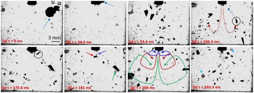
CAV2021
                                    11th International Symposium on Cavitation
                                          May 10-13, 2021, Daejon, Korea

     In the present work, experiments were conducted, to study the cavitation erosion mechanism of free-
floating primary Al3Zr intermetallic in de-ionised water upon exposure to high power ultrasound for
different treatment durations. The micro-fragmentation behaviour of the crystal was elucidated by means
of high-speed imaging and post-mortem SEM observations. The results revealed that ultrasonic treatment
of floating crystals lead to severe erosion and breakdown into small size fragments that can act as
favourable sites facilitating heterogeneous nucleation and promoting microstructural refinement in real
alloy melts.

2. Materials and Methods
     Primary Al3Zr crystals were initially extracted from an Al-3 wt% Zr alloy using the process described
in Priyadarshi et al. [10]. Single crystals with dimensions in the range of 4.5 ± 1 mm × 3.3 ± 1 mm × 0.06 ±
0.01 mm were then exposed to ultrasonic irradiation using a transducer (UP200S, Hielscher Ultrasonics)
operating at a frequency of 24 kHz in a transparent vessel of size 25 mm × 10 mm × 45 mm (L×W×H)
containing 4 ml of de-ionised water. The ultrasound was supplied using a titanium built sonotrode (Ø = 3
mm) submerged 10 mm below the liquid surface under ambient conditions. The in-situ fragmentation of
an extracted crystal as mentioned above was captured in real-time using a high-speed camera (Photron SA-
Z Fast Cam) in-combination with a Navitar 12x adapter lens providing a focussed view of intermetallic
disintegration at a working distance of 165 mm. The imaging was carried out at an optimum frame rate of
3000 frames per second (fps) to record the whole sequence of events at resolution of 640× 512 pixels, under
a visible light background illumination supplied by a GS Vitec Multi LED flash lamp. Figure 1 shows the
high speed imaging setup used for capturing the crystal breakdown under the ultrasonic horn. The
acoustically induced crystal break-up sequence was repeated with at least five such separate single crystals
to check the consistency of results.
     Further, post-mortem microscopic analysis of the fragmented crystals obtained after ultrasonic
treatment for 3 s, 6 s, and 9 s was conducted using SEM. After each treatment duration, the fragmented
crystals were separated from the sonicated liquid using a 2.5 μm filter paper. The crystal fragments were
then dried in ambient air and preserved for SEM examination.

Figure 1. Schematic representation of an in-situ high-speed imaging experimental setup.

* Corresponding Author: Abhinav Priyadarshi, abhinav.priyadarshi-2018@brookes.ac.uk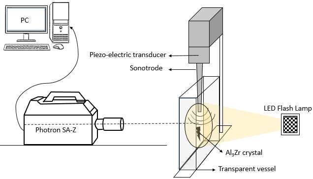
CAV2021
11th International Symposium on Cavitation
May 10-13, 2021, Daejon, Korea
3. Results and discussion
3.1. Observation of crystal fragmentation
Figure 2 shows the fragmentation sequence of a free-floating crystal obtained using in-situ imaging. Figure
2a shows a free-floating single intermetallic crystal with dimension as given in section 2 at t = 0 ms. The
introduction of ultrasonic waves into the liquid drives the crystal towards the vibrating source as shown
in Figure 2b due to developed recirculating acoustic flow [12]. As soon as the crystal enters the cavitation
zone, it seemingly instantaneously breaks into several small pieces and the fragments are pushed away
into the bulk liquid by acoustic streaming (Figure 2c). Acoustic streaming then causes the fragments to
recirculate along the marked route as illustrated in Figure 2d. For instance, the fragmented crystal encircled
in the black dotted circle can be seen moving back into the intense cavitation zone near the sonotrode in
the next frame (Figure 2e). Some of the broken crystals subsequently re-fragment when coming in close
proximity of the vibrating probe, where the emitted shock waves are strongest thus causing the fragments
to recirculate in a repetitive pattern. The recirculating path varies depending on the momentum acquired
by the fragmented crystals (marked in Figure 2f) from the ultrasonic source as shown in Figure 2g. This
process then keeps on repeating and the crystal continues to disintegrate via layer-by layer erosion and
fragmentation as will be discussed in section 3.2. It is evident from these observations that acoustic
streaming is the driver inducing the localised flow causing the crystal that to feed back into the cavitation
zone expediting the treatment process. In a real Al melt environment, assuming the acoustic streaming
patterns resemble those of the water experiment [6], it is expected that breakdown of these floating primary
crystals will be much faster owing to the strong dynamic behaviour of cavitation environment, about 4
times more intense than in water [13], and the aggressiveness of the cavitating field [14,15]. On the other
hand, the higher density of the liquid Al will prevent rapid sedimentation of intermetallics and may further
facilitate the fragmentation. However, this reasoning is based on preliminary results and is the subject of
further research. It has been shown that these crystal fragments will then act as a heterogeneous nucleating
sites for Al dendrites ultimately inducing grain refinement in the solidified metallic alloy as will be
discussed in the next section [16].
Figure 2. Series of in-situ high-speed images (3000 fps) of ultrasonically induced free-floating crystal breakdown.
3.2. SEM examination of disintegrated primary crystals
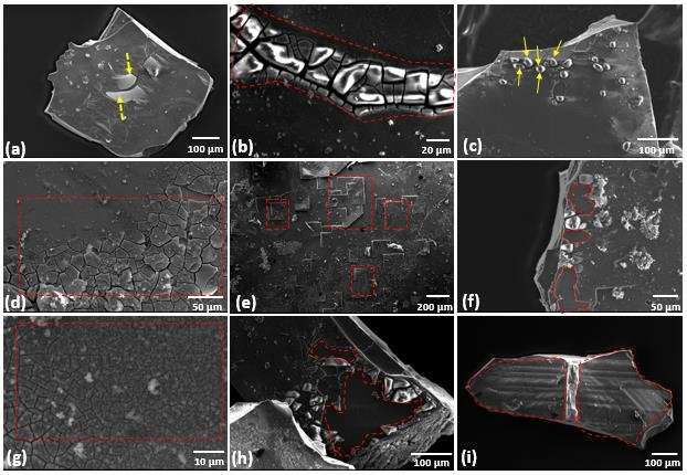
Figure 3 shows the morphology of the fragmented intermetallic particles after exposure to the ultrasonic
cavitating environment for 3 s, 6 s and 9 s. As shown by our previous research, fragmentation is a result of
multiple interactions of shock waves [10] and liquid jets [17] caused by the collapsing bubbles. After 3 s of
* Corresponding Author: Abhinav Priyadarshi, abhinav.priyadarshi-2018@brookes.ac.ukCAV2021
11th International Symposium on Cavitation
May 10-13, 2021, Daejon, Korea
treatment, the crystal surface starts to delaminate along the formed crack network structure with crack
sizes ranging from 10 to 100 μm as shown in Figure 3a (marked with arrows). It is interesting to note that
these fragmented particles show the formation of micron sized crack network along the edges of pre-
existing micro crack present on the uppermost layer of the crystal as shown in Figure 3b. Additionally, the
fragmented crystals also exhibit various small round shaped blisters projecting out of the surface as evident
in Figure 3c. After 6 s, the fragmented crystal undergoes severe cavitation erosion of its upper layer with
relatively finer crack network spreading across the whole surface resembling the profile of cracked dried
land (Figure 3d). In addition, delamination of the upper layer at specific regions along the surface can be
clearly seen in Figure 3e. Furthermore, there is decrease in the density of formed upper layer projections as
the majority erode away revealing a crater underneath (Figure 3f). With further increase in the ultrasound
exposure i.e. to 9 sec, the crack network becomes even more dense and finer with grid sizes going down to
1-10 μm (Figure 3g). There are almost no visible projections with longer treatment times since the majority
must have eroded from the upper layer exposing a large part of the layer beneath (Figure 3h). The top layer
is then completely chipped off exposing the layer underneath for further breakdown and disintegration
(Figure 3i). As observed from Figure 3, it is obvious that fragmentation of crystal is expedited as exposure
time increases. Longer treatment times cause the intermetallic crystal to disintegrate into numerous small
fragments in the range of a few microns ideal for promotingheterogeneous nucleation substrates in a real
melt [6].
Figure 3. SEM images of fragmented crystal after exposure to ultrasonic cavitation for 3 s (a,b,c), 6 s (d,e,f), and 9 s
(g,h,i).
4. Conclusions
Cavitation erosion behaviour of a freely floating single Al 3Zr crystal was qualitatively discussed based on
the microstructural examination of the fragmented crystals exposed to different treatment times and in-
situ imaging of the entire crystal disintegration dynamics. Exposure to ultrasound first leads to generation
of a micron sized hierarchal crack network followed by delamination and fracture in a layer-by-layer
manner. Chipped off and protruding fragments are of the size preferable for heterogeneous nucleation in
real melts.
* Corresponding Author: Abhinav Priyadarshi, abhinav.priyadarshi-2018@brookes.ac.ukCAV2021
11th International Symposium on Cavitation
May 10-13, 2021, Daejon, Korea
Acknowledgments: This research work has been funded by the UK Engineering and Physical Sciences Research
Council (EPSRC) under the project UltraMelt2 (grant EP/R011001/1, EP/R011095/1 and EP/R011044/1).
References
1. Ramirez A, Qian M, Davis B, Wilks T, StJohn DH. Potency of high-intensity ultrasonic treatment for grain
refinement of magnesium alloys. Scr Mater. 2008, 59(1),19–22.
2. Stjohn DH, Qian M, Easton MA, Cao P. The Interdependence Theory: The relationship between grain
formation and nucleant selection. Acta Mater. 2011,59(12),4907–21.
3. Eskin GI, Eskin DG. Production of natural and synthesized aluminum-based composite materials with the aid
of ultrasonic (cavitation) treatment of the melt. Ultrason Sonochem. 2003,10(4–5),297–301.
4. Taghavi F, Saghafian H, Kharrazi YHK. Study on the ability of mechanical vibration for the production of
thixotropic microstructure in A356 aluminum alloy. Mater Des. 2009,30(1),115–21.
5. Metan V, Eigenfeld K, Räbiger D, Leonhardt M, Eckert S. Grain size control in Al-Si alloys by grain refinement
and electromagnetic stirring. J Alloys Compd. 2009,487(1–2),163–72.
6. Eskin DG, Tzanakis I, Wang F, Lebon GSB, Subroto T, Pericleous K, et al. Fundamental studies of ultrasonic
melt processing. Ultrason Sonochem. 2019,52,455–67.
7. Qian M, Ramirez A, Das A, Stjohn DH. The effect of solute on ultrasonic grain refinement of magnesium
alloys. J Cryst Growth. 2010,312(15),2267–72.
8. Tzanakis I, Xu WW, Eskin DG, Lee PD, Kotsovinos N. In situ observation and analysis of ultrasonic capillary
effect in molten aluminium. Ultrason Sonochem. 2015,27,72–80.
9. Wang F, Eskin D, Mi J, Wang C, Koe B, King A, et al. A synchrotron X-radiography study of the fragmentation
and refinement of primary intermetallic particles in an Al-35 Cu alloy induced by ultrasonic melt processing.,
Acta Materialia. 2017, Vol. 141,142–53.
10. Priyadarshi A, Khavari M, Subroto T, Conte M, Prentice P, Pericleous K, et al. On the governing fragmentation
mechanism of primary intermetallics by induced cavitation. Ultrason Sonochem. 2021,70,105260-75.
11. Swallowe GM, Field JE, Rees CS, Duckworth A. A photographic study of the effect of ultrasound on
solidification. Acta Metall. 1989,37(3),961–7.
12. Lebon GSB, Tzanakis I, Pericleous K, Eskin D, Grant PS. Ultrasonic liquid metal processing: The essential role
of cavitation bubbles in controlling acoustic streaming. Ultrason Sonochem. 2019,55,243–55.
13. Tzanakis I, Lebon GSB, Eskin DG, Pericleous K. Comparison of cavitation intensity in water and in molten
aluminium using a high-temperature cavitometer. J Phys Conf Ser. 2015,656(1).
14. Tzanakis I, Lebon GSB, Eskin DG, Pericleous KA. Characterisation of the ultrasonic acoustic spectrum and
pressure field in aluminium melt with an advanced cavitometer. J Mater Process Technol. 2016,229,582–6.
15. Lebon GSB, Tzanakis I, Pericleous K, Eskin D. Experimental and numerical investigation of acoustic pressures
in different liquids. Ultrasonics Sonochemistry. Vol. 42, 2018,411–21.
16. Atamanenko TV, Eskin DG, Zhang L, Katgerman L. Criteria of Grain Refinement Induced by Ultrasonic Melt
Treatment of Aluminum Alloys Containing Zr and Ti. Metall Mater Trans A [Internet]. 2010,41(8),2056–66.
17. Wang F, Tzanakis I, Eskin D, Mi J, Connolley T. In situ observation of ultrasonic cavitation-induced
fragmentation of the primary crystals formed in Al alloys. Ultrason Sonochem. 2017,39,66–76.
* Corresponding Author: Abhinav Priyadarshi, abhinav.priyadarshi-2018@brookes.ac.ukYou can also read