Comparison of Mean and Centroid of Surgically Induced Astigmatism After Standard Cataract Surgery - Frontiers
←
→
Page content transcription
If your browser does not render page correctly, please read the page content below
ORIGINAL RESEARCH
                                                                                                                                                published: 04 June 2021
                                                                                                                                        doi: 10.3389/fmed.2021.670337

                                              Comparison of Mean and Centroid of
                                              Surgically Induced Astigmatism After
                                              Standard Cataract Surgery
                                              Kazutaka Kamiya 1*, Kei Iijima 2 , Wakako Ando 2 and Nobuyuki Shoji 2
                                              1
                                               Visual Physiology, School of Allied Health Sciences, Kitasato University, Kanagawa, Japan, 2 Department of Ophthalmology,
                                              School of Medicine, Kitasato University, Kanagawa, Japan

                                              Purpose: To compare the arithmetic mean of surgically induced astigmatism (M-SIA) and
                                              the centroid of surgically induced astigmatism (C-SIA) after standard cataract surgery.
                                              Methods: We retrospectively examined 200 eyes of 100 consecutive patients
                                              undergoing bilateral cataract surgery through a 2.8 mm temporal clear corneal
                                              incision. We quantitatively measured the magnitude and axis of corneal astigmatism
                                              preoperatively and 3 months postoperatively using an automated keratometer
                                              (TONOREFF-II, Nidek). We assessed the M-SIA, the C-SIA, and the double angle plots
                                              for the display of the individual SIA distributions.

                           Edited by:
                                              Results: For bilateral data analysis, the magnitude of corneal astigmatism significantly
                  Ravirajsinh Jadeja,         increased from 0.66 ± 0.39 D preoperatively to 0.74 ± 0.46 D postoperatively (paired
     Augusta University, United States        t-test, p = 0.012). The M-SIA was 0.50 ± 0.36 D. On the other hand, the C-SIA was
                         Reviewed by:         0.18 ± 0.60 D at an axis of 97◦ . For unilateral analysis, we obtained similar outcomes
       Juan Alvarez De Toledo Elizalde,
Clínica Oftalmológica Barraquer, Spain        between the right and left eye groups.
             Francesco Maria D’Alterio,
                                              Conclusions: According to our experience, standard cataract surgery induces the
      Imperial College Healthcare NHS
                 Trust, United Kingdom        M-SIA by approximately 0.5 D. The magnitude of the C-SIA largely decreased to
                 *Correspondence:             approximately 40% of the M-SIA, and the direction of the C-SIA showed a tendency
                    Kazutaka Kamiya           toward with-the-rule astigmatism. It should be noted that the M-SIA was considerably
              kamiyak-tky@umin.ac.jp
                                              different from the C-SIA, especially when selecting the appropriate toric IOL model
                    Specialty section:        and power.
          This article was submitted to
                                              Keywords: surgically induced astigmatism, corneal astigmatism, mean, centroid, vector analysis, temporal
                        Ophthalmology,
                                              incision, clear corneal incision, cataract surgery
                a section of the journal
                   Frontiers in Medicine

         Received: 21 February 2021
            Accepted: 12 May 2021
                                              BACKGROUND
           Published: 04 June 2021
                                              Toric intraocular lens (IOL) implantation has been widely acknowledged as a safe and effective
                            Citation:         means for the treatment of cataract patients with corneal astigmatism (1–3). Although modern
      Kamiya K, Iijima K, Ando W and
                                              sophisticated cataract surgery does not largely induce astigmatism due to a 2.0 to 3.0 mm-
  Shoji N (2021) Comparison of Mean
   and Centroid of Surgically Induced
                                              incision size as well as the unnecessity for sutures to the wound, it is essential to accurately
 Astigmatism After Standard Cataract          predict surgically induced astigmatism (SIA) to maximize visual function and subsequent patient
      Surgery. Front. Med. 8:670337.          satisfaction, especially in toric IOL-implanted eyes. The arithmetic mean (M-SIA) calculation
     doi: 10.3389/fmed.2021.670337            is based on the magnitude of astigmatism, but does not include the direction of astigmatism.

Frontiers in Medicine | www.frontiersin.org                                         1                                             June 2021 | Volume 8 | Article 670337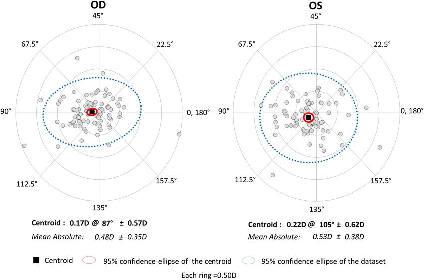
Kamiya et al.                                                                                                                  Mean and Centroid of SIA

By contrast, the centroid of SIA (C-SIA) is determined by both           TABLE 1 | Preoperative demographic data of the study population through a
the magnitude and the direction of astigmatism. Therefore, it            2.8 mm temporal clear corneal incision.

is reasonable that the use of the C-SIA is beneficial to grasp           Patient demographics
the overall SIA trends of cataract surgery, and thus the C-SIA,          Age (years)                          73.5 ± 8.1 years (range, 53 to 92 years)
instead of the M-SIA, should be theoretically applied in toric           Gender (M:F)                                  Male: 40, Female: 60
IOL calculation software and might be somewhat different from            Bilateral data
the M-SIA. To the best of our knowledge, a detailed comparison
                                                                         Flat keratometry (K1)               43.82 ± 1.44 D (range, 39.75 to 48.00 D)
between the M-SIA and the C-SIA has not so far been conducted.
                                                                         Steep keratometry (K2)              44.49 ± 1.46 D (range, 40.50 to 48.25 D)
Moreover, the bilateral differences in these two SIAs between
                                                                         Mean keratometric readings (D)      44.16 ± 1.44 D (range, 40.13 to 48.13 D)
the right and left eyes have not been fully understood. It may
                                                                         Corneal astigmatism (D)               0.66 ± 0.39 D (range, 0.00 to 2.00 D)
give us intrinsic insights into the current understanding of
                                                                         Axial length (mm)                 23.76 ± 1.12 mm (range, 21.93 to 26.86 mm)
astigmatic correction, especially when using a toric IOL model
                                                                         Central corneal thickness (µm)      548.6 ± 31.3 µm (range, 470 to 616 µm)
in daily practice.
                                                                         LogMAR UCVA                            0.72 ± 0.42 (range, −0.08 to 2.00)
   The purpose of the present study is 2-fold; to retrospectively
                                                                         LogMAR BSCVA                           0.26 ± 0.34 (range, −0.08 to 2.00)
compare the M-SIA and the C-SIA, and to assess the bilateral
                                                                         Manifest sphere                    −0.51 ± 3.10 D (range, −10.00 to 5.50 D)
differences in the two SIAs between the right and left eyes, after
                                                                         Manifest cylinder                     0.89 ± 0.82 D (range, 0.00 to 3.50 D)
standard cataract surgery.
                                                                         Unilateral data (right eye)
                                                                         Flat keratometry (K1)               43.83 ± 1.42 D (range, 40.00 to 47.50 D)
                                                                         Steep keratometry (K2)              44.50 ± 1.45 D (range, 40.50 to 47.75 D)
MATERIALS AND METHODS
                                                                         Mean keratometric readings (D)      44.16 ± 1.42 D (range, 40.25 to 47.63 D)
Study Population                                                         Corneal astigmatism (D)               0.67 ± 0.41 D (range, 0.00 to 2.00 D)
The study protocol was registered with the University                    Axial length (mm)                 23.77 ± 1.10 mm (range, 21.93 to 26.71 mm)
Hospital Medical Information Network Clinical Trial Registry             Central corneal thickness (µm)      547.9 ± 31.2 µm (range, 471 to 612 µm)
(000043349). A total of 200 eyes of 100 consecutive patients             LogMAR UCVA                             0.74 ± 0.44 (range, 0.05 to 2.00)
(mean age ± standard deviation, 73.5 ± 8.1 years), who                   LogMAR BSCVA                           0.27 ± 0.37 (range, −0.08 to 2.00)
underwent bilateral standard phacoemulsification with IOL                Manifest sphere                    −0.56 ± 3.16 D (range, −10.00 to 5.25 D)
implantation through a 2.8 mm temporal clear corneal incision,           Manifest cylinder                     0.88 ± 0.77 D (range, 0.00 to 3.00 D)
and who completed a 3 month follow-up at Kitasato University             Unilateral data (left eye)
Hospital, were enrolled in the current study. This retrospective         Flat keratometry (K1)               43.82 ± 1.47 D (range, 39.75 to 48.00 D)
review of the clinical charts was approved by the Institutional          Steep keratometry (K2)              44.48 ± 1.48 D (range, 40.50 to 48.25 D)
Review Board at Kitasato University Hospital (B20-302) and               Mean keratometric readings (D)      44.15 ± 1.46 D (range, 40.13 to 48.13 D)
followed the tenets of the Declaration of Helsinki. Institutional        Corneal astigmatism (D)               0.66 ± 0.37 D (range, 0.00 to 1.50 D)
review board approval was not required for this review.                  Axial length (mm)                 23.75 ± 1.14 mm (range, 21.94 to 26.86 mm)
                                                                         Central corneal thickness (µm)      549.3 ± 31.6 µm (range, 470 to 616 µm)

Inclusion and Exclusion Criteria                                         LogMAR UCVA                            0.71 ± 0.40 (range, −0.08 to 1.70)

Inclusion criteria were 20 ≤ age < 95 years, eyes undergoing             LogMAR BSCVA                           0.25 ± 0.31 (range, −0.08 to 1.70)
bilateral cataract surgery by one experienced surgeon, and no            Manifest sphere                     −0.46 ± 3.06 D (range, −9.50 to 5.50 D)
history of any trauma or ocular surgery. Exclusion criteria were         Manifest cylinder                     0.90 ± 0.88 D (range, 0.00 to 3.50 D)
eyes with concomitant corneal diseases, such as keratoconus,
pellucid marginal degeneration, irregular corneal astigmatism, or
severe dry eye, eyes developing intraoperative or postoperative
complications, eyes requiring a wound enlargement, or eyes
                                                                         Assessment of Corneal Astigmatism and
requiring some sutures to the wound. Written informed consent
for cataract surgery was obtained from all patients.                     Surgically Induced Astigmatism
                                                                         Preoperatively and 3 months postoperatively, we quantitatively
                                                                         assessed the magnitude and the axis of corneal astigmatism
Cataract Surgical Procedures                                             using an automated keratometer (TONOREFF-II, Nidek, Aichi,
Standard phacoemulsification was performed by capsulorhexis,             Japan). We measured this at least 5 times immediately after
nuclear and cortex extraction, and a non-toric IOL (KS-1, STAAR          blinking according to the manufacturer’s instructions and used
Japan, Chiba, Japan) implantation in the capsular bag through            the average value for statistical analysis. We assessed the M-
a 2.8 mm temporal clear corneal single-plane incision (4). All           SIA, the C-SIA by vector analysis, and the double angle
surgeries were performed by one experienced surgeon using the            plots for the display of the individual SIA distributions (5),
same technique. Postoperatively, steroidal (0.1% betamethasone),         by using the astigmatism double angle plot tool available
antibiotic (levofloxacin), diclofenac sodium medications were            on the American Society of Cataract and Refractive Surgery
topically administered 4 times daily for 2 weeks, with the dose          website (https://ascrs.org/tools/astigmatism-double-angle-plot-
being steadily reduced thereafter.                                       tool) (6).

Frontiers in Medicine | www.frontiersin.org                          2                                            June 2021 | Volume 8 | Article 670337
Kamiya et al. Mean and Centroid of SIA TABLE 2 | Visual and refractive outcomes of the study population undergoing standard cataract surgery through a 2.8 mm temporal clear corneal incision. Demographics Preoperative Postoperative P-value Bilateral Data LogMAR UCVA 0.72 ± 0.42 (range, −0.08 to 2.00) 0.23 ± 0.34 (range, −0.30 to 1.00)
Kamiya et al.                                                                                                                             Mean and Centroid of SIA

 FIGURE 1 | Graph showing the magnitude of corneal astigmatism preoperatively and 3 months postoperatively using bilateral and unilateral data. The bar represents
 standard deviation. D, diopters. *Indicates statistical significance.

   We assume that both SIAs were small in amount,                                    SIA trends of cataract surgery, and thus that the C-SIA,
but not necessarily negligible, since modern cataract                                instead of the M-SIA, should be applied for entering the
surgery is deemed as one of the refractive surgeries that                            SIA data in the manufacturer’s toric IOL software during
aims to correct both spherical and cylindrical errors as                             preoperative planning.
much as possible. We believe that this information may                                  There are several limitations to this study. Firstly, we
be simple, but clinically helpful, not only for cataract                             employed the autokeratometer for the assessment of corneal
surgeons but also for IOL manufacturers, in order                                    astigmatism, since it is still most widely utilized for astigmatic
to further improve the astigmatic outcomes of toric                                  evaluation in daily practice. Accordingly, we did not evaluate
IOL implantation.                                                                    posterior corneal astigmatism in this study. Although the amount
   Standard cataract surgery through a 3.0 mm temporal corneal                       of posterior corneal astigmatism is far smaller than that of
incision has been reported to induce corneal astigmatism                             anterior corneal astigmatism, total corneal astigmatism using
by ∼0.5 D with a WTR shift in astigmatism (7–11). We                                 a corneal tomographer would be beneficial for understanding
also reported that phakic IOL implantation through the                               the precise SIA of cataract surgery (13, 14). Secondly, we only
same 3.0 mm temporal incision induced similar astigmatism                            examined the patients undergoing cataract surgery through
in magnitude and direction (12). These previous findings of                          a 2.8 mm clear corneal incision. We await further studies
the SIA were in good accordance with our current results                             on the SIA using different incision sizes as well as different
of the M-SIA, but the C-SIA has not been meticulously                                incisional techniques such as corneoscleral incision. Thirdly,
investigated in their studies. We are of the opinion that                            we did not assess the effect of other contributing factors,
the C-SIA is theoretically beneficial to grasp the overall                           such as corneal diameter, the type of the incision, and the

Frontiers in Medicine | www.frontiersin.org                                      4                                            June 2021 | Volume 8 | Article 670337
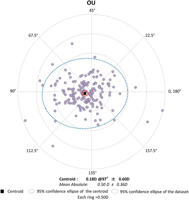
Kamiya et al.                                                                                                                        Mean and Centroid of SIA

 FIGURE 2 | Graph showing the double angle plots of the individual surgically induced astigmatism using bilateral data.

intrastromal length of the incision on these SIAs in this study                       indicating that the corneal diameter should always be measured
population. Theodoulidou et al. reported that a change >0.5                           preoperatively when planning cataract surgery and accounted
D of corneal astigmatism at 1 and 6 months postoperatively                            for in cases of large and small corneas (15). On the other
was significantly lower in eyes with a corneal diameter of                            hand, Zhang et al. demonstrated that the horizontal corneal
12.0 to 12.2 mm and ≥12.3 mm in comparison with eyes                                  diameter had minimal effects on the SIA in uncomplicated
with a corneal diameter of ≤11.6 mm and 11.7 to 11.9 mm,                              small-incisional cataract surgery through a 2.2 mm clear corneal

Frontiers in Medicine | www.frontiersin.org                                       5                                       June 2021 | Volume 8 | Article 670337
Kamiya et al.                                                                                                                         Mean and Centroid of SIA

 FIGURE 3 | Graph showing the double angle plots of the individual surgically induced astigmatism using unilateral data.

incision, indicating that the corneal diameter does not play a                        DATA AVAILABILITY STATEMENT
vital role in eyes undergoing small-incisional cataract surgery
(16). Fourthly, we did not actually evaluate the astigmatic                           The original contributions presented in the study are included
outcomes of toric IOL implantation. We are currently conducting                       in the article/supplementary material, further inquiries can be
a new comparative study on the astigmatic correction in                               directed to the corresponding author/s.
toric IOL-implanted eyes by the use of the M-SIA and
the C-SIA.                                                                            ETHICS STATEMENT

CONCLUSIONS                                                                           The studies involving human participants were reviewed
                                                                                      and approved by the Institutional Review Board at Kitasato
In summary, our findings showed that standard cataract surgery                        University Hospital (B20-302). Written informed consent for
through a 2.8 mm temporal corneal incision induced the M-SIA                          participation was not required for this study in accordance with
by ∼0.5 D and that the magnitude of the C-SIA considerably                            the national legislation and the institutional requirements.
decreased to ∼40% of the M-SIA, with a WTR astigmatic shift
in direction. Based on the fact that the M-SIA was largely                            AUTHOR CONTRIBUTIONS
different from the C-SIA in magnitude, the C-SIA, instead of
the M-SIA, may be recommended to be applied, especially                               KK and NS were involved in the design and conducted the
when we select the appropriate toric IOL model and power.                             study. KI and WA were involved in the collection, management,
This information will be helpful not only for cataract surgeons                       analysis, and interpretation of data. KK, KI, WA, and NS
during preoperative planning, but also for IOL manufacturers,                         were involved in preparation, review, and final approval of the
in order to further improve the astigmatic outcomes of toric                          manuscript. All authors contributed to the article and approved
IOL implantation.                                                                     the submitted version.

Frontiers in Medicine | www.frontiersin.org                                       6                                        June 2021 | Volume 8 | Article 670337
Kamiya et al. Mean and Centroid of SIA
REFERENCES scleral tunnel self-sealing incisions. J Cataract Refract Surg. (1997) 23:347–53.
doi: 10.1016/S0886-3350(97)80178-2
1. Shimizu K, Misawa A, Suzuki Y. Toric intraocular lenses: correcting 11. Rainer G, Menapace R, Vass C, Annen D, Findl O, Schmetterer
astigmatism while controlling axis shift. J Cataract Refract Surg. (1994) K. Corneal shape changes after temporal and superolateral
20:523–6. doi: 10.1016/S0886-3350(13)80232-5 3.0 mm clear corneal incisions. J Cataract Refract Surg. (1999)
2. Holland E, Lane S, Horn JD, Ernest P, Arleo R, Miller KM, et al. The AcrySof 25:1121–6. doi: 10.1016/S0886-3350(99)00132-7
toric intraocular lens in subjects with cataracts and corneal astigmatism: a 12. Kamiya K, Shimizu K, Aizawa D, Igarashi A, Komatsu M. Surgically induced
randomized, subject-masked, parallel-group, 1-year study. Ophthalmology. astigmatism after posterior chamber phakic intraocular lens implantation. Br
(2010) 117:2104–11. doi: 10.1016/j.ophtha.2010.07.033 J Ophthalmol. (2009) 93:1648–51. doi: 10.1136/bjo.2009.160044
3. Miyake T, Kamiya K, Amano R, Iida Y, Tsunehiro S, Shimizu K, et al. Long- 13. Canovas C, Alarcon A, Rosén R, Kasthurirangan S, Ma JJK, Koch DD,
term clinical outcomes of toric intraocular lens implantation in cataract et al. New algorithm for toric intraocular lens power calculation considering
cases with preexisting astigmatism. J Cataract Refract Surg. (2014) 40:1654– the posterior corneal astigmatism. J Cataract Refract Surg. (2018) 44:168–
60. doi: 10.1016/j.jcrs.2014.01.044 74. doi: 10.1016/j.jcrs.2017.11.008
4. Kamiya K, Shimizu K, Ohmoto F, Amano R. Time course 14. Holladay JT, Pettit G. Improving toric intraocular lens calculations using total
of corneal biomechanical parameters after phacoemulsification surgically induced astigmatism for a 2.5 mm temporal incision. J Cataract
with intraocular lens implantation. Cornea. (2010) 29:1256– Refract Surg. (2019) 45:272–83. doi: 10.1016/j.jcrs.2018.09.028
60. doi: 10.1097/ICO.0b013e3181d9284b 15. Theodoulidou S, Asproudis I, Kalogeropoulos C, Athanasiadis A, Aspiotis
5. Holladay JT, Dudeja DR, Koch DD. Evaluating and reporting astigmatism M. Corneal diameter as a factor influencing corneal astigmatism after
for individual and aggregate data. J Cataract Refract Surg. (1998) 24:57– cataract surgery. Cornea. (2016) 35:132–6. doi: 10.1097/ICO.0000000000
65. doi: 10.1016/S0886-3350(98)80075-8 000668
6. Abulafia A, Koch DD, Holladay JT, Wang L, Hill WE. Editorial. 16. Zhang W, Pasricha ND, Kuo AN, Vann RR. Influence of corneal diameter
Pursuing perfection in IOL calculations IV: astigmatism analysis, SIA on surgically induced astigmatism in small-incision cataract surgery. Can J
and double angle plots. J Cataract Refractive Surg. (2018) 44:1169– Ophthalmol. (2019) 54:556–9. doi: 10.1016/j.jcjo.2018.12.013
74. doi: 10.1016/j.jcrs.2018.07.027
7. Vass C, Menapace R. Computerized statistical analysis of corneal topography Conflict of Interest: The authors declare that the research was conducted in the
for the evaluation of changes in corneal shape after surgery. Am J Ophthalmol. absence of any commercial or financial relationships that could be construed as a
(1994) 118:177–84. doi: 10.1016/S0002-9394(14)72897-1 potential conflict of interest.
8. Long DA, Monica ML. A prospective evaluation of corneal curvature changes
with 3.0- to 3.5-mm corneal tunnel phacoemulsification. Ophthalmology. Copyright © 2021 Kamiya, Iijima, Ando and Shoji. This is an open-access article
(1996) 103:226–32. doi: 10.1016/S0161-6420(96)30712-4 distributed under the terms of the Creative Commons Attribution License (CC BY).
9. Masket S, Tennen DG. Astigmatic stabilization of 3.0 mm temporal clear The use, distribution or reproduction in other forums is permitted, provided the
corneal cataract incisions. J Cataract Refract Surg. (1996) 22:1451–5. original author(s) and the copyright owner(s) are credited and that the original
doi: 10.1016/S0886-3350(96)80146-5 publication in this journal is cited, in accordance with accepted academic practice.
10. Oshima Y, Tsujikawa K, Oh A, Harino S. Comparative study of intraocular No use, distribution or reproduction is permitted which does not comply with these
lens implantation through 3.0 mm temporal clear corneal and superior terms.
Frontiers in Medicine | www.frontiersin.org 7 June 2021 | Volume 8 | Article 670337You can also read