Chandelier endoillumination in Vitreoretinal Surgery
←
→
Page content transcription
If your browser does not render page correctly, please read the page content below
cover story
   eyetube.net

              Chandelier
           Endoillumination
            in Vitreoretinal
                Surgery
                   This method of illumination improves visualization in challenging cases
                               and can be used in many different situations.

                                               By Yusuke Oshima, MD

   O
               n the heels of widespread adoption of wide-
               angle viewing systems and new light sources
               for small-gauge vitrectomy, a variety of chan-
               delier lighting systems have been developed
    to provide stationary, wide-angle and uniform endoil-
    lumination for obtaining adequate visualization of the
    retina during surgery. During the past several years,
    Synergetics, DORC, and Alcon Laboratories Inc. have
    manufactured a variety of chandelier lighting systems,
    including a single-fiber system available in 25-gauge
    (Figure 1) and 27-gauge (Figure 2) formats and a sepa-
    rated 2-fiber system in a 27-gauge (Figure 3) or 29-gauge
    model (Figure 4).1-4 In some models, the tip of the chan-
    delier light probes can be placed into the cannula, while
    others require a separate needle to create an additional
    sclerotomy for inserting the fiber tip into the vitreous
    cavity. Generally, chandelier endoillumination with           Figure 1. A 25-gauge cannula-guided single-fiber chandelier
    2 optic fibers,1 first described by Eckardt as the twin-      probe (Alcon Laboratories Inc.). It is simple to set and easy to
    light chandelier, is more useful than a single fiber system   self-retain transconjunctivally.
    for obtaining homogeneous and more widespread
    illumination. The 2-fiber system eliminates the need          Basic roles and Techniques for
    to reposition the fiber and minimizes the shadow seen         Chandelier Endoillumination
    with single-fiber chandelier endoillumination because           The basic advantages of using chandelier endoillumi-
    the illumination comes from 2 different directions.2-4,5      nation have been described in several articles.1-7 When

 68 Retina Today january/february 2013
cover story

A                               B

C                               D

                                                                   Figure 3. The 27-gauge twin-light chandelier fiber designed
Figure 2. A new 27-gauge single fiber chandelier illumina-         by Eckardt, which is compatible with a xenon light illumina-
tion system (27G-VIVID chandelier fiber) from Synergetics          tor (Bright Star, DORC).
(A). The tip of chandelier light probes can be placed into a
specially designed cannula (B). The distal end of the fiber        ual manipulation during surgery. In retinal detachment
can be connected with mercury vapor (C) or xenon (D) light         cases, I can perform scleral indentation and achieve more
sources. Sufficient endoillumination can be obtained for           controlled and smooth peripheral vitreous base shaving
wide-angle fundus observation.                                     without the need for an assistant (Figure 6). For membrane
                                                                   dissections in challenging cases, such as diabetic tractional
considering retinal phototoxicity, the working distance for        retinal detachment or proliferative vitreoretinopathy,
light irradiation is important, and holding the light probe        the freed hand is helpful for holding forceps to grasp the
as far away from the retina as possible increases safety.8 For     membranes for separation from the retina or for dissection
this reason, I use the chandelier fiber for most of my cases. In   using scissors or a cutter (Figure 7). For cases in which I use
simple cases such as macular surgery, I hold the chandelier        a self-retaining chandelier system, I prefer to set up the fiber
probe with 1 hand in a manner similar to which I would             superiorly—eg, a single fiber at 12 o’clock or dual fibers at
use a light pipe to control the illuminating direction during      2 and 10 o’clock—to make the instrument shadow appear
surgery (Figure 5). In addition to the safety advantage, the       anteriorly and not interfere with the working area view. Not
self-retaining nature of chandelier endoilluminators frees up      only is it easy to adjust the optic fiber tips from this angle,
my hand from holding a light probe, allowing true biman-           but illumination is optimized and glare from the tips of the
                                                                                                   instruments is minimized. The
 A                                               B                         C
                                                                                                   direction of illumination can
                                                                                                   be changed from the poste-
                                                                                                   rior pole to the periphery by
                                                                                                   changing the curvature of the
                                                                                                   chandelier fiber outside the
                                                                                                   orbit (Figure 8).

                                                                                                  Improving
                                                                                                  Anterior Chamber
                                                                                                  Visualization for
                                                                                                  Phaco-vitrectomy
Figure 4. A 29/30-gauge dual-chandelier fiber system (Synergetics). The cannulas are helpful      Case 1: Corneal Opacity
for fiber positioning and to avoid missing the small wounds in cases of inadvertent removal of      A 71-year-old woman with
the fiber during surgery (A). Chandelier endoillumination with two separated ultra–small-gauge    cornea opacity and dense
fibers provides homogeneous and more widespread illumination than a single fiber. It can be       cataract had a total retinal
connected with the xenon light sources featured in the new generation machines (Constellation     detachment in her only see-
Vision System [Alcon Laboratories, Inc.; B], Stellaris PC [Bausch + Lomb; C]).                    ing right eye (Figure 9A, 9B).

                                                                                         january/february 2013 Retina Today 69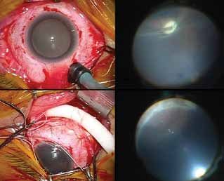
cover story
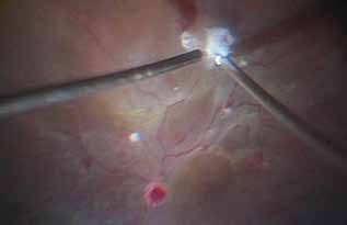
Figure 5. In simple cases, the cannula-guided chandelier Figure 6. Self-retaining chandelier endoillumination can free
probe can be held in a manner similar to a light pipe and 1 hand for scleral indentation, and thereby surgeons will
used to control the illuminating direction during surgery. achieve more controlled and smooth peripheral vitreous base
shaving by themselves without the need for an assistant.
Although crystalline lens removal
eyetube.net
is preferable to improve the fundus
visualization for safer vitrectomy,
capsulorrhexis and phacoemulsifica-
tion through a hazy cornea are very
challenging under the conventional eyetube.net/?v=Swalo
microscopic illumination because of
poor visibility of the anterior capsule and crystalline lens.
To overcome the difficulty to perform phaco in eyes with
corneal haze, retroillumination generated by a chandelier
lighting system inserted transconjunctivally into the vitreous
cavity is a helpful illumination technique for clearly visualiz-
ing the crystalline lens for safer phacoemulsification surgery
(Figure 9C, 9D).9 Once the lens is removed, vitrectomy can Figure 7. In more challenging cases, the freed hand is help-
be performed sequentially with the chandelier illumination ful for holding forceps to grasp membranes for separation
as is (scan QR code for video). from the retina or for dissection using scissors or a cutter in a
bimanual procedure. Setting the chandelier fiber superiorly
Case 2: Dense Vitreous Hemorrhage is helpful, making the shadow go down to the inferior area,
eyetube.net
A 61-year-old man with a cataract which can help in avoiding the instrument shadow coming
had dense vitreous hemorrhage into the working zone.
and suspicion of traction retinal
detachment due to proliferative 10).10 Once the lens is safely removed, vitrectomy can be
diabetic retinopathy in the right eye. eyetube.net/?v=Hiril carried out sequentially under wide-angle fundus viewing
A phaco-vitrectomy is preferable in with chandelier illumination as is (scan QR code for video).
this patient. However, phacoemulsification surgery may be
somewhat challenging because severe vitreous hemorrhage Scleral buckling under wide-angle fundus
often obscures the red reflex from the fundus and interferes viewing with chandelier illumination
with clear visualization of the crystalline lens structure and Scleral buckling is a widely prevalent treatment option
capsule during cataract surgery. Similar to Case 1, the use of for primary rhegmatogenous retinal detachment, and it has
a chandelier lighting system to generate retroillumination in usually been carried out with the use of binocular ophthal-
this case can improve visualization of the cataractous lens moscopy via the aid of a condensing lens. Although most
and its capsule, thereby facilitating safer cataract surgery in buckling procedures are performed sequentially under sur-
selected patients with dense vitreous hemorrhage (Figure gical microscopic viewing, repeated wearing and removal of
70 Retina Today january/february 2013
cover story
A B the binocular ophthalmoscope for fundus
examination is a routine procedure dur-
ing surgery. The recent widespread use
of chandelier illumination in conjunction
with a wide-angle viewing system offers
wide, excellent visibility of the fundus to
achieve safer surgical manipulation during
pars plana vitrectomy. The whole surgical
procedure can be sequentially performed
with viewing through the surgical micro-
scope without the burden of repeated
Figure 8. The direction of the illumination focusing on the posterior pole (A) or wearing and removal of the binocular
periphery (B) can be optimized easily by changing the curvature of the chande- ophthalmoscope usually needed during
lier fiber outside the eyeball with this flexible type of chandelier fiber. scleral buckling. In addition, adjusting the
viewing focus and magnification under
the surgical microscope may be more helpful to identify
A B preoperatively unrecognized tears during surgery. To enjoy
the advantages seen in vitrectomy, scleral buckling can also
be carried out under wide-angle fundus viewing with chan-
A B
C D
C D
E F
E F
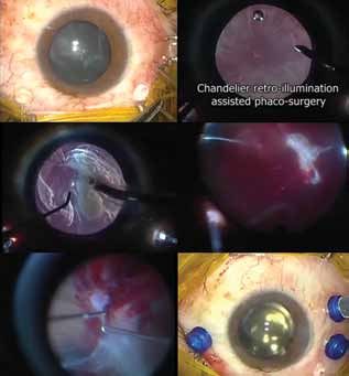
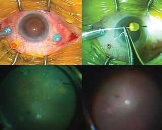
Figure 9. A 71-year-old woman had a cornea opacity and
dense cataract obscuring fundus visibility (A). B-scan echog-
raphy suggested a retinal detachment occurred in her only
seeing eye (B). Intraoperative views of chandelier retroillumi-
nation-assisted phacoemulsification surgery (C, D). The self- Figure 10. A 61-year-old man with a cataract had dense vit-
retaining chandelier stays in the inferotemporal pars plana reous hemorrhage obscuring the red reflex from the fundus.
region (C). Retroillumination by chandelier endoillumination A 27-gauge twin-light chandelier fiber was set followed by
from the posterior side offers sufficient lighting to view starting phaco-vitrectomy (A). Retroillumination from the
the crystalline lens clearly without obstruction by the hazy chandelier lighting clearly illustrated the anterior capsule to
cornea. Phacoemusfication surgery was performed with a perform capsulorrhexis (B) and phacoemulscification with
bimanual chopping technique as usual (D). Pars plana vitrec- a bimanual chopping technique (C). Vitrectomy to remove
tomy was sequentially performed to treat a myopic macular the dense hemorrhage was carried out sequentially after
hole-induced retinal detachment (E). Postoperative fundus uneventful lens removal (D). Fibrovascular membrane dis-
photography through a hazy cornea (F). Retina was attached section was performed bimanually under chandelier endoil-
with visual acuity improvement from counting fingers to lumination (E). Finally, an intraocular lens was implanted to
20/200 after surgery. conclude microinsicion phaco-vitrectomy (F).
january/february 2013 Retina Today 71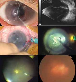
cover story
delier illumination (Figure 11; scan QR my opinion, careful disinfection of the ocular surface by
eyetube.net
code for video).11,12 The quality and repeated irrigation with diluted povidone-iodine and the
angle of view of the fundus through a use of a cannula-compatible smaller gauge fiber would be
surgical microscope with chandelier preferable in this scenario.
endoillumination is at least equal to
or much better than that observed eyetube.net/?v=retej Summary
through the conventional binocular The utility and efficacy of chandelier endoillumination in
ophthalmoscope via the condensing lens. The theoretical a variety of situations during vitreoretinal surgery has been
concerns of the current procedure may include bacterial described herein based on personal experiences and pref-
inoculation into the vitreous cavity during transconjunc- erences. It is clear, however, that there are many different
tival insertion of the chandelier fiber tip and vitreous surgical situations in which chandelier endoillumination is
incarceration to the sclerotomy after the fiber removal. In beneficial for improving intraocular visibility and thereby
achieving favorable surgical outcomes. Nevertheless, sur-
A B geons must still bear in mind that the final goal of illumi-
nation is to enhance the efficiency of surgery while main-
taining safety. Similar to the introduction of xenon and
mercury vapor bulbs in our field, new light-emitting diode
light sources (Figure 12) have recently been developed with
unique potential. The evolution of next-generation chan-
delier illumination systems continues and looks promising
C D for the future. n
Yusuke Oshima, MD is an Associate Professor
of Ophthalmology at the Osaka University
Graduate School of Medicine in Suita, Japan,
and an Honorary Director of the Vitreoretinal
Division at the Tianjin Eye Hospital, Tianjin,
China. He is a member of the Retina Today Editorial
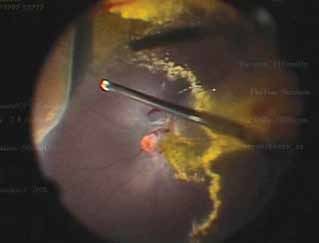
Figure 11. A 27-year-old man underwent scleral buckling Board. Dr. Oshima is a consultant to Topcon Medical
to treat primary rhegmatogenous retinal detachment. A Laser Systems and Synergetics. He has received lecture fees
25-gauge Awh chandelier fiber (Synergetics) was settled and/or travel support from Alcon Laboratories, Bausch
in the pars plana region opposite the region with retinal and Lomb, Carl Zeiss Meditec, DORC International,
tears (A). Cryoretinopexy was carried out under chandelier Novartis Pharmaceuitical Inc., and Synergetics, when he
endo-illumination observed through a wide-angle viewing spoke at sponsored seminars, but he received no propri-
system (B). After suturing the scleral buckle (C), the position etary interests or royalties from any companies in relation
of scleral indentation with the buckle was again examined to any products mentioned in this article. Dr. Oshima may
through the wide-angle viewing system under chandelier be reached at yusukeoshima@gmail.com.
endoillumination (D).
1. Eckardt C. Twin lights: a new chandelier illumination for bimanual surgery. Retina. 2003;23:893-894.
2. Oshima Y, Awh CC, Tano Y. Self-retaining 27-gauge transconjunctival chandelier endoillumination for panoramic
viewing during vitreous surgery. Am J Ophthalmol. 2007;143:166-167.
3. Eckardt C, Eckert T, Eckardt U. 27-gauge Twinlight chandelier illumination system for bimanual transconjunctival
vitrectomy. Retina. 2008;28:518-519.
4. Sakaguchi H, Oshima Y, Nishida K, Awh CC. A 29/30-gauge dual-chandelier illumination system for panoramic
viewing during microincision vitrectomy surgery. Retina. 2011;31:231-1233.
5. Chow DR. Tips on improving your use of endoillumination. Retinal Physician. 2011:8:43-46.
6. Sakaguchi H, Oshima Y. Considering the illumination choices in vitreoretinal surgery: continual improvements
allow for better, safer outcomes. Retinal Physician. 2012;3:20-25.
7. Witmer MT, Chan P. Chandelier lighting during vitreoretinal surgery. Retina Today. 2012:7:35-37.
8. Charles S. Illumination and phototoxicity issues in vitreoretinal surgery. Retina. 2008;28:1-4.
9. Oshima Y, Shima C, Maeda N, Tano Y. Chandelier retroillumination-assisted torsional oscillation for cataract
surgery in patients with severe corneal opacity. J Cataract Refract Surg. 2007:33;2018-2022.
10. Jang SY, Choi KS, Lee SJ. Chandelier retroillumination-assisted cataract extraction in eyes with vitreous hemor-
rhage. Arch Ophthalmol. 2010;128:911-4.
Figure 12. A light-emitting diode light source (DORC) devel- 11. Venkatesh P, Garg S. Endoillumination-assisted scleral buckling: a new approach to retinal detachment repair.
Retinal Physician. 2012;9:34-37.
oped for illumination during vitrectomy, which is a new plat- 12. Aras C, Ucar D, Koytak A, Yetik H. Scleral buckling with a non-contact wide-angle viewing system. Ophthalmo-
form of illuminator for safer endoillumination. logica. 2012; 227:107-110.
72 Retina Today january/february 2013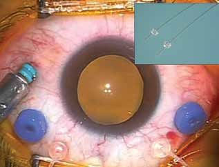
You can also read