Simple partial status epilepticus presenting with jargon aphasia and focal hyperperfusion demonstrated by ictal pulsed arterial spin labeling MRI ...
←
→
Page content transcription
If your browser does not render page correctly, please read the page content below
Neurology Asia 2018; 23(1) : 77 – 83

Simple partial status epilepticus presenting with jargon
aphasia and focal hyperperfusion demonstrated by
ictal pulsed arterial spin labeling MRI
1,2
   Hana Maizuliana MBBS MMed, 1Hitoshi Ikeda MD, 1Toshio Hiyoshi MD, 1Takuji Nishida MD,
1
  Kazumi Matsuda MD, 1Inoue Yushi MD PhD
1
    National Epilepsy Center, Shizuoka Institute Epilepsy and Neurological Disorders, Shizuoka, Japan.
2
    Universiti Sains Islam Malaysia, Kuala Lumpur, Malaysia

Abstract

We report a case of 74-year-old lady, presented with recurrent jargon aphasia as simple partial
status epilepticus (SPSE) which lasted for a few days to a few weeks, following a brain abscess
removal from the left temporo-parieto-occipital region at the age of 71 years. The ictal activity on
electroencephalogram was documented at left posterior quadrant, where marked hyperperfusion was
clearly visualized by perfusion image acquired with magnetic resonance imaging (MRI) using pulsed
arterial spin-labeling (PASL). Jargon aphasia as a primary feature of simple partial status epilepticus
is so uncommon that only few cases have been reported. Furthermore, this report suggests that MRI
using PASL is a promising method not only to localize the seizure foci but also to follow up the
corresponding regional cerebral blood flow changes noninvasively.

Keywords: Jargon aphasia; non-convulsive seizure; simple partial status epilepticus, pulsed arterial
spin-labeling-MRI

INTRODUCTION                                          left posterior quadrant of the brain, resulting in the
                                                      right homonymous superior quadrantanopia. She
The commonest cause of acute onset aphasia is
                                                      could not speak properly, walk or carry out daily
vascular insult, however when the symptoms
                                                      activities during the episodes. Her husband noted
of aphasia occurs episodically or fluctuates, it
                                                      that the patient just suddenly had a speech arrest
may be suggestive of seizure. Jargon aphasia
                                                      and later she uttered meaningless simple syllables
as the primary manifestation of focal epilepsy
                                                      and kept repeating it. These episodes lasted
is uncommon, more so presenting as status
                                                      for several days to a week and spontaneously
epilepticus. It has been accepted that interictal
                                                      ceased. She had been treated with valproate
regional hypoperfusion visualized by pulsed
                                                      and levetiracetam, which failed to prevent the
arterial spin-labeling (PASL)-MRI has diagnostic
                                                      recurrence.
value in localization of focus in epilepsy. Ictal
                                                          She was first seen at our hospital when she
regional hyperperfusion on PASL-MRI should
                                                      was 73 years old and asymptomatic. The inter-
have more localizing value. However, there still
                                                      ictal EEG showed slow waves of delta range at
has been only a few reports of ictal PASL-MRI.
                                                      left temporal, occipital and parietal regions (T3-
We report an elderly lady who presented with
                                                      P3-O1). MRI revealed post-operative changes
jargon aphasia as simple partial status epilepticus
                                                      such as enlarged dorsal horn of left lateral ventricle
(SPSE), during which PASL-MRI revealed ictal
                                                      in addition to diffuse cerebral atrophy. Interictal
regional hyperperfusion over temporal lobe and
                                                      PASL-MRI showed significant hypoperfusion at
posterior cortices of language dominant side. We
                                                      the left posterior quadrant (Figure 1).
discuss ictal jargon aphasia and validity of ictal
                                                          Three months later, she was admitted to our
PASL-MRI.
                                                      hospital presenting with strange speech for eleven
                                                      days. The EEG on the day of admission (Day 12)
CASE REPORT
                                                      revealed ictal pattern at the left posterior regions
A 74-year-old right-handed Japanese lady began        repeating every 5 minutes (Figure 2). The ictal
to have paroxysmal episodes of ‘strange speech’ at    speech was characterized by meaningless fluent
the age of 71 years. Six months prior to this, she    words and phrases such as “Jubemu-Atamaikan-
underwent surgical evacuation of brain abscess at     Shakkin-Biehun”, “Nehryan-Nehzan-Nehzan-ne”.

                                                                                                         77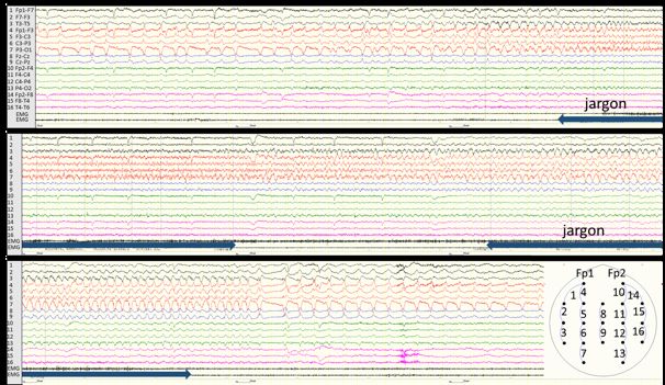
Neurology Asia March 2018
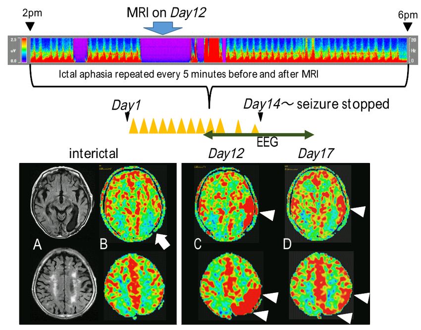
Figure 1. Colored density spectral array represents 4-hour EEG record from 2pm to 6pm on Day 12 of SPSE (upper).
Interictal, ictal and periictal PASL-MRI (lower). (A) FLAIR images showed post-operative changes,
enlarged dorsal horn of left lateral ventricle in addition to diffuse cerebral atrophy and ischemic change.
(B) Hypoperfusion at the left posterior quadrant on interictal PASL-MRI (arrow) (C) Hyperperfusion at
left posterior cortices on ictal PASL-MRI on Day 12 when aphasic seizures repeated every five minutes.
(arrowheads) (D) Persistent but milder hyperperfusion at the same regions on Day 17, three days after
aphasic seizure has ceased (arrowheads)
The prosody of her speech was preserved. In hyperperfusion over the left posterior cortices
between bouts of seizures, she tried to explain (Figure 1).
haltingly how difficult situation she was in, Intravenous midazolam was only temporarily
using non-fluent, brief and meaningful words and effective to abort the seizure. Intravenous
phrases. Her spontaneous speech was significantly phenytoin loading finally stopped the cycling
reduced. The jargon aphasia was associated with pattern of the seizures and there was no recurrence
clumsiness of her right hand. She could not hold of ictal pattern on EEG after day-14. PASL-MRI
her chopsticks properly when epileptic discharges was repeated at Day 17 when no ictal discharge
were present. She could skillfully use them again, was seen on EEG performed just before MRI,
soon after the discharge disappeared. still showing hyperperfusion predominantly over
PASL-MRI was obtained in the middle of the left temporal and parietal lobes, which was
repetitive ictal discharges appearing every five relatively reduced in intensity compared to the
minutes on Day12. The images were acquired ictal PASL imaging on Day12.
using MRI GE Health care, Signa HDxt Optima She developed another episode of SPSE in the
Edition TwinSpeed 1.5T (Ver23) 8-ch, Brain following month triggered by decreased level of
Array Coil. The following parameters of PASL phenytoin. This time, the EEG showed continuous
was used: 36 slices; 4 mm thickness, FOV: 24 ictal discharges predominantly over the similar
cm, TE: 9.8ms, TV: 4955ms, NEX: 3.0, points: regions for five days, which later developed
512, arms: 8, bandwidth: 62.5, post-label delay into periodic lateralized epileptiform discharges
1525ms. The ictal PASL-MRI showed marked (PLEDs) for the following three days (Figure 3).
78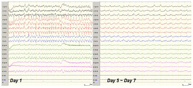
Figure 2. Ictal EEG (longitudinal montage)on Day 12. Focal ictal discharges over the posterior quadrant and the evolution in the frequency of the discharges.

79
80
                                                                                                                                                                                  Neurology Asia

     Figure 3. The EEG (longitudinal montage) showed continuous ictal discharges predominantly over the left temporal, occipital and parietal regions (T3-P3-O1) for five days,
               and the periodic discharges over the same regions for the subsequent three days.
                                                                                                                                                                                  March 2018
Both PASL-MRI and N-isopropyl- p-[(123) as the predominant feature of SPSE has rarely
I] iodoamphetamine single photon emission been reported.1,2 The diagnostic criteria for ictal
computed tomography (IMP-SPECT) were aphasia were first defined by Rosenbaum et al.3
performed on Day 7, when PLEDs still existed and then was modified further; (i) The patient
on EEG, which revealed mild hyperperfusion at has language production during the seizure. (ii)
the left temporoparietal region (Figure 4). Language production shows aphasic features. (iii)
The combination of phenytoin, valproate and Consciousness is preserved. (iv) The seizures are
clobazam has made her free from SPSE since correlated with the aphasia, as documented by
then, except minor brief episodes of aphasia EEG monitoring and behavioral testing. (v) The
triggered by her missing drugs. Her speech became aphasia resolves, or nearly so, concurrent with
comprehensible enough for daily conversation, successful treatment of the seizures.4 Our patient
though some paraphasic errors remained and fulfilled the criteria as above.
some difficulties in reading and writing words Jargon aphasia is described as fluent,
persisted. non-effortful flow of sounds and words but
incomprehensible without meaning5,6 and it is
DISCUSSION divided into undifferentiated jargon, asemantic
jargon, paraphasic jargon, and circumlocutory
Our patient’s ictal presentation was jargon aphasia anomic speech.5 Our patient uttered meaningless
with preserved consciousness, accompanied by speech fluently during seizure. The prosody of
ictal discharges on EEG. Ictal jargon aphasia
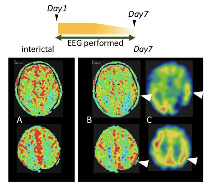
Figure 4. Periictal PASL-MRI (B) and IMP-SPECT (C) on Day 7 in the second SPSE, when left periodic discharges
on EEG continued. (A) Interictal PASL-MRI. (B and C) Hyperperfusion at left temporal and parietal
lobes was seen on both modalities. (arrowheads)
81Neurology Asia March 2018
her speech was preserved. The type of aphasia hyperperfusion shown in periictal PASL-MRI
observed during her seizures can be categorized was equivalent to the IMP-SPECT’s finding. A
into asemantic jargon, a fluent aphasia of Wernicke few authors also have described the concordance
type, which consists of phonologically nonsense between ASL and SPECT, the standard method for
syllables or neologisms, however, it may sound evaluation of CBF in localizing the epileptogenic
grammatically organized foreign language zone.17,20
due to its preservation of function words. Her Although jargon aphasia presenting as SPSE
speech consisted of a copious flow of complex, is rare and it is difficult to recognize, regional
reiterative, phonologically related neologisms. hyperperfusion on PASL-MRI would prompt
The iterative pattern of phonemic variation is physicians to consider SPSE and perform EEG
named as stereotypic pattern of alliteration and for differential diagnosis. This report also suggests
assonance by Green 19697 which is considered that PASL-MRI is useful in assessing CBF during
characteristic for the neologistic jargon.6 seizures and it is as reliable as SPECT. It is a non-
Bell et al. hypothesized that ictal neologistic invasive technique requiring no radioactive tracer,
speech automatism may implicate a focus in thus it can be repeated over time to evaluate the
the hemisphere dominant for propositional progression of the cerebral perfusion pattern of
language and the word’s sounds are disconnected the evolving seizure.
from word’s meaning, producing neologism.8
Posterior ictal aphasia can manifest either as ACKNOWLEDGEMENT
fluent neologistic speech or as neologistic speech
The author would like to thank the doctors and
automatisms.8 Other studies also noted that
staffs of National Epilepsy Center, Shizuoka
their patients had lesion at left dominant side
Institute Epilepsy and Neurological Disorders,
particularly posterior quadrant.9,10 Our patient has
Shizuoka, Japan for taking care of the patient.
a post-operative lesion for brain abscess at the
left posterior quadrant where sensory language
area is close to. REFERENCES
PASL is a developing technique of MRI 1. Patil B, Oware A. De-novo simple partial status
which is non-invasive and needs no radioactive epilepticus presenting as Wernicke’s aphasia. Seizure
tracer. It has been used to measure cerebral blood 2012; 21(3):219-22.
2. Inoue Y, Mihara T, Matsuda K, Tottori T, Seino M.
flow (CBF) which is helpful in the diagnosis
Evolution of ictal dysphasia in relation to intracranial
of neurological diseases, such as stroke and EEG expressions. High Brain Funct Res 1990;
cerebrovascular diseases, epilepsy, tumour, 10:157-64 (Japanese).
infection, neurodegenerative and neuropsychiatric 3. Rosenbaum D, Siegel M, Barr W, Rowan A. Epileptic
disorders.11-13 Quantitative nature of PASL is aphasia. Neurology. 1986; 36(6):822-5.
capable of quantifying local CBF by measuring the 4. Grimes DA, Guberman A. De novo aphasic status
magnetically labelling inflow of the arterial blood epilepticus. Epilepsia. 1997; 38(8):945-9.
5. Alajouanine T. Verbal realization in aphasia. Brain.
at a certain point into the target area and it has 1956; 79:1-28.
been used for lateralization of the epileptic foci.11,12 6. Perecman E, Brown J. Phonemic jargon: A case
The PASL perfusion scans are able to show report. In: Brown JW, ed: Jargonaphasia. New York:
interictal hypoperfusion, which corresponding Academic Press: 177-258..
to hypoperfusion in inter-ictal positron emission 7. Green E. Phonological and grammatical aspects
topography (PET).14,15 Meanwhile, there had been of jargon in an aphasic patient: a case study. Lang
reports identifying the localizing value in ictal Speech 1969; 12(2):103-18.
8. Bell WL, Horner J, Logue P, Radtke RA. Neologistic
hyperperfusion of PASL-MRI while patients are speech automatisms during complex partial seizures.
having ongoing seizure16-18 and up to five hours Neurology 1990; 40:49-52.
post-ictallly.19 9. Inoue Y, Mihara T, Fukao K, Kudo T, Watanabe Y,
In our case, PASL-MRI was performed during Yagi K. Ictal paraphasia induced by language activity.
the sequence of repetitive seizures in an interval Epilepsy Res 1999; 35(1):69-79.
of 5 minutes, which was confirmed on EEG just 10. Bell WL, Horner J, Logue P, Radtke RA. Neologistic
speech automatisms during complex partial seizures.
before and after MRI. One or two seizures were
Neurology 1990; 40(1):49-52.
likely to occur during PASL-MRI, as acquisition 11. Wolf RL, Detre JA. Clinical neuroimaging using
of PASL-MRI needed 4 minutes and 22 seconds. arterial spin-labeled perfusion magnetic resonance
Marked hyperperfusion at left posterior region imaging. Neurotherapeutics 2007; 4(3):346-59.
was likely correlated with the concurrent epileptic 12. Grade M, Tamames JAH, Pizzini FB, Achten E,
activity. In the second episode of SPSE, regional Golay X. A neuroradiologist ’ s guide to arterial spin
82labeling MRI in clinical practice. Neuroradiology
2015; 57:1181-202.
13. Deibler AR, Pollock JM, Kraft RA, Tan H, Burdette
JH, Maldjian JA. Arterial spin-labeling in routine
clinical practise, part 3: hyperperfusion patterns. Am
J Neuroradiol 2008; 29(8):1428-35.
14. Pendse N, Wissmeyer M, Altrichter S, et al. Interictal
arterial spin-labeling MRI perfusion in intractable
epilepsy. J Neuroradiol 2010; 37(1):60-3.
15. Blauwblomme T, Boddaert N, Chémaly N, et al.
Arterial spin labeling MRI: A step forward in non-
invasive delineation of focal cortical dysplasia in
children. Epilepsy Res 2014; 108(10):1932-39.
16. Kanazawa Y, Morioka T, Arakawa S, Furuta Y,
Nakanishi A, Kitazono T. Nonconvulsive partial
status epilepticus mimicking recurrent infarction
revealed by diffusion-weighted and arterial spin
labeling perfusion magnetic resonance images. J
Stroke Cerebrovasc Dis 2015; 24(4):731-8.
17. Matsuura K, Maeda M, Okamoto K, et al. Usefulness
of arterial spin-labeling images in periictal state
diagnosis of epilepsy. J Neurol Sci 2015; 359(1-
2):424-9.
18. Galazzo IB, Storti SF, Del Felice A, et al. Patient-
specific detection of cerebral blood flow alterations
as assessed by arterial spin labeling in drug-resistant
epileptic patients. PLoS One 2015; 10(5):1-24.
19. Pizzini FB, Farace P, Manganotti P, et al. Cerebral
perfusion alterations in epileptic patients during
peri-ictal and post-ictal phase: PASL vs DSC-MRI.
Magn Reson Imaging. 2013; 31(6):1001-5.
20. Sierra-Marcos A, Carreño M, Setoain X, et al.
Accuracy of arterial spin labeling magnetic
resonance imaging (MRI) perfusion in detecting
the epileptogenic zone in patients with drug-
resistant neocortical epilepsy: Comparison with
electrophysiological data, structural MRI, SISCOM
and FDG-PET. Eur J Neurol 2016; 23(1):160-7.
83You can also read