Unusual Presentation of Emphysematous Pyelonephritis (EPN): "Double trouble" A Case Report
←
→
Page content transcription
If your browser does not render page correctly, please read the page content below
Asian Journal of Case Reports in Surgery

                            7(1): 8-13, 2021; Article no.AJCRS.63896

                 Unusual Presentation of Emphysematous
             Pyelonephritis (EPN): "Double trouble" A Case
                                                    Report
                          Abdul Malek Mohamad1*, Sarmukh Singh A/L Charanjit Singh2
                                                                 and Azmi Hassan2
    1
     Sultan Ahmad Shah Medical Centre @IIUM, Jalan Sultan Ahmad Shah, 25200 Kuantan, Pahang,
                                                                                     Malaysia.
                2
                 Hospital Sultan Hj Ahmad Shah, Jalan Maran, 28000 Temerloh, Pahang, Malaysia.

                                                                                            Authors’ contributions

       This work was carried out in collaboration among all authors. Author AMM designed the study,
      performed the statistical analysis, wrote the protocol, and wrote the first draft of the manuscript.
  Authors SS A/L CS and AH managed the analyses of the study. Author AMM managed the literature
                                         searches. All authors read and approved the final manuscript.

                                                                                                 Article Information

                                                                                                                 Editor(s):
                                             (1) Dr. Ashish Anand, GV Montgomery Veteran Affairs Medical Center, USA.
                                                                                                               Reviewers:
                                                     (1) Grigore T. Popa, University of Medicine and Pharmacy, Romania.
                                                          (2) Rawnak Afrin , Dhaka Medical College Hospital, Bangladesh.
                                            Complete Peer review History: http://www.sdiarticle4.com/review-history/63896

                                                                                     Received 10 November 2020
  Case Study                                                                          Accepted 18 January 2021
                                                                                     Published 09 February 2021

ABSTRACT

 Introduction: Emphysematous Pyelonephritis (EPN) is a possibly life-threatening condition that is
 usually present as sepsis. Delay in identifying the disease may lead to detrimental outcomes, even
 mortality. The present study reports a case of incidental finding of an EPN in a perforated viscus
 patient. Studies also advocate the minimally invasive approach of percutaneous drainage with
 antibiotics in asymptomatic EPN patients. Historically, EPN was managed by nephrectomy or open
 drainage along with antimicrobial therapy but resulted in high mortality of 40-50%. Introduction of
 percutaneous drainage had shown better outcome compared to nephrectomy.
 Case Presentation: A 63-year-old gentleman, with underlying Diabetes Mellitus (DM), initially
 presented with a sudden onset of severe generalized abdominal pain and distension. Examination
 revealed peritonitic abdomen. Erect CXR revealed air under the diaphragm suggestive of
 perforated hollow viscus. Patient underwent exploratory laparotomy with intraoperative findings of
_____________________________________________________________________________________________________

*Corresponding author: E-mail: drabdmalek@iium.edu.my;
Mohamad et al.; AJCRS, 7(1): 8-13, 2021; Article no.AJCRS.63896
perforated prepyloric gastric ulcer that managed with modified Graham patch. Besides, there was
also huge retroperitoneal mass. Post operatively, patient subjected for abdominal and pelvic CT
that revealed right EPN. It was managed with antibiotic and percutaneous drainage. Patient was
responded and recovered.
Conclusion: EPN is a fatal disease that requires early detection with a high index of suspicion
particularly in patients with signs of sepsis and pyelonephritis. Although it is rare, in subjects with
pneumoperitoneum and the presence of pathology over renal area, EPN should be one of the
differential diagnosis. In this case, it is possible that the presence of EPN poses stress to the
patient leading to development of perforated viscus.
Keywords: Emphysematous pyelonephritis; diabetes mellitus; nephrectomy; mortality.
1. INTRODUCTION fixed, non-pulsating with no obvious signs of
inflammation.
EPN is defined as a severe gas forming infection
of renal parenchyma and its neighbouring Postoperatively, the patient had a speedy
structure [1]. It is a possible life-threatening recovery and was discharged home well.
condition that is usually present as sepsis.
Delay in identifying the disease may lead to He was then planned for contrast enhanced
detrimental outcomes, even mortality. However, abdominal and pelvic CT to investigate the
in rare cases, its presentation diverts its normal retroperitoneal mass. Abdomen and pelvic CT
clinical pictures, masquerading the true revealed features suggestive of right EPN with
pathology. We present a case of incidental large perinephric abscess measuring
finding of an EPN in a perforated viscus patient. 9.2x6.7x10.9cm and bilateral ureteric calculi as
We advocate the minimally invasive approach of portrayed in Fig. 2. We proceeded with
percutaneous drainage with antibiotics in ultrasound guided percutaneous drainage and
asymptomatic EPN patients. However, serial thick pus was aspirated and sent for cultures.
radiological reassessment should be carried out The pus culture grew Extended Spectrum β-
to see the resolution of the abscess collection. Lactamase Klebsiella Pneumoniae. He had
uneventful recovery afterwards, however the
2. CASE REPORT pigtail catheter meant for pus drainage
prematurely dislodged. A repeat ultrasound
A 63-year-old gentleman, with underlying revealed residual decreasing size collection. He
Diabetes Mellitus (DM), initially presented with a was planned for conservative management with
sudden onset of severe generalized abdominal antibiotics (initially he was started with
pain and distension. He was tachycardic and intravenous cefoperazone and metronidazole
dehydrated. Abdominal examination revealed and completed for one week). It was later
distended abdomen with peritonism. Laboratory changed to cefuroxime based on its pus culture
investigation showed leucocytosis with WBC of and sensitivity for another 2 weeks. He had
23.9 x 10^9/L and acute kidney injury (AKI) frequent visits to surgical outpatient clinic. His
evidenced by elevated urea of 11.8 mmol/L and latest visit was unremarkable and showing good
creatinine of 131 umol/L. The urine examination recovery.
showed red blood cells 3+, leucocyte 3+. We
proceeded with an Erect chest X-ray showed air 3. DISCUSSION
under the diaphragm. Our initial diagnosis was
perforated hollow viscus with differential EPN is defined by severe necrotizing and gas
diagnosis of perforated gastric ulcer (PGU) forming infection of the renal parenchyma and its
/perforated duodenal ulcer (PDU). His diabetes surrounding structure namely perinephric tissue
was managed with insulin infusion (actrapid and collecting system [1,2]. The first documented
infusion) which was later replaced with basal case of EPN was described in 1898 and the term
bolus insulin after the blood sugar was optimized. EPN coined in 1962 to correlate between acute
He was then subjected for operation where he kidney infection with gas forming condition [1].
underwent exploratory laparotomy with modified
Graham Patch repair for perforated prepyloric The incidence for female is more common than
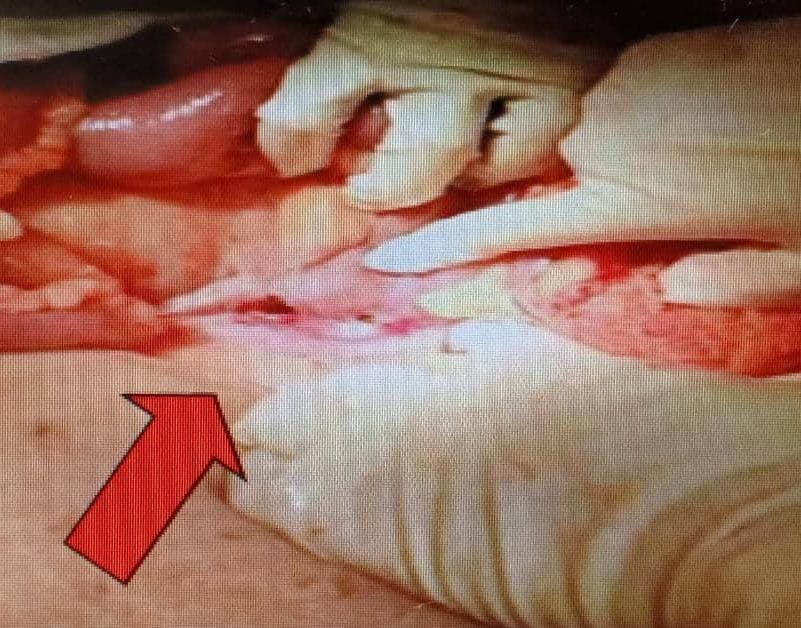
gastric ulcer measuring 2cm as shown in Fig. 1. men, with a mean age of 40-50 years old [2]. It
Intraoperatively, there was a huge retroperitoneal had been reported that left kidney is affected
mass measuring 8cm x 8cm over the right side, more than right without apparent explanation
9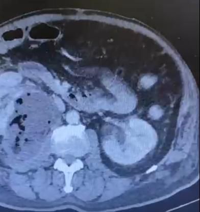
Mohamad et al.; AJCRS, 7(1): 8-13, 2021;; Article no.AJCRS.63896
                                                                                                   no.

[2].Usually, it presented with sepsis and                  accounts for more than 95% of all EPN cases
pyelonephritis signs such as fever with rigor,             [6,7]. Besides, urinary tract obstruction also
lumbar pain and urinary tract infection symptoms           predispose patients to develop EPN [6].
[3].However, in some extreme cases, peritonitis
with pneumoperitoneum suggested
                             ggested of perforated         Since 1984, multiple classification systems
viscus had been reported [4,5]. This results from          developed in order to aid in managing the
the extension of the disease into the peritoneum.          disease as illustrated in Table 1. The latest
This unique presentation is a red -herring                 composition by Huang et al had provided
resulting in a different approach in managing the          objective means in treating EPN [7].
disease. However, in our case the patient
presented with peritonitis secondary to                    EPN is a devastating condition that bears high
perforated gastric ulcer with incidental finding of        risk of morbidity and mortality mainly due to
EPN. This caused a diagnostic dilemma and                  septic complications. Variety of risk factors
difficulty in managing this case.                          identified to prognosticate the outcome of the
                                                           illness. The meta -analysis
                                                                                   analysis had concluded
Most      EPN        is     associated       with          several factors including patient who was treated
immunocompromised state particularly diabetes              conservatively, type 1 EPN, PN, bilateral EPN and
mellitus where it occurrence in diabetic patients          patient with low platelet (thrombocytopenia) [8].

                             Fig. 1.. Perforated prepyloric gastric ulcer

                         Fig. 2.. Right EPN with large perinephric abscess

                                                      10
Mohamad et al.; AJCRS, 7(1): 8-13, 2021;; Article no.AJCRS.63896
                                                                                               no.

                               Table 1. Classification of EPN

Author               Radiological modality    Class
Michaeli et al [4]   Plain x-ray
                             ray with         I : Gas in the parenchyma or perinephric tissue
                     intravenous urography    II : Gas in the kidneys and it surroundings
                                              III : Gas through fascia or bilateral kidney
                                              involvement
Wan et al [5]        CT scan                  I : Renal necrosis with presence of gas but no fluid
                                              II: Renal or perinephric fluid collection associated
                                              with gas in the collecting system.
Huang et al [6]      CT scan                  1. Gas only in collecting system
                                              2. Gas only in renal parenchyma
                                              3A. Perinephric space involvement
                                              3B. Pararenal space involvement
                                              4. Bilateral kidney involvement or solitary kidney
                                              with EPN

Fig. 3. Management algorithm of EPN. (Risk factors: Diabetes, thrombocytopenia, acute kidney
                            injury, altered sensorium, shock) [7]

                                              11
Mohamad et al.; AJCRS, 7(1): 8-13, 2021; Article no.AJCRS.63896
The diagnosis of EPN is radiological supported particularly in patients with signs of sepsis and
by the presenting history and pyelonephritis. Although it is rare, in subjects with
laboratory investigations [5]. Although plain pneumoperitoneum and the presence of
radiograph may show presence of air near the pathology over renal area, EPN should be one of
kidney, the gold standard modality in diagnosing the differential diagnosis. In this case, it is
EPN is undoubtedly CT scan [9]. CT scan yields possible that the presence of EPN poses stress
100% accuracy compared to plain to the patient leading to development of
radiograph which only positive up till 69% of the perforated hollow viscus.
cases [9].
CONSENT
Historically, EPN was managed by nephrectomy
or open drainage along with antimicrobial therapy As per international standard or university
but resulted in high mortality of 40-50% [4]. After standard, patients’ written consent has been
the introduction of minimally invasive collected and preserved by the authors.
percutaneous drainage, the management shifted
towards percutaneous drainage, and this ETHICAL APPROVAL
method proved to yield better outcome. A
systematic review advocated percutaneous It is not applicable.
drainage as the initial management of
EPN whereas it is associated with reduced
COMPETING INTERESTS
mortality compared to emergency nephrectomy
or medical management however, nephrectomy
Authors have declared that no competing
may be required in some cases not
interests exist.
responding to the aforementioned
treatment [9]. Fig. 3 demonstrates the
management algorithm extracted from Huang et REFERENCES
al. [7].
1. Schultz EH, Klorfein EH. Emphysematous
Referring to our case, as the patient was pyelonephritis. J Urol. 1962;87(6):762–6.
asymptomatic, percutaneous drainage with 2. Misgar R, Mubarik I, Wani A, Bashir M,
medical management were the treatment of Ramzan M, Laway B. Emphysematous
choice. Radiological imaging repeated after one pyelonephritis: A 10-year experience with
month showed improvement. Chen et al had 26 cases. Indian J Endocrinol Metab.
concluded that imaging should be repeated after 2016;20(4):475–80.
4-7 weeks and from their cohort, the treatment 3. Tang HJ, CM Li, Yen MY, Chen YS, Wann
lasted for approximately 3 months [10]. Based on SR, Lin HH, Lee YCL SS. Clinical
study by Chen MT et al, their patient was characteristics of emphysematous
followed up till 10 years with mean of 5 years pyelonephritis. J Microbiol Immunol Infect.
(follow up till clinically and radiologically 2001;34(2):125–30.
resolved) showed no recurrence or complications 4. Michaeli J, Mogle P, Perlberg S, Heiman
[10]. Our patient is still on regular follow up and S, Caine M. Emphysematous
planned to be followed up till complete clinical pyelonephritis. J Urol. 1984;131(2):203–8.
and radiological resolution. In cases which do not 5. Wan YL, Lee TY, Bullard MJ, Tsai CC.
respond to percutaneous drainage, nephrectomy Acute gas-producing bacterial renal
should be considered [6]. As EPN is a rare infection: Correlation between imaging
occurrence, there is limited consensus pertaining findings and clinical outcome. Radiology.
to the treatment of incidental finding of EPN 1996;198(2):433–8.
during an exploratory laparotomy. However, a 6. Jeng-Jong Huan C-CT. Emphysematous
safer approach must be undertaken to avoid pyelonephritis. Japanese J Clin Urol.
causing more harm to the patients particularly in 2011;65(1):23–9.
asymptomatic patients. Open drainage with 7. Ubee SS, McGlynn L, Fordham M.
posterior approach (flank incision) is one of the Emphysematous pyelonephritis. BJU Int.
methods of choice [11]. 2011;107(9):1474–8.
8. Falagas ME, Alexiou VG, Giannopoulou
4. CONCLUSION KP, Siempos II. Risk Factors for Mortality
in Patients With Emphysematous
EPN is a fatal disease that requires early Pyelonephritis: A Meta-Analysis. J Urol.
detection with a high index of suspicion 2007;178(3):880–5.
12Mohamad et al.; AJCRS, 7(1): 8-13, 2021; Article no.AJCRS.63896
9. Somani BK, Nabi G, Thorpe P, Hussey J, emphysematous pyelonephritis: 10-
Cook J, N’Dow J. Is percutaneous Year experience. J Urol.
drainage the new gold standard in the 1997;157(5):1569–73.
management of emphysematous 11. Hayashi T, Yanaihara H, Kaguyama H,
pyelonephritis? evidence from a systematic Hanashima F, Sakamoto H, Nakahira Y et
review. J Urol. 2008;179(5):1844–9. al. Emphysematous pyelonephritis with
10. Chen MT, Huang CN, Chou YH, Huang successful renal preservation using open
CH, Chiang CP, Liu GC. Percutaneous drainage surgery: A case report. Urol Case
drainage in the treatment of Reports. 2018;17:76–8.
_________________________________________________________________________________
© 2021 Mohamad et al.; This is an Open Access article distributed under the terms of the Creative Commons Attribution
License (http://creativecommons.org/licenses/by/4.0), which permits unrestricted use, distribution, and reproduction in any
medium, provided the original work is properly cited.
Peer-review history:
The peer review history for this paper can be accessed here:
http://www.sdiarticle4.com/review-history/63896
13You can also read