Unusual Location of a Posterior Lipoma Originating From the Left Atrial Roof: Case Report and Review of the Literature
←
→
Page content transcription
If your browser does not render page correctly, please read the page content below
The Heart Surgery Forum #2020-2933 Online address: http://journal.hsforum.com
23 (2), 2020 [Epub March 2020]
doi: 10.1532/hsf.2933
Unusual Location of a Posterior Lipoma Originating From the Left Atrial Roof:
Case Report and Review of the Literature
Ovidiu Stiru, MD, PhD, Roxana Carmen Geana, MD, Liana Valeanu, MD, Adrian Tulin, MD, PhD,
Laura Raducu, MD, PhD, Vlad Anton Iliescu, MD, PhD
Prof. Dr. C. C. Iliescu Emergency Institute for Cardiovascular Diseases, Bucharest, Romania
ABSTRACT CLs are fat-containing encapsulated tumors with unknown
etiology [Italiano 2008].
Lipomas are the most common type of soft tissue tumors.
They mainly are located in subcutaneous tissue in the body,
including the heart. The cardiac location of lipomas is rare,
mostly asymptomatic, and can cause life-threatening compli-
cations by rapid growth. The clinical symptoms, when pres-
ent, occur in evolution with the growth in size, depending
upon the location and degree of invasion in the endocardium.
We present the case of a 63-year-old male patient with a
large intrapericardial lipoma with an unusual location, origi-
nating from the left atrial roof. The initial symptoms of the
patient were shortness of breath, dizziness, and mild dyspnea.
Transthoracic echocardiography (TTE) was the first line
diagnosis method, followed by computed tomography (CT);
both showed a large posterior intrapericardial mass. Resec-
tion of a 12/8/5 cm lipomatous tumor mass was performed via
median sternotomy, under cardiopulmonary bypass (CPB) on
the beating heart. Histopathologic examination revealed the
presence of diffuse proliferation of large- and medium-sized
mature adipocytes consistent with the diagnosis of pericardial Figure 1. Thoracic CT scan. The large, ovoid tumor, 11.8/9.3/5.4 cm
lipoma. The patient was discharged at home on the seventh located in the posterior medi-astinum along the left border of the heart,
postoperative day, with a marked improvement of his clini- toward the base of the heart.
cal state and effort tolerance. He did not present evidence of
recurrence at his 1-year follow up.
INTRODUCTION
Primary tumors of the heart account for less than 0.2-0.4%
of all cardiac tumors [McAllister 1999]. The most common
primary cardiac tumors in adults are myxomas [Iliescu 2008],
followed by papillary fibroelastomas and lipomas [McAllister
1978]. Lipomas account for 8.4% to 10% cases of primary
heart neoplasms and approximately 14% of benign cardiac
masses [McAllister 1978]. Since clinical findings often are
nonspecific, many cardiac lipomas (CL) are found in imaging
studies performed for other reasons. However, when clinical
features appear, they depend on the size and tumor location.
Received February 18, 2020; accepted March 6, 2020.
Correspondence: Ovidiu Stiru, Cardiac surgery department. Prof. Dr. C. C.
Iliescu Emergency Institute for Cardiovascular Diseases Sos. Fundeni 258,
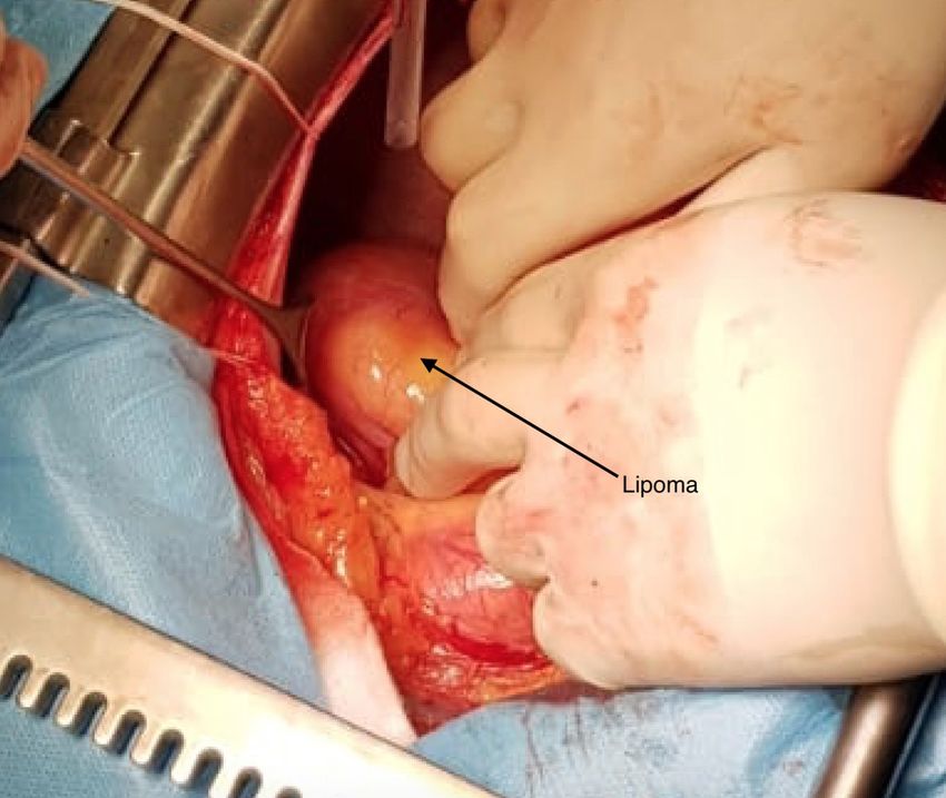
sector 2, 022328 Bucharest, Romania; fax: +400722207286; +400722207286 Figure 2. Intraoperative aspect-in-situ incapsulated lipomatous tumor
(e-mail: ovidiu_stiru@yahoo.com). attached to the left atrial roof.
E140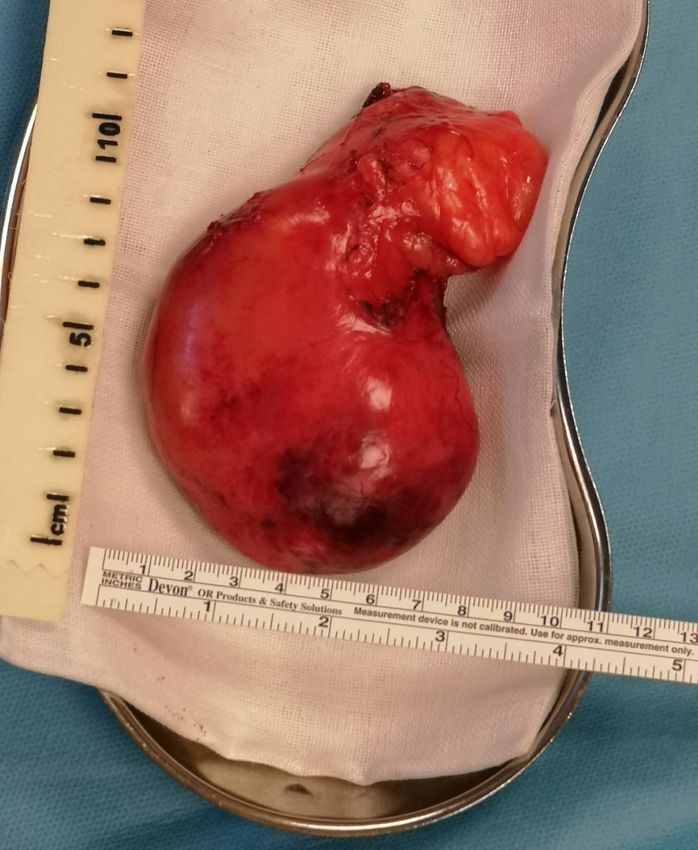
Unusual Location of a Posterior Lipoma Originating From the Left Atrial Roof: Case Report and Review of the Literature—Stiru et al

                                                                                     Figure 4. Histopathologic examination revealed the presence of diffuse
                                                                                     proliferation of large and medium sized mature adipocytes consistent with
                                                                                     the diagnosis of pericardial lipoma (H&E staining; magnification × 10).

                                                                                 and left ventricle (LV). This mass exerted no compression
                                                                                 against any of the heart chambers, at least while the patient
                                                                                 was at rest. The contractility was normal. Following the
                                                                                 TTE examination, a thoracic CT scan thorax confirmed the
                                                                                 presence of an intrapericardial mass with a fat-like density,
                                                                                 measuring 11.8/9.3/5.4 cm in the posterior mediastinum,
                                                                                 located along the posterior surface of the heart from dia-
                                                                                 phragm level to pulmonary artery bifurcation level (Figure
                                                                                 1). In our patient, both TTE and CT failed to determine
                                                                                 the site of origin of the tumor. Cardiothoracic surgery was
                                                                                 consulted for tumoral resection, and it was decided that no
   Figure 3. Postexcision-aspect of incapsulated lipomatous tumor.               further imaging, such as cardiac magnetic resonance images
                                                                                 (MRI) or coronary angiography, were needed. Consider-
CASE REPORT                                                                      ing the lack of guidelines regarding this clinical entity, the
                                                                                 management of the case is mainly lead by the symptoms
   A 64-year-old man was admitted to our clinic with short-                      tied to the mass effect of the tumor. The presence of dys-
ness of breath that worsened over the course of a year,                          pnea as early as the lowest level of effort, required surgical
evolving to a NYHA class III congestive heart failure. In                        removal of the pericardial mass. During the surgery, the inci-
addition to sex and age, severe arterial hypertension, dyslip-                   sion was median sternotomy; the thymus and pericardial fat
idemia, and obesity accounted for an almost complete chart                       pad were found to be normal. After the pericardiotomy, it
of cardiovascular risk factors in this case. Other important                     was noted that a soft, encapsulated epicardial tumor did not
medical and surgical history points are permanent atrial                         adhere to the pericardium and had a stalk measuring approx-
fibrillation, nephrolithiasis, 3B stage chronic kidney dis-                      imately 5 cm in diameter, attached to the left atrial roof
ease, hepatic steatosis, repaired inguinal hernia, and cho-                      (Figure 2). The pedicle was extending posteriorly toward the
lecystectomy. Despite this rather rich medical and surgical                      left atrioventricular groove. The amount of traction needed
history, the overall clinical state of the patient was good.                     to expose the entire origin of the tumor was unsafe, and it
Physical examination was normal, except for faint heart                          was impossible to dissect without CPB support. The aorta
sounds and irregular tachycardia (110 bpm), without marks                        and right appendage were cannulated so that the heart could
of lipomatosis under the skin. Electrocardiogram (ECG)                           be drained and safely mobilized. The entire mass measuring
showed atrial fibrillation (AF) with low amplitude QRS                           12/8/5 cm was removed with CPB on beating heart using
complexes in the frontal leads and poor R wave progression                       electric coagulation, (Figure 3) and CPB was easily weaned.
in precordial leads. Laboratory tests demonstrated a normal                      The patient quickly was extubated, and his condition was
complete blood count, normal electrolytes and liver func-                        uneventful in the intensive care unit. Histopathologic exami-
tion tests, with renal dysfunction, serum creatinine 1.55                        nation revealed the presence of diffuse proliferation of large
mg/dL (local laboratory reference range 0.7 to 1.2mg/dL),                        and medium-sized mature adipocytes consistent with the
total cholesterol 250 mg/dL, LDL cholesterol 190 mg/dL.                          diagnosis of pericardial lipoma (Figure 4). The patient was
The cardiothoracic ratio on a chest standard posteroante-                        discharged from the hospital on the sixth postoperative day.
rior radiograph exceeded 50%, revealing an enlargement                           The patient currently is asymptomatic with complete reso-
of the cardiac size. Transthoracic echocardiography (TTE)                        lution of dyspnea with a marked improvement of his clinical
revealed a large echolucent 48/76 mm intrapericardial mass                       state and effort tolerance and has not presented with evi-
was lying lateral and posteriorly to the left atrium (LA)                        dence of recurrence at the 1-year follow up.

© 2020 Forum Multimedia Publishing, LLC                                                                                                                   E141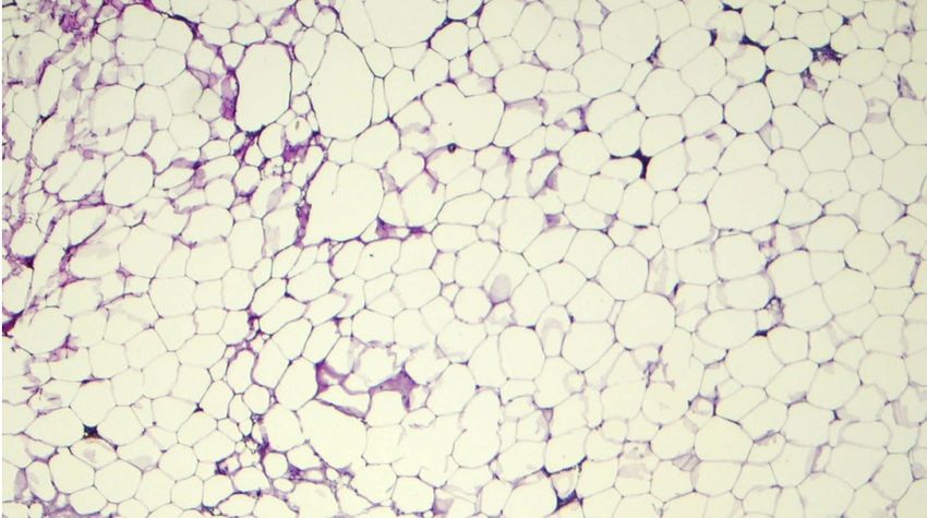
The Heart Surgery Forum #2020-2933

 DISCUSSION                                                         CONCLUSION

    Various lipomatous tumors may be found around the                 CL originating from the left atrial roof is a very uncom-
heart. Some of these lesions are discovered and have no            mon situation. It can be diagnosed at any age, regardless of
consequence; others may require additional evaluation and          gender, and can be associated with a wide range of symptoms
management. CL can occur at any age and affect both gen-           that depend on the size, invasion of adjacent structures, and
ders equally. Typically, CL includes the right atrium and the      cardiac location. The first diagnostic tool for cardiac lipoma,
left ventricle location, and they have origin either from the      even in asymptomatic cases, remains TTE. However, CT and
subendocardial (approximately 50%), subpericardial (25%),          MRI provide a better image, allowing the surgeon to evaluate
or from the myocardial layer (25%) [Fine 1968]. Most CLs           more accurately the size, composition, and invasion of adja-
are asymptomatic, as the slow growth often delays diagno-          cent structures. However, these sometimes fail to determine
sis. When present, symptoms are variable, depending on the         the tumor’s site of origin. Surgical resection is mandatory for
location or invasion of the underlying structures. The clinical    symptomatic CL, but should be performed only in cardiac
findings are nonspecific and include dyspnea, fatigue, angina,     centers, with the use of CPB in posterior localizations.
palpitations, arrhythmias, atrioventricular block, precordial-
gia, syncope, left or right heart failure, pericardial constric-
tion, interference in the electrical conduction system and          REFERENCES
even sudden death [Heath 1968; Mourad 2009; Valencia               Da Silva, De Campos AMF, Vieira Baldo RC, et al. 2017. Pericardial
2011; Noly 2016].                                                  lipoma: Diagnosed unexpectedly during heart failure investigation – Case
    TTE, CT, and cardiac MRI are imaging methods allow-            Report. Curr Res Cardiol 4(4):55-57.
ing an analysis of the relationship between tumoral mass and       Dorobantu LF, Stiru O, Prodea A, et al. 2011. Unique case of primary
adjacent structures and play a significant role in establishing    malignant fibrous histiocytoma of the right ventricle with moderator
a diagnosis. Attention should be focused on the localization       band involvement. Heart Surg Forum 14(4):E245–E248.
of these lesions and extent of the disease, including invasion
                                                                   Fine G. 1968. Neoplasms of the pericardium and heart. In: Gould SE,
of adjacent structures, involvement of the coronary arteries,      editor. Pathology of the Heart and Blood Vessels, 3rd. Springfield:
compression of cardiac chambers and pericardial effusion           Charles C. Thomas. 865.
[Noly 2016]. Cardiac MRI also provides information about
                                                                   Heath D. 1968. Pathology of cardiac tumors The American Journal of
tissue composition [Dorobantu 2011] (difference with lipo-
                                                                   Cardiology 21(3):315-327.
sarcoma) and hemodynamic significance [Noly 2016]. In
some occasions, it allows full characterization of tumoral mass    Iliescu VA, Dorobantu LF, Stiru O, et al. 2008. Second recurrence of car-
features, hypodense tissue with attenuation of 20 to 50 HU on      diac myxoma 7 years after the initial operation. Chirurgia 103(2):239-241.
CT images being characteristic for fat tissues [Steger 2011].      Italiano A, Ebran N, Attias R, et al. 2008. NFIB rearrangement in super-
In the reported case, an intrathoracic giant lipomatous tumor      ficial, retroperitoneal, and colonic lipomas with aberrations involving
was diagnosed from these imagery findings, but the origin          chromosome band 9p22. Genes Chromosomes Cancer 47(11):971-7.
from either the thymus, pericardial fat pad, or epicardial heart   McAllister, Hall RJ, Cooley DA. 1999. Tumors of the heart and pericar-
could not be determined. Although rare, there is a consen-         dium. Curr Probl Cardiol 24(2):57-116.
sus that surgical resection should be attempted in all symp-       McAllister HA, Fenoglio JJ. 1978. Tumors of the cardiovascular system
tomatic patients. Total resection is preferred for encapsulated    atlas of tumor pathology. 2nd series. Fascicle 15. Washington DC: Armed
tumors to prevent local recurrence, although some authors          Forces Institute of Pathology 52-8.
consider that the rate of recurrence of subtotal resection of
                                                                   Mourad OMA, Andrade FM, Abrahao P. et al. 2009. Massa mediasti-
cardiac lipoma is quite low [Steger 2011]. In asymptomatic
                                                                   nal gigante assintomática: um raro caso de timolipoma. J Bras Pneumol
patients with incidentally small tumors, close monitoring is       35(10):1049-52.
prudent. Surgical intervention may be considered, depending
on the appearance of symptoms. Lipomas should be differen-         Noly PE, Mongeon FP, Rochon A, et al. 2016. Pericardial Constriction
                                                                   Caused by a Giant Lipoma Circulation 133:1709-1712.
tiated from liposarcomas not only by cardiac MRI features,
but also histologically. CLs are made up of mature adipocytes      Steger C. M. 2011. Intrapericardial giant lipoma displacing the heart.
that are limited by a collagenous capsule, while liposarcomas      ISRN cardiology. 243637.
predominately are made up of mature fat cells [Steger 2011;        Valencia A, Lombo M, Correa J. 2011. Lipoma intrapericárdico gigante
Da Silva 2017].                                                    silente. Rev Colomb Cir 26:222-5.

E142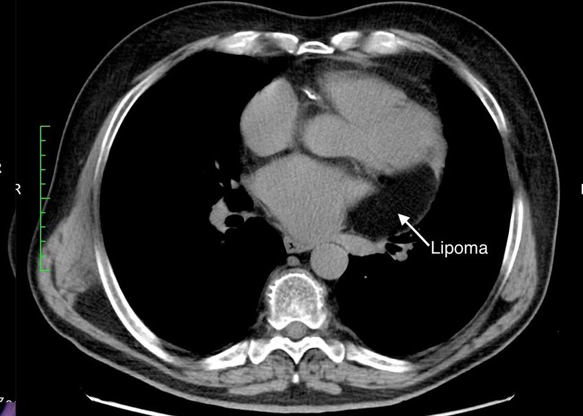
You can also read