AIUM-ACR-SPR-SRU Practice Parameter for the Performance of an Ultrasound Examination for Detection and Assessment of Developmental Dysplasia of ...
←
→
Page content transcription
If your browser does not render page correctly, please read the page content below
PRACTICE GUIDELINES

AIUM–ACR–SPR–SRU Practice
Parameter for the Performance of an
Ultrasound Examination for Detection
and Assessment of Developmental
Dysplasia of the Hip

                                                   Preamble

                                                   T        he American Institute of Ultrasound in Medicine (AIUM) is a
                                                           multidisciplinary association dedicated to advancing the safe
                                                           and effective use of ultrasound in medicine through
                                                   professional and public education, research, development of
                                                   parameters, and accreditation.
                                                        The AIUM represents the entire range of clinical and basic
                                                   science interests in medical diagnostic ultrasound, and, with hun-
                                                   dreds of volunteers, the AIUM has promoted the safe and effective
                                                   use of ultrasound in clinical medicine since 1952. This document
                                                   and others like it will continue to advance this mission.
                                                        Practice parameters of the AIUM are intended to provide the
                                                   medical ultrasound community with parameters for the perfor-
                                                   mance and recording of high-quality ultrasound examinations. The
                                                   parameters reflect what the AIUM considers the minimum criteria
                                                   for a complete examination in each area but are not intended to
                                                   establish a legal standard of care. AIUM-accredited practices are
                                                   expected to generally follow the parameters with recognition that
                                                   deviations from these parameters will be needed in some cases,
                                                   depending on patient needs and available equipment. Practices are
                                                   encouraged to go beyond the parameters to provide additional ser-
                                                   vice and information as needed.

                                                   Introduction
                                                   The clinical aspects contained in specific sections of this practice
                                                   parameter (Introduction, Indications/Contraindications and Tim-
                                                   ing, Specifications of the Examination, and Equipment Specifica-
                                                   tions) were revised collaboratively by the AIUM, the American
                                                   College of Radiology (ACR), the Society for Pediatric Radiology
                                                   (SPR), and the Society of Radiologists in Ultrasound (SRU). Rec-
doi:10.1002/jum.14829                              ommendations for Qualifications and Responsibilities of Person-
                                                   nel; Written Request for the Examination; Documentation; and

    © 2018 by the American Institute of Ultrasound in Medicine | J Ultrasound Med 2018; 9999:1–5 | 0278-4297 | www.aium.org
Quality Control and Improvement, Safety, Infection          hip ultrasound examinations are usually not performed
Control, and Patient Education vary among the orga-         on patients younger than 6 weeks of age unless indi-
nizations and are addressed by each separately.             cated on the basis of an abnormal finding on physical
     This practice parameter is intended to assist prac-    examination.4
titioners performing ultrasound studies for detection
and assessment of developmental dysplasia of the hip
(DDH). Adherence to the following recommenda-               Qualifications and Responsibilities of
tions will maximize the probability of detecting most       Personnel
of the abnormalities that relate to acetabular mor-
phology, position of the femoral head, and stability.       See www.aium.org for AIUM Official Statements,
     When available, ultrasound imaging is the pre-         including Standards and Guidelines for the
ferred method for diagnostic imaging of the immature        Accreditation of Ultrasound Practices and relevant Phy-
hip.1,2 It affords direct visualization of the cartilagi-   sician Training Guidelines.5
nous and other components of the hip joint and per-
mits a dynamic examination that can be used to
assess hip stability. The value of ultrasound dimin-        Written Request for the Examination
ishes as the femoral head ossifies; therefore, radiogra-
phy is preferable for patients 6 months of age or           The written or electronic request for an ultrasound
older, unless the relationship of the femoral head to       examination should provide sufficient information to
the acetabulum (including the triradiate cartilage) is      allow for the appropriate performance and interpreta-
adequately visualized with ultrasound.                      tion of the examination. The request for the examina-
                                                            tion must be originated by a physician or another
                                                            appropriately licensed health care provider or under
Indications/Contraindications and Timing                    the physician’s or provider’s direction. The accompa-
                                                            nying clinical information should be provided by a
Two of the strongest risk factors for DDH are a             physician or other appropriate health care provider
female neonate in a frank breech presentation at birth      familiar with the patient’s clinical situation and should
and a history of a parent and/or a sibling with DDH.3       be consistent with relevant legal and local health care
                                                            facility requirements.
Accepted indications for ultrasound of the infant hip
include but are not limited to:
1. Abnormal or equivocal findings of hip instability         Specifications of the Examination6,7
   on physical examination of the hip;
2. Any family history of DDH;                               Both hips should be examined. The diagnostic exami-
3. Breech presentation at birth;                            nation for DDH incorporates 2 orthogonal planes: a
4. Neuromuscular conditions; and                            coronal view in the standard plane at rest and a trans-
5. Monitoring infants with DDH undergoing                   verse view of the flexed hip with and without stress.
   treatment.                                               This enables an assessment of hip position, stability,
Relative indications for ultrasound of the infant hip       and acetabular morphology.
include but are not limited to:                                 If position, stability, and/or morphology cannot
                                                            be assessed when attempting to perform a complete
1. Oligohydramnios; and
                                                            examination, the report should note the portion not
2. Other intrauterine causes of postural molding.
                                                            performed. It is acceptable to perform the examina-
    There are no absolute contraindications to ultra-       tion with the infant in a supine or in each lateral
sound of the infant hip for DDH, but as discussed           decubitus position separately.
above, the study becomes less reliable compared to              Morphology is assessed at rest. The stress
radiography as ossification of the femoral head pro-         maneuver (posterior push maneuver) is performed to
gresses. Because of the presence of physiologic laxity,     evaluate for hip instability with the hip and knee

2                                                                                       J Ultrasound Med 2018; 9999:1–5
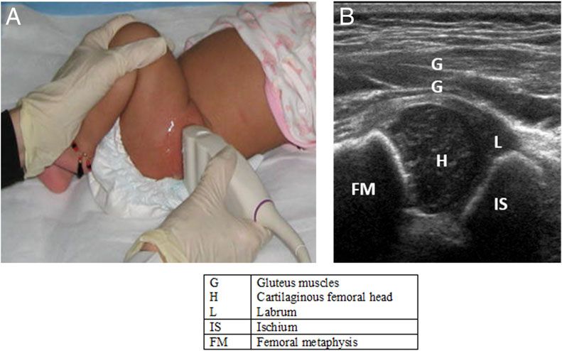
flexed and the thigh adducted (Barlow maneuver). If                          Acetabular morphology is assessed in the coronal neu-
the femoral head is subluxated, subluxable, dislocated,                     tral view and may be validated by measuring the acetab-
or dislocatable, reducibility can be assessed by abduct-                    ular alpha angle (normally ≥60 ). Validation by angle
ing and externally rotating the hip (Ortolani maneu-                        and femoral head coverage measurement is optional.
ver). If the examiner chooses, additional views and                         Performance of stress in this plane is also optional.
maneuvers can be obtained. It is important that the                               The examination is performed with the hip flexed
infant be relaxed when hips are assessed for instabil-                      at 90 . The transverse plane is the anatomic trans-
ity. Feeding the infant during the examination can                          verse or axial plane with respect to the body, similar
increase comfort and cooperation.8 Stress maneuvers                         to the plane of an axial computed tomographic image
are not performed when the patient is immobilized in                        (Figure 2). With the transducer nearly parallel to the
a Pavlik harness or splint unless specifically requested                     femoral shaft, the femoral shaft is seen anteriorly, ter-
by the referring orthopedic surgeon.9                                       minating in the femoral head, which rests on the
                                                                            ischium. The hip is tested for position at rest with
Coronal View                                                                passive abduction and adduction. The transducer is
The anatomic coronal plane is approximately parallel                        kept parallel to the femoral shaft placed in a postero-
to the infant’s body. If the superior edge of the trans-                    lateral position so that imaging can be accomplished
ducer is rotated 10 to 15 (usually posteriorly) into                      while the hip is abducted and adducted (Ortolani and
an oblique coronal plane, the ilium will appear                             Barlow maneuvers). Next, gentle stress is applied to
straight. After adjustment to ensure that the imaging                       assess stability. If the relationship of the femoral head
plane extends through the deepest part of the acetab-                       with the posterior acetabulum changes with gentle
ulum (including visualization of the triradiate cartilage                   stress, the hip is unstable. Again, application of stress
and the ischium), the resulting image will be a coro-                       is omitted when hips are being examined in a Pavlik
nal view in the standard plane.                                             harness or splint device unless otherwise requested by
     The standard plane is defined by identifying a                          the orthopedic surgeon.
straight iliac line, the tip of the acetabular labrum, and
the transition from the os ilium to the triradiate carti-
lage (Figure 1). The coronal view in the standard plane                     Modification of the Diagnostic Examination
can be obtained with the hip in the physiologic neutral                     The supervising physician may modify the examina-
position (15 –20 flexion) or in the flexed position.                        tion depending on clinical circumstances, such as dur-
The femoral head position and displacement are noted.                       ing or after treatment for DDH.

Figure 1. A, Coronal view: the ultrasound transducer is placed parallel to the lateral aspect of the infant’s hip.

J Ultrasound Med 2018                                                                                                              3
Figure 2. A, Transverse flexion view: the hip and knee are flexed 90 , and the ultrasound transducer is placed perpendicular to the lateral
aspect of the infant’s hip nearly parallel to the femoral shaft. B, Transverse ultrasound image.

Documentation                                                          transducer that permits penetration of the soft tissues.
                                                                       Total ultrasound exposure should be kept as low as
Adequate documentation is essential for high-quality                   reasonably achievable (ALARA) while optimizing
patient care. There should be a permanent record of                    diagnostic information.
the ultrasound examination and its interpretation. A
comparison with prior relevant imaging studies may
prove helpful. Images of all appropriate areas, both
normal and abnormal, should be recorded. Variations                    Quality Control and Improvement, Safety,
from normal size should generally be accompanied by                    Infection Control, and Patient Education
measurements. Images should include the patient
identification, facility identification, examination date,               Policies and procedures related to quality control,
hip being imaged, image orientation, and whether                       patient education, infection control, and safety should
stress is being applied. An official interpretation (final               be developed and implemented in accordance with the
report) of the ultrasound examination should be                        AIUM Standards and Guidelines for the Accreditation of
included in the patient’s medical record, indicating                   Ultrasound Practices.
acetabular morphology, the position of the femoral                          Equipment performance monitoring should be in
head, and stability.9                                                  accordance with the AIUM Standards and Guidelines
     Retention of the ultrasound images should be                      for the Accreditation of Ultrasound Practices.
consistent both with clinical needs and with relevant
legal and local health care facility requirements.
     Reporting should be in accordance with the
AIUM Practice Parameter for Documentation of an
Ultrasound Examination.10
                                                                       ALARA Principle
                                                                       The potential benefits and risks of each examination
                                                                       should be considered. The ALARA principle should be
Equipment Specifications                                                observed by adjusting controls that affect the acoustic
                                                                       output and by considering transducer dwell times. Fur-
A hip ultrasound examination for detecting DDH                         ther details on ALARA may be found in the AIUM
should be performed with a high-frequency linear                       publication Medical Ultrasound Safety, Third Edition.11

4                                                                                                        J Ultrasound Med 2018; 9999:1–5
Acknowledgments                                            Isabelle Wilkins, MD

This parameter was revised by the AIUM in collabo-         Original copyright 2003; revised 2018, 2013, 2008
ration with the ACR, the SPR, and the SRU accord-          Renamed 2015
ing to the process described in the AIUM Clinical
Standards Committee Manual.                                References

                                                            1. Roposch A, Wright JG. Increased diagnostic information and
Collaborative Committee                                        understanding disease: uncertainty in the diagnosis of developmen-
                                                               tal hip dysplasia. Radiology 2007; 242:355–359.
Members represent their societies in the initial ver-       2. Smergel E, Losik SB, Rosenberg HK. Sonography of hip dysplasia.
sion and final revision of this practice parameter.             Ultrasound Q 2004; 20:201–216.
                                                            3. Bache CE, Clegg J, Herron M. Risk factors for developmental dys-
                                                               plasia of the hip: ultrasonographic findings in the neonatal period.
           ACR                           AIUM
                                                               J Pediatr Orthop B 2002; 11:212–218.
Terry L. Levin, MD, chair        Brian D. Coley, MD         4. Mulpuri K, Song KM, Gross RH, et al. The American Academy of
Maria A. Calvo-Garcia, MD        T. Robin Goodman, MBBS
                                                               Orthopaedic Surgeons evidence-based guideline on detection and
Monica S. Epelman, MD            Harriet J. Paltiel, MD
Henrietta K. Rosenberg, MD                                     nonoperative management of pediatric developmental dysplasia of the
                                                               hip in infants up to six months of age. J Bone Joint Surg Am 2015; 97:
         SPR                                SRU                1717–1718.
                                                            5. American Institute of Ultrasound in Medicine. AIUM physician
Richard D. Bellah, MD               Lynn A. Fordham, MD
                                                               training guidelines. American Institute of Ultrasound in Medicine website.
Christian L Carlson, MD              Valerie L. Ward, MD
Rachelle Goldfisher, MD                                         https://www.aium.org/resources/ptGuidelines.aspx. Accessed June
Jonathan R. Wood, MD                                           8, 2018.
                                                            6. Harcke HT, Grissom LE. Performing dynamic sonography of the
                                                               infant hip. AJR Am J Roentgenol 1990; 155:837–844.
                                                            7. Morin C, Harcke HT, MacEwen GD. The infant hip: real-time
AIUM Clinical Standards Committee
                                                               US assessment of acetabular development. Radiology 1985; 157:
                                                               673–677.
John Pellerito, MD, chair
                                                            8. Grissom LE, Harcke HT, Kumar SJ, Bassett GS, MacEwen GD.
Bryann Bromley, MD, vice chair
                                                               Ultrasound evaluation of hip position in the Pavlik harness. J Ultra-
Sandra Allison, MD                                             sound Med 1988; 7:1–6.
Anil Chauhan, MD                                            9. Roposch A, Moreau NM, Uleryk E, Doria AS. Developmental dys-
Stamatia Destounis, MD                                         plasia of the hip: quality of reporting of diagnostic accuracy for
Eitan Dickman, MD, RDMS                                        US. Radiology 2006; 241:854–860.
Beth Kline-Fath, MD                                        10. American Institute of Ultrasound in Medicine. AIUM practice
Joan Mastrobattista, MD                                        parameter for documentation of an ultrasound examination. Amer-
Marsha Neumyer, BS, RVT                                        ican Institute of Ultrasound in Medicine website. http://www.
Tatjana Rundek, MD, PhD                                        aium.org/resources/guidelines/documentation.pdf. Accessed June
Khaled Sakhel, MD                                              10, 2018.
James Shwayder, MD, JD                                     11. American Institute of Ultrasound in Medicine. Medical Ultrasound
Ants Toi, MD                                                   Safety. 3rd ed. Laurel, MD. American Institute of Ultrasound in
Joseph Wax, MD                                                 Medicine; 2014.

J Ultrasound Med 2018                                                                                                                  5
You can also read