Severe Orbital Myiasis Caused by Chrysomya bezziana: A Case Report
←
→
Page content transcription
If your browser does not render page correctly, please read the page content below
Ng ve ark. Chrysomya bezziana’nın Neden Olduğu Orbita Miyazisi
DOI: 10.4274/tjo.galenos.2020.00225
Turk J Ophthalmol 2021;51:62-65
Case Report
Severe Orbital Myiasis Caused by Chrysomya bezziana:
A Case Report
Yu Siang Ng*, Yuen Keat Gan*, Leni Tupang**
*Ministry of Health Malaysia, Hospital Keningau, Department of Ophthalmology, Sabah, Malaysia
**Entomology & Pest Unit, Public Health Keningau, Ministry of Health Malaysia, Sabah, Malaysia
Abstract
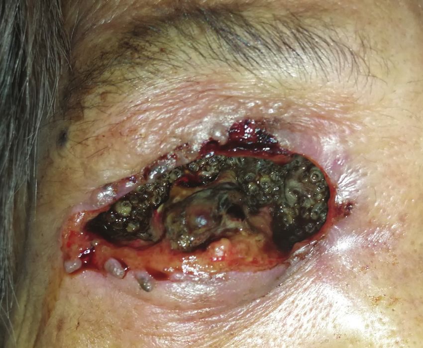
An 88-year-old woman was brought to the hospital immediately after her neighbours noticed that she was bleeding from her right eye.
On examination, her right eye was phthisic with maggot infestation of her right orbit. Over a hundred live maggots were extracted
using forceps. Computed tomography scan revealed the infestation was confined to the right orbit. The patient underwent exenteration
of the right orbit under general anaesthesia. The species was identified by an entomologist as Chrysomya bezziana, which has aggressive
larvae that eat living tissue. This case report demonstrates that orbital myiasis caused by C. bezziana poses a very real risk of intracranial
invasion as they feed on living tissues. Adjacent tissue destruction can be very rapid and definitive treatment involves urgent removal
of its larvae via surgical debridement. To our knowledge, we are reporting the first case of orbital myiasis from a patient in Malaysia.
Therefore, our case report may be helpful in the management of similar case of orbital myiasis.
Keywords: Chrysomya bezziana, orbital myiasis, maggots, exenteration
Introduction Sabah. She lived with another equally infirm relative with poor
social support due to their place of residence in the thick jungle.
Myiasis, derived from the Greek word mya which means fly, She had presented to the Hospital Keningau Eye clinic in 2016
is a condition caused by infestation of dipterous larvae in humans with eyelash loss and a dark pigmented growth with central
or vertebrate animals. It is a rare condition, mainly seen among ulceration over the right upper eyelid that bled on contact.
elderly, infirm, and immobile patients that are poorly cared for.1 Malignancy of the right eyelid was suspected at the time but
Eggs are deposited on an exposed wound or in this case on the she refused excisional biopsy of the lesion and was lost to follow-
eyelid by an adult fly. The larvae can feed on necrotic or living up. Apart from that, she had no known comorbidity such as
tissues, leading to tissue destruction of the orbit. Identification diabetes mellitus or immunosuppression. Three years later, she
of the species responsible for a case of myiasis is not commonly was brought to the emergency department by her neighbors
done.2 We report a case of orbital myiasis in a geriatric patient due to bleeding from her right eye. On examination, there were
who was immobile and emaciated in the interior parts of Sabah, numerous live maggots in her right orbit with the orbital bone
Malaysia. exposed (Figure 1). The right eye was phthisic and surrounded
with necrotic tissue. The fellow eye was erythematous around the
Case Report eyelids with no maggots seen. She was treated for right orbital
An 88-year-old indigenous Murut woman who was bed- myiasis with left preseptal cellulitis. She also presented with
bound with underlying chronic lung disease resided in rural acute kidney injury that eventually improved with treatment.
Address for Correspondence: Yu Siang Ng, Ministry of Health Malaysia, Hospital Keningau, Department of Ophthalmology, Sabah, Malaysia
Phone: +60172211019 E-mail: ysng2003@hotmail.com ORCID-ID: orcid.org/0000-0002-4206-2977
Received: 16.05.2020 Accepted: 23.10.2020
Cite this article as: Ng YS, Gan YK, Tupang L. Severe Orbital Myiasis Caused by Chrysomya bezziana: A Case Report. Turk J Ophthalmol 2021;51:62-65
©Copyright 2021 by Turkish Ophthalmological Association
Turkish Journal of Ophthalmology, published by Galenos Publishing House.
62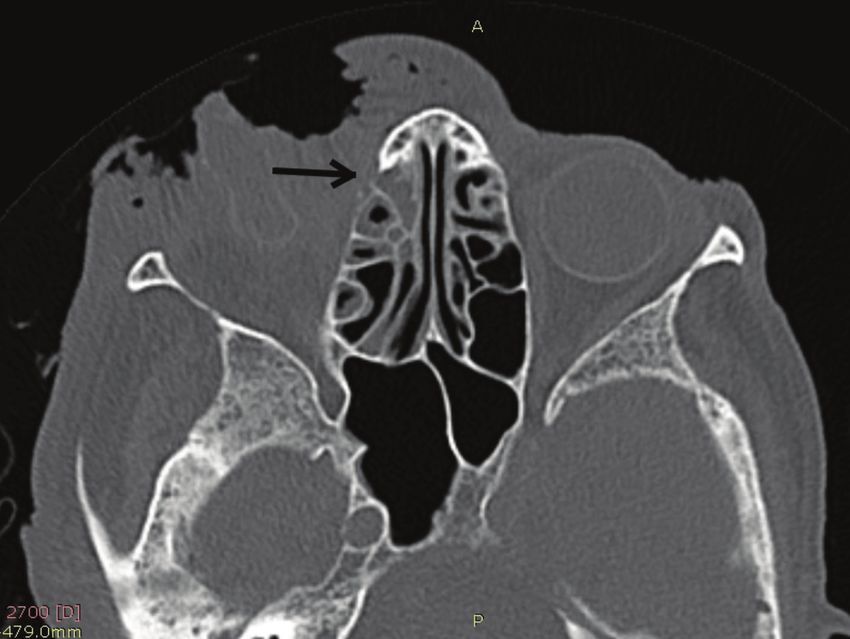
Ng et al. Orbital Myiasis Caused by Chrysomya bezziana
Urgent computed tomography showed the myiasis was confined worldwide), followed by C. bezziana (15 cases), Cochliomyia
to the right orbit, with no brain or paranasal sinuses involved hominivorax (14 cases), and Wohlfartia magnifica (12 cases).4
(Figures 2, 3). Nasoendoscopy and otoscopy showed no myiasis Adult female flies usually lay eggs directly onto necrotic,
in the sinuses and auditory canal. Intravenous ceftriaxone 1 hemorrhagic, or suppurative tissue. The larvae emerge and feed
g once a day and intravenous metronidazole 500 mg 3 times on organic matter, which can lead to complications such as
a day were administered for 2 weeks. Daily extraction of live cornea ulcer, orbital cellulitis, globe invasion, endophthalmitis,
maggots from the wound using forceps was carried out in the blindness, disfigurement, and even death.1,2
ward. More than a hundred maggots were removed bedside The adult C. bezziana fly is green or blue-green in color and
prior to surgery. Patient eventually underwent an exenteration feeds on decaying matter, excreta, and flowers. Approximately
of the right orbit under general anesthesia. Intraoperatively 150-200 eggs at a time are laid by the female adult on exposed
there was bony erosion at the right lamina papyracea and greater wounds and mucous membranes of the mouth, ears, and nose.
wing of the right sphenoid bone, but no tumorous tissue or
After 24 hours, the eggs hatch and the larvae burrow deep into
regional lymphadenopathy were apparent. Live maggots were
living tissue in a screw-like fashion, completing their development
removed intraoperatively on sight. Postoperatively, the right
while feeding on host tissue for 5 to 7 days. Thereafter they
orbital wound was clean with no remaining maggots detected.
Unfortunately, patient succumbed to pneumonia 2 weeks after fall to the ground to pupate. The pupal stage is temperature-
surgery. dependent, with sexual maturation in approximately 1 week to
The maggots were identified by an entomologist 2 months. Thus it takes 2 to 3 months to complete their life
as Chrysomya bezziana. At the entomology & pest unit,
macroscopic examination of the larvae revealed worm-like bodies
of fully-grown third instars, each measuring approximately 13
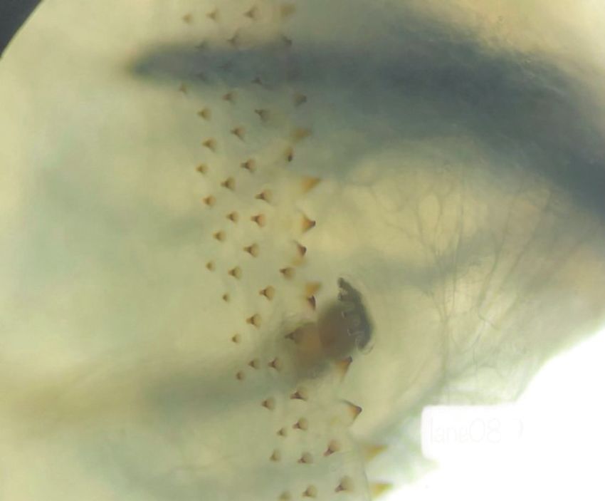
mm in length (Figure 4). They had smooth, broad bodies, with
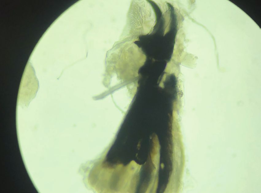
cuticular spines along the body segment (Figure 5). Examination
was focused on the characteristics of the cephaloskeleton (Figure
6), anterior spiracle with 5 papillae (Figure 5), and a posterior
spiracle (Figure 7). The cuticular spines between the first and
second thoracic segments were thorn-like black spines with
single teeth. All these characteristics were unique to C. bezziana,
the Old World screwworm fly.3
Discussion
Orbital myiasis is a rare form of eye disease which has been
reported in both developed as well as developing countries.
A review of the literature revealed that human myiasis is
most commonly caused by Oestrus ovis (32 cases reported
Figure 2. Computed tomography image showing bony erosion of the greater wing
of sphenoid of the right orbit caused by Chrysomya bezziana larvae
Figure 1. An 88-year-old woman from the interior division of Sabah, Malaysia, Figure 3. Computed tomography image showing bony erosion of the lamina
presenting with massive maggot infestation of the right orbit papyracea of the right orbit caused by Chrysomya bezziana larvae
63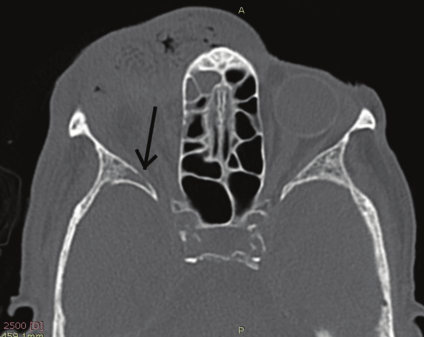
Turk J Ophthalmol 51; 1: 2021

cycle.5 In the present case, the larvae were creamy white in color,                  In contrast to many other fly larvae that feed on necrotic
12-18 mm long, with a worm-like appearance that gradually                            tissue, C. bezziana larvae feed on living tissue of warm-blooded
tapered at the anterior end. The anterior spiracles were palmate                     mammals, and hence is capable of deep tissue penetration and
in shape, each being composed of 4 to 6 lobes arranged in a single                   rapid destruction. It buries deep in the tissue and remains firmly
row located at dorso-posterior margin on each side of prothorax.                     attached using its cuticular spines. Upon contact with forceps
The posterior spiracles consisted of 3 oblique slits encircled by a                  during removal, the larvae tend to escape by burrowing deeper
dark, thick peritreme that is incomplete ventro-medially around                      into the tissue, making removal challenging.
the compressed button. With the help of available keys in the                            In orbital myiasis, the extent of infestation and tissue
literature, the maggots were identified as third instar larvae of                    destruction should be delineated to exclude intracranial spread
C. bezziana on the basis of anterior and posterior spiracles and the                 using computed tomography or magnetic resonance imaging. If
cephalophyaryngeal apparatus.5,6                                                     orbital involvement is localized and less extensive, non-invasive
     C. bezziana is an obligate parasite of mammals, commonly                        mechanical removal of the maggots can be done.3 Non-tooth
found in Asia, tropical Africa, India, and Papua New Guinea.                         forceps are reported to be suitable for larvae removal to avoid
                                                                                     breaking the larvae, which may lead to foreign body or allergic
                                                                                     reactions.7 We found epilation forceps especially useful due to
                                                                                     their broad tips for a firmer grasp. We also recommend making
                                                                                     circular motions during removal of deeply burrowed larvae, as
                                                                                     it eases removal and prevents them from breaking midway. All
                                                                                     larvae were removed in toto using this method.

Figure 4. Third instar of Chrysomya bezziana larva removed from the patient’s
right orbit wound
                                                                                     Figure 6. Cephaloskeleton of a Chrysomya bezziana larva (x40 magnification)

Figure 5. Anterior spiracle (5 fingers) and body spine on the thoracic segments of   Figure 7. Peritreme of the posterior spiracle of a Chrysomya bezziana larva (x40
a Chrysomya bezziana larva (x40 magnification)                                       magnification)

64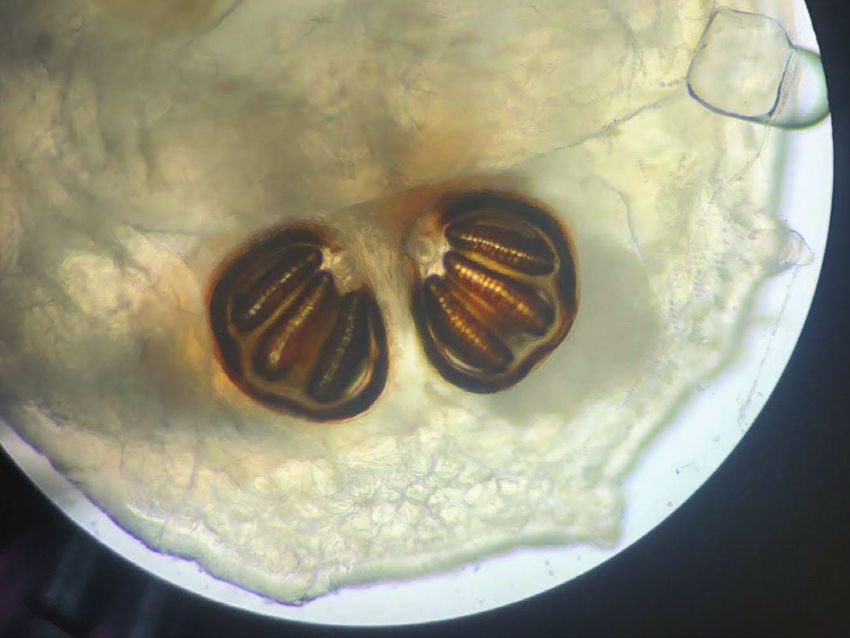
Ng et al. Orbital Myiasis Caused by Chrysomya bezziana

     It has also been reported that materials such as petroleum              Conflict of Interest: No conflict of interest was declared by
jelly or pork fat helps in asphyxiating larvae, forcing them to          the authors.
protrude to the surface intermittently.8 This makes the extraction           Financial Disclosure: The authors declared that this study
easier.                                                                  received no financial support.
     The degree of orbital involvement was extensive in our
patient, and an exenteration and debridement of necrotic tissue               References
was done to prevent intracranial extension.1                             1.   Yeung JC, Chung CF, Lai JS. Orbital myiasis complicating squamous cell
     There are anecdotal reports that oral or topical ivermectin              carcinoma of eyelid. Hong Kong Med J. 2010;16:63-65.
is beneficial in the treatment of severe orbital myiasis by              2.   Spradbery JP. Identification Of Larvae. Old World Screw-worm fly: A
C. hominivorax because it paralyzes the larvae.9 However, overall             Diagnostic Manual (3rd ed). Animal Health Australia; 2017:37-44.
these are insufficient evidence to support the use of ivermectin in      3.   Francesconi F, Lupi O. Myiasis. Clin Microbiol Rev. 2012;25:79-105.
                                                                         4.   Singh A, Singh Z. Incidence of myiasis among humans-a review. Parasitol Res.
C. bezziana orbital myiasis.3                                                 2015;114:3183-3199.
     In conclusion, infestation by this fly species causes rapid         5.   Sukontason KL, Piangjai S, Boonsriwong W, Bunchu N, Ngern-klun R,
tissue destruction and requires urgent definitive treatment via               Vogtsberger RC, Sukontason K. Observations of the third instar larva and
surgical debridement.                                                         puparium of Chrysomya bezziana (Diptera: Calliphoridae). Parasitol Res.
     Informed Consent: As the patient is deceased, informed                   2006;99:669-674.
                                                                         6.   Erzinclioglu YZ. Studies on morphology and Taxonomy of the immature
consent to report this case was obtained from the patient’s next-             stages of Calliphoridae, with analysis of phylogenetic relationships within
of-kin. This case report has been approved by National Institutes             the family, and between it and other groups in the Cyclorrhapha (Diptera),
of Health Malaysia for publication. NMRR ID: NMRR-19-                         Durham E-Thesis, Durham University; 1984:63-89.
3321-51464.                                                              7.   Hochedez P, Caumes E. Common skin infections in travelers. J Travel Med.
     Peer-review: Externally peer reviewed.                                   2009;15:252-262.
                                                                         8.   Diaz JH. The epidemiology, diagnosis, management, and prevention of
    Authorship Contributions                                                  ectoparasitic diseases in travelers. J Travel Med. 2006;13:100-111.
    Surgical and Medical Practices: Y.S.N., Y.K.G., Concept:             9.   Osorio J, Moncada L, Molano A, Valderrama S, Gualtero S, Franco-Paredes
Y.K.G., Design: Y.S.N., Data Collection or Processing: Y.S.N.,                C. Role of Ivermectin in the Treatment of Severe Orbital Myiasis Due to
                                                                              Cochliomyia hominivorax. Clin Infect Dis. 2006;43:57.
Y.K.G., L.T., Analysis or Interpretation: Y.S.N., Y.K.G., Literature
Search: Y.S.N., Y.K.G., L.T., Wrting: Y.S.N., Y.K.G., L.T.

                                                                                                                                                      65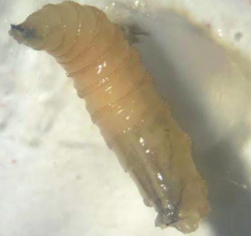
You can also read