Clinical and immunohistochemical findings of splenic mast cell tumour in a cat: A case report
←
→
Page content transcription
If your browser does not render page correctly, please read the page content below
Case Report                                                                       Veterinarni Medicina, 66, 2021

                                                                      https://doi.org/10.17221/11/2021-VETMED

Clinical and immunohistochemical findings of splenic
mast cell tumour in a cat: A case report
Seokwoo Lee1, Min Soo Kang2, Yunho Jeong1, Yoonhwan Kim1,
Ho-Hyun Kwak3, Eun Wha Choi4, Sooyoung Choi5, Inchul Park5,
Jin-Young Chung1, Jung Hoon Choi2, Jin-Ok Ahn1*
1
  Department of Veterinary Internal Medicine and Institute of Veterinary Science, College
  of Veterinary Medicine, Kangwon National University, Chuncheon, Republic of Korea
2
  Department of Anatomy, College of Veterinary Medicine, Kangwon National University,
  Chuncheon, Republic of Korea
3
  Department of Veterinary Surgery, College of Veterinary Medicine, Kangwon National
  University, Chuncheon, Republic of Korea
4
  Department of Veterinary Clinical Pathology, College of Veterinary Medicine, Kangwon
  National University, Chuncheon, Republic of Korea
5
  Department of Veterinary Diagnostic Imaging, College of Veterinary Medicine, Kangwon
  National University, Chuncheon, Republic of Korea
Seokwoo Lee and Min Soo Kang contributed equally to this work
*Corresponding author: joahn@kangwon.ac.kr

Citation: Lee S, Kang MS, Jeong Y, Kim Y, Kwak HH, Choi EW, Choi S, Park I, Chung JY, Choi JH, Ahn JO (2021): Clinical
and immunohistochemical findings of splenic mast cell tumour in a cat: A case report. Vet Med-Czech 66.

Abstract: A 6-year-old, spayed, female, domestic shorthair cat presented with a 4-month history of chronic inter-
mittent vomiting and anorexia. The haematologic results indicated moderate anaemia and a circulating mast cell
population. The abdominal radiography revealed a markedly enlarged spleen. The cytological analysis of the spleen
showed a uniform population of mast cells, and a diagnosis of systemic mastocytosis (splenic mast cell tumour
with mastocytaemia) was made. This diagnosis was subsequently confirmed by the histopathological examina-
tion of the spleen. The immunohistochemistry for KIT showed KIT pattern II (focal cytoplasmic expression).
A splenectomy and chemotherapy with vinblastine and prednisolone resulted in remission of the anaemia and
other clinical signs. This case report highlights the importance of cytological evaluations of peripheral blood
smears and/or aspirates of enlarged spleens for diagnosing splenic mast cell tumours and for quickly initiating
the appropriate treatment.

Keywords: KIT; mastocytaemia; prednisolone; spleen; splenectomy; vinblastine

  Unlike mast cell tumours (MCTs) in dogs, which            2013). A visceral MCT is more common in cats than
are mostly cutaneous/subcutaneous in nature,                in dogs, with approximately 50% of feline MCTs oc-
MCTs in cats can be classified anatomically into            curring at extracutaneous sites. Although a splenic
cutaneous, splenic, or intestinal forms with some           MCT is one of the most common splenic tumours
overlap between these variants (Henry and Herrera           in cats, it is still a relatively rare diagnosis, and

Supported by 2019 Research Grant from Kangwon National University.

1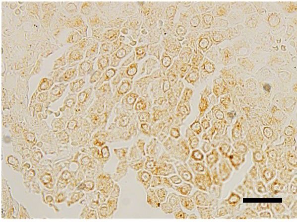
Case Report                                                                      Veterinarni Medicina, 66, 2021

                                                                    https://doi.org/10.17221/11/2021-VETMED

the literature characterising the disease is sparse,       no significant findings. Mast cells were observed
consisting mainly of case reports and small case           in the peripheral blood smears (Figure 1A).
series (Allan et al. 2000; Evans et al. 2018).               Splenomegaly was detected on the X-ray and so-
  Systemic mastocytosis involves neoplastic mast           nographic examinations of the abdomen. A com-
cell proliferation in multiple visceral organs, espe-      puted tomography (CT) scan revealed an enlarged
cially the spleen and liver, and in the bone marrow.       spleen and multiple enlarged lymph nodes in the
Systemic mastocytosis is rarely observed in cats           cranial, mediastinal, sternal, splenic, sub-lumbar,
(Piviani et al. 2013); however, when present, pa-          and mesenteric regions (Figure 1B).
tients commonly suffer from lethargy, anorexia,              The initial diagnostic evaluation included an ul-
weight loss, and intermittent vomiting (Allan et al.       trasound-guided fine-needle aspiration (FNA)
2000). These clinical signs are nonspecific, making        of the spleen for the cytological examination.
this diagnosis clinically challenging.                     Diff-Quik-stained smears of the spleen contained
  The aetiology of feline MCTs is currently un-            numerous, well-differentiated mast cells cover-
known; however, it is evident that cats with MCTs          ing the entire specimen, with basophilic granules
possess somatic activating mutations in the KIT
(Isotani et al. 2006; Sabattini et al. 2016). The            Figure 1
KIT proto-oncogene encodes the receptor tyros-
ine kinase, KIT, which regulates the proliferation,
differentiation, and migration of normal mast
cells (Okayama and Kawakami 2006). However,
the prognostic relevance of aberrant cytoplasmic
KIT protein localisation has not yet been clearly
elucidated for feline splenic MCTs.
  This study aimed at determining the clinical
signs and immunohistochemical findings in a cat
with a splenic MCT.
                                                             (A)

Case description

  A 6-year-old, spayed, female, domestic shorthair
cat (weight: 4.2 kg) was referred to our hospital
with a 4-month history of chronic intermittent
vomiting, anorexia, and weight loss. Before being
admitted to our hospital, the cat was taken to a lo-
cal hospital, where she was tentatively diagnosed
with inflammatory bowel disease and administered
prednisolone and antiemetic drugs until her symp-
toms became more severe.
  On physical examination, the cat was lethargic
and dehydrated with pale mucous membranes. The
abdominal cavity was largely distended with a firm,
palpable organ on the left side. The haematological
                                                              (B)
findings included a normal white blood cell count
(14.96 × 10⁹/l; reference interval, 2.87–17.02 × 10⁹/l),   Figure 1. Light microscopy findings of the peripheral
a decreased red blood cell count (4.66 × 10¹²/l; ref-      blood smear (A) and post-contrast computed tomogra-
erence interval, 6.54–12.20 × 10¹²/l), a decreased         phy (CT) findings (B)
red blood cell specific volume (24.8%; reference           (A) A mast cell (black arrow) was observed in the peripheral
interval, 30.3–52.3%), and decreased platelets             blood smear (Diff-Quik, scale bar = 10 µm). (B) CT images
(112 × 10 9/l; reference interval, 151–600 × 10 9/l).      revealed an enlarged spleen (white asterisk) and multiple
The serum biochemistry and urinalysis revealed             enlarged mesenteric lymph nodes (white arrows)

2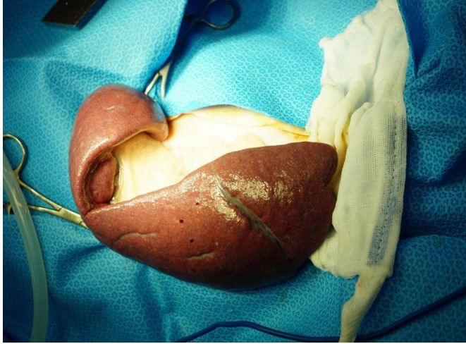
Case Report Veterinarni Medicina, 66, 2021
https://doi.org/10.17221/11/2021-VETMED
in the background (Figure 2A). Based on these find- The histopathological analysis showed that the
ings, a tentative diagnosis of a splenic MCT was native parenchyma of the spleen had largely
made, and the cat underwent a total splenectomy been replaced by sheets of neoplastic mast cells
within a few days (Figure 2B). (Figure 3A–B). These cells were round with distinct
Figure 2
(A) (B)
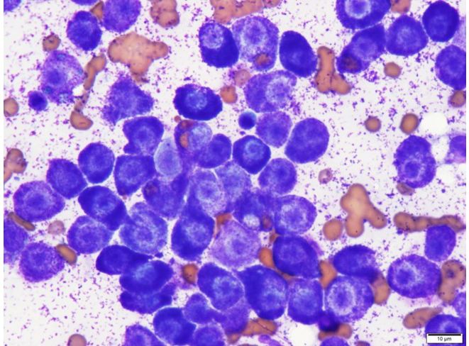
Figure 2. Light microscopy findings of the splenic aspirate (A) and the enlarged and firm spleen (B) of a cat with
a splenic mast cell tumour
(A) Cells in the splenic aspirate were almost entirely composed of well-differentiated mast cells (Diff-Quik, scale bar =
10 µm). (B) A markedly enlarged spleen in a cat with a splenic mast cell tumour
Figure 3
(A) (B)
(C) (D)
Figure 3. Histopathological (A–C) and immunohistochemical (D) images of a feline splenic mast cell tumour
(A, B) Histopathology of the splenic mast cell tumour showed well-differentiated mast cells on haematoxylin and eosin
staining. (C) Granulated mast cells interspersed in the splenic parenchyma on toluidine blue staining. (D) Immunohis-
tochemistry for c-KIT in a feline splenic mast cell tumour. Scale bar: A = 50 µm, B–D = 20 µm
3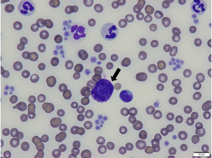
Case Report                                                                  Veterinarni Medicina, 66, 2021

                                                                 https://doi.org/10.17221/11/2021-VETMED

cell borders and an abundant, poorly granulated,         absence of apparent splenomegaly or if fine-needle
basophilic cytoplasm. Mild anisocytosis and an-          aspirates of the affected internal organs are not ex-
isokaryosis were observed. The nuclei were round         amined (Woldemeskel et al. 2017). Mastocytaemia
with coarsely clumped chromatin and prominent            can be detected by examination of a peripheral
nucleoli. Toluidine blue staining revealed large         blood smear or the buffy coat (Piviani et al. 2013).
round cells with characteristic metachromatic            A cytological evaluation of the peripheral blood
granules of mast cells (Figure 3C). An immuno-           smear or the buffy coat should be performed in cats
histochemical staining for KIT was performed             with weight loss and vomiting of unknown aetiology
on sections of the spleen [Dako rabbit polyclonal        to rule out mastocytaemia.
anti-CD117 (KIT) antibody diluted 1 : 150; Dako            In the current case, there was mild anisocytosis
North America, Inc., Carpinteria, CA, USA]. The          and anisokaryosis, the mitotic rate was low, and
KIT protein expression was classified based on the       neither uninucleated nor multinucleated neoplastic
patterns described for canine MCTs (Kiupel et al.        giant cells were observed, which is most consistent
2004). The immunohistochemistry for KIT showed           with the well-differentiated form of MCT in cats.
positive dark brown staining in the mast cells,          Staging procedures for feline MCTs are far less
mostly with a focal cytoplasmic expression (KIT          standardised than for dogs, and their utility is still
pattern II) (Figure 3D).                                 being debated, probably due to the lower frequency
  A month after the splenectomy, the cat’s symp-         of MCTs in cats and because the biological behav-
toms, including the anaemia, vomiting, and anorex-       iour of these tumours is considered less aggressive
ia, had almost completely disappeared, and the cat       (Sabattini et al. 2017).
was in an overall better clinical condition. However,      In canine MCTs, juxtamembrane domain KIT
mast cells were still found in the peripheral blood      mutations and aberrant cytoplasmic KIT protein
smear; therefore, chemotherapy with vinblastine          localisation have been associated with increased
and prednisolone was initiated. The patient re-          cellular proliferation and reduced progression-free
ceived vinblastine (Vinblastine; Teva-Handok,            and overall survival (Webster et al. 2006). However,
Seoul, Republic of Korea) 10 mg/m 2 intravenous-         the prognostic relevance of aberrant cytoplasmic
ly every two weeks and prednisolone (Solondo;            KIT protein localisation in feline MCTs has not
Yuhan, Seoul, Republic of Korea) 5 mg/cat orally         yet been clearly elucidated. Aberrant cytoplasmic
once a day. Two weeks after the chemotherapy, the        KIT immunohistochemical expression has been
mast cells in the peripheral blood smears had dis-       reported in 29–67% of feline cutaneous MCTs, and
appeared without evidence of myelosuppression.           a higher frequency has been observed in tumours
After the fourth chemotherapy session, the owner         with unfavourable outcomes (Sabattini and Bettini
reported that the cat appeared to be in good over-       2010; Mallett et al. 2013). Previous studies have
all health. At the last follow-up, the cat was doing     reported that aberrant cytoplasmic KIT protein lo-
well without recurrence of MCT after treatment           calisation does not appear to be strictly correlated
(for 22 months).                                         with the biological behaviour or prognostic rel-
                                                         evance (Sabattini et al. 2013; Sabattini et al. 2017).
                                                           In this case, the symptoms almost completely dis-
DISCUSSION AND CONCLUSIONS                               appeared and the clinical signs became more stable
                                                         after the patient underwent splenectomy. Though
   This case involved a cat with a splenic MCT and       there is a lack of a standardised treatment approach
mastocytaemia. In cats, systemic mastocytosis is al-     for this disease, a splenectomy is considered the
most exclusively associated with visceral MCTs and       treatment of choice for splenic MCTs, even in cats
is frequently associated with splenic MCTs (Allan        with systemic involvement (Evans et al. 2018).
et al. 2000; Woldemeskel et al. 2017). When mast         Although the role of chemotherapy is not yet cer-
cells are identified in the peripheral blood of a cat,   tain, it is known that prednisolone and vinblastine
an MCT, and especially a splenic MCT with sys-           multi-agent protocols can partially confer a high
temic mastocytosis, should be considered (Skeldon        treatment response in dogs and cats with mast cell
et al. 2010). Because splenic MCTs in cats can pre-      disease. Furthermore, some partial responses have
sent with nonspecific clinical signs of a systemic       been observed with vinblastine treatment in cats
illness, this disease may not be recognised in the       with splenic MCTs (Kraus et al. 2015). After our

4
Case Report Veterinarni Medicina, 66, 2021
https://doi.org/10.17221/11/2021-VETMED
patient’s first chemotherapy session, her masto- as prognostic tools for canine cutaneous mast cell tumors.
cytaemia disappeared and was well maintained. Vet Pathol. 2004 Jul;41(4):371-7.
Following treatment, intermittent restaging, which Kraus KA, Clifford CA, Davis GJ, Kiefer KM, Drobatz KJ.
involves monitoring of the enlarged lymph nodes Outcome and prognostic indicators in cats undergoing
and mastocytaemia, is required to identify any dis- splenectomy for splenic mast cell tumors. J Am Anim
ease recurrence or progression (Piviani et al. 2013). Hosp Assoc. 2015 Jul-Aug;51(4):231-8.
This case report highlights the importance of cy- Mallett C, Northrup N, Saba C, Rodriguez C, Rassnick K,
tological evaluations of peripheral blood smears Gieger T, Childress M, Howerth E. Immunohistochemi-
and/or aspirates of enlarged spleens for diagnos- cal characterization of feline mast cell tumors. Vet Pathol.
ing splenic MCT-associated systemic mastocyto- 2013 Jan;50(1):106-9.
sis in cats and for initiating appropriate treatment Okayama Y, Kawakami T. Development, migration, and sur-
in a timely manner. Twenty-two months after treat- vival of mast cells. Immunol Res. 2006 Feb;34(2):97-115.
ment, this cat was doing well without any recurrence Piviani M, Walton RM, Patel RT. Significance of mastocy-
of an MCT. In this case, no prognostic value was temia in cats. Vet Clin Pathol. 2013 Mar;42(1):4-10.
associated with the KIT staining pattern; therefore, Sabattini S, Barzon G, Giantin M, Lopparelli RM, Da-
further studies are necessary to assess the utility casto M, Prata D, Bettini G. Kit receptor tyrosine kinase
of the immunohistochemical analysis of the KIT dysregulations in feline splenic mast cell tumours. Vet
protein in cats with a splenic MCT. Comp Oncol. 2017 Sep;15(3):1051-61.
Sabattini S, Bettini G. Prognostic value of histologic and
immunohistochemical features in feline cutaneous mast
Conflict of interest cell tumors. Vet Pathol. 2010 Jul;47(4):643-53.
Sabattini S, Frizzon MG, Gentilini F, Turba M, Capitani O,
The authors declare no conflict of interest. Bettini G. Prognostic significance of kit receptor tyrosine
kinase dysregulations in feline cutaneous mast cell tu-
mors. Vet Pathol. 2013 Sep;50(5):797-805.
REFERENCES Sabattini S, Giantin M, Barbanera A, Zorro Shahidian L,
Dacasto M, Zancanella V, Prata D, Trivigno E, Bettini G.
Allan R, Halsey T, Thompson K. Splenic mast cell tumour Feline intestinal mast cell tumours: Clinicopathological
and mastocytaemia in a cat: Case study and literature characterisation and kit mutation analysis. J Feline Med
review. N Z Vet J. 2000 Aug;48(4):117-21. Surg. 2016 Apr;18(4):280-9.
Evans BJ, O’Brien D, Allstadt SD, Gregor TP, Sorenmo KU. Skeldon NC, Gerber KL, Wilson RJ, Cunnington SJ. Mas-
Treatment outcomes and prognostic factors of feline tocytaemia in cats: Prevalence, detection and quantifica-
splenic mast cell tumors: A multi-institutional retrospec- tion methods, haematological associations and potential
tive study of 64 cases. Vet Comp Oncol. 2018 Mar;16 implications in 30 cats with mast cell tumours. J Feline
(1):20-7. Med Surg. 2010 Dec;12(12):960-6.
Henry C, Herrera C. Mast cell tumors in cats: Clinical up- Webster JD, Yuzbasiyan-Gurkan V, Kaneene JB, Miller R,
date and possible new treatment avenues. J Feline Med Resau JH, Kiupel M. The role of c-kit in tumorigenesis:
Surg. 2013 Jan;15(1):41-7. Evaluation in canine cutaneous mast cell tumors. Neo-
Isotani M, Tamura K, Yagihara H, Hikosaka M, Ono K, plasia. 2006 Feb;8(2):104-11.
Washizu T, Bonkobara M. Identification of a c-kit exon 8 Woldemeskel M, Merrill A, Brown C. Significance of cyto-
internal tandem duplication in a feline mast cell tumor logical smear evaluation in diagnosis of splenic mast cell
case and its favorable response to the tyrosine kinase in- tumor-associated systemic mastocytosis in a cat (Felis
hibitor imatinib mesylate. Vet Immunol Immunopathol. catus). Can Vet J. 2017 Mar;58(3):293-5.
2006 Nov 15;114(1-2):168-72.
Kiupel M, Webster J, Kaneene J, Miller R, Yuzbasiyan-Gur- Received: February 1, 2021
kan V. The use of kit and tryptase expression patterns Accepted: May 11, 2021
5You can also read