A Case of Extra-adrenal Retroperitoneal Ganglioneuroma in a 9-year-old Female: A New Case Report with a Comprehensive Literature Review
←
→
Page content transcription
If your browser does not render page correctly, please read the page content below
Clinical Case Report
                                                                                                                                Case Report
                                                                                                         Central Eur J Paed 2020;16(2):xx-xx
                                                                                                                                       DOI

A Case of Extra-adrenal Retroperitoneal Ganglioneuroma in a 9-year-old Female:
A New Case Report with a Comprehensive Literature Review

Zlatan Zvizdic1, Nedzad Rustempasic2, Irmina Sefic Pasic3, Faruk Skenderi4, Semir Vranic5
1
 Clinic of Pediatric Surgery, University Clinical Center Sarajevo, Sarajevo, Bosnia and Herzegovina, 2 Clinic of Cardiovascular
Surgery, University Clinical Center Sarajevo, Sarajevo, Bosnia and Herzegovina, 3 Department of Radiology, University Clinical
Center Sarajevo, Sarajevo, Bosnia and Herzegovina,4 Department of Pathology, University Clinical Center Sarajevo, Sarajevo,
Bosnia and Herzegovina and 5 College of Medicine, QU Health, Qatar University, Doha, Qatar
Correspondence: semir.vranic@gmail.com or svranic@qu.edu.qa: Tel.: + 974 4403 7873; Fax.: + 974 4403 3344; ORCID:
 http://orcid.org/0000-0001-9743-7265
Received: April 25, 2020 Accepted: May 21, 2020

Abstract
Objective – We present herein a new case and survey comprehensively literature on this rare condition. Case report – A 9-year-
old girl with a medical history of surgical correction of clubfoot three months earlier presented to our department with an inci-
dentally detected abdominal mass during diagnostic workup for orthopedic surgery. A CT scan revealed a solid right extra-adrenal
mass measuring 7×6 cm. It was compressing/involving the infrahepatic part of inferior vena cava, right renal vein and artery with
an incomplete encasing of the abdominal aorta. The surgery was successfully performed. The histopathological analysis confirmed
GN. Conclusions – Although pediatric extra-adrenal retroperitoneal ganglioneuroma (GN) are rare, their propensity for envelop-
ing major blood vessels is not. GNs should be considered in the differential diagnosis of any circumscribed retroperitoneal mass.
These tumors can be successfully treated with surgery that leads to an excellent outcome, even in case of incomplete resections
with tumor residuals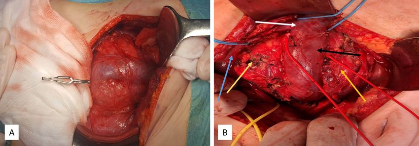
Zlatan Zvizdic et al.: ■ Pediatric Retroperitoneal Ganglioneuroma

is the treatment of choice whenever possible and          adrenal hypoechogenic, somewhat heterogeneous
it usually gives an excellent outcome (7). Unlike         solid mass, occupying the right aspect of the retro-
smaller GNs that are commonly diagnosed in child-         peritoneum, measuring 7 cm in its largest diameter.
hood, large GNs tumors are mainly diagnosed after             Subsequent contrast-enhanced CT scan con-
the age of ten. To date, only a few cases of giant        firmed a vividly enhancing solid right-sided extra-
extra-adrenal retroperitoneal GNs were reported in        adrenal mass measuring 7.4×6.6×4.2 cm that was
pediatric patients, especially below 10 years of age.     compressing/involving the infrahepatic part of
    We present herein a case affecting a 9-year-old       inferior vena cava (IVC), right renal vein and ar-
girl, discuss the therapeutic approach and briefly        tery with an incomplete encasing of the abdomi-
review literature on this rare pediatric condition.       nal aorta (Fig. 1A-B). IVC was markedly dilated. A
                                                          provisional diagnosis of non-functioning, probably
                                                          benign, right extra-adrenal tumor was made. MRI
Case Report
                                                          was also performed to further characterize the mass
A 9-year-old girl, with a medical history of surgi-       (not shown). An ultrasonography-guided percuta-
cal correction of clubfoot three months earlier,          neous fine-needle biopsy (FNAB) of the tumor was
presented to our department with an incidentally          performed revealing a cellular neoplasm composed
detected abdominal mass during diagnostic work-           of ganglion cells and spindle-shaped cells with “ci-
up for orthopedic surgery. The physical examina-          gar-shaped nuclei”. Morphological findings indi-
tion revealed a painless palpable mass on the right       cated a ganglioneuroma (GN).
side of her abdomen. Blood pressure was within                After obtaining parental consent, the patient
the normal range. Family history was negative for         underwent surgery. The lesion was approached
neurofibromatosis and other cancer syndromes.             transperitoneally through an extended transverse
Laboratory investigations (complete blood count,          supra-umbilical incision. After mobilization of the
serum electrolytes, immunoglobulins, and urine)           right colon, an encapsulated right-sided extra-ad-
and endocrine tests (cortisol, adrenocorticotropic        renal retroperitoneal tumor was carefully dissected
hormone levels, and 24-hour urinary catechol-             away from the surrounding adherent structures in-
amines) were all within normal ranges. Routine            cluding IVC, right renal vein and artery, abdomi-
tumor markers were also negative. Abdominal ul-           nal aorta, and the right kidney, but patency of the
trasound examination revealed a right-sided extra-        compressed vessels were maintained (Fig. 2A-B).

Fig. 1A-B. Contrast Enhanced Coronal (a) and Axial (b) CT scan Showing a Large Heterodense Mass in the Right Upper
Abdomen (white Arrows), Compressing Inferior Vena Cava with Incomplete Encasing of Abdominal Aorta.
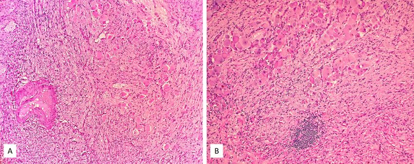
Central Eur J Paed 2020;16(2):x-xx

Fig. 2A-B. Intraoperative Images of the Tumor (yellow Arrows) Arising from Retroperitoneum That Was Adherent to the
Infrahepatic Vena Cava (black Arrow), Right Renal Artery and Vein (white Arrow), and the Right Kidney before (blue
Arrow) (a) and after Mobilization and Exposure of Large Blood Vessels (b).

Fig. 3A-B. Hematoxylin and Eosin (H&E) Slides of the Moderately Cellular Neoplasm at Low Power (40´) Magnification
(a); the Tumor Was Composed of Admixture of Schwann Cells and Scattered Nests and Clusters of Ganglion Cells within
the Fibrous Stroma. The Multinucleated Ganglion Cells Were Numerous Exhibiting Only Mild to Moderate Atypia (b)
(100× Magnification).

Enlarged retroperitoneal lymph nodes were not de-          Discussion
tected. The small cuff of tumor capsule about 0.5
cm was left behind the fascia transversalis, as it was     Ganglioneuromas (GNs), well-differentiated, slow-
not possible to safely dissect it from the paraver-        growing, usually non-secreting and asymptomatic
tebral space and the underlying spinal nerves. The         benign tumors, belong to the group of neuroblas-
tumor specimen was submitted to histopathology             tic tumors originating from neural crest cells that
analysis that confirmed the mass to be a GN (Fig.          comprise a spectrum of both benign tumors like
3A-B). The patient had an uneventful postopera-            GN and malignant tumors such as (ganglio)neuro-
tive recovery and was discharged on the seventh            blastomas (8). Although GNs are often localized in
day. One-year follow-up consisted of visits every 3        the retroperitoneum, they account for only 1% of
months revealing no recurrence of the tumor.               primary retroperitoneal tumors and approximately
Zlatan Zvizdic et al.: ■ Pediatric Retroperitoneal Ganglioneuroma

half of the reported cases are extra-adrenal (2). The                     of the tumor. In our case, the preoperative diag-
differential diagnosis of solid retroperitoneal mass                      nosis of GN was obtained by FNAB and was fur-
includes various tumors with neurogenic/neuro-                            ther confirmed by histopathological analysis. In the
blastic origins (ganglioneuroma, ganglioneuroblas-                        current literature, reports of GN that are reliably
toma intermixed, neuroblastoma, neurofibroma,                             diagnosed by FNAB and its cytological appearance
schwannoma, and pheochromocytoma), adrenal                                are scarce. A literature search revealed only a few
gland tumors (adrenocortical adenoma and carci-                           cases diagnosed by FNAB at this location (24, 25).
noma), various lymphomas (usually non-Hodgkin                             GNs on FNAB can be difficult to differentiate from
type), soft tissue sarcomas (e.g. liposarcoma), and                       other neurogenic/neuroblastic tumors, peripher-
germ cell tumors (e.g. teratoma) (9).                                     al nerve sheath tumors, such as schwannomas or
    Extra-adrenal retroperitoneal ganglioneuromas                         neurofibromas and a meticulous search for mature
in pediatric population are very rare. Our literature                     ganglion cells is critical for making an accurate di-
survey (PubMed/MEDLINE, Scopus, and Web of                                agnosis. Taken together, a broad differential diag-
Science) revealed a few reported cases and small                          nosis and subtle morphological details give FNAB
case series (1, 4, 7, 10-20) with very few cases re-                      a limited clinical utility (26).
ported in the patients < 10 years (summarized in                               Retroperitoneal GNs may clinically remain si-
Table 1). Scherer et al. (2001) reported the largest                      lent for a long time and the diagnosis is often inci-
case series of 5 patients (4 patients under the age of                    dental. Despite their benign nature, retroperitoneal
10) depicting the radiological (CT and MRI) fea-                          GNs show the tendency to partially or completely
tures of retroperitoneal GNs. The tumor size varied                       surround large blood vessels without compromis-
between 4 and 11 cm and girls were predominantly                          ing the lumen in most cases, making their surgical
affected (4/5). Although GNs may present as an                            excision extremely challenging (27).
isolated finding as confirmed in our case, these tu-                           The optimal treatment approach for most pa-
mors may be occasionally seen in association with                         tients with retroperitoneal GN is a complete surgi-
neurofibromatosis type 1 (NF1) (21-23).                                   cal excision (7). However, this approach has certain
    The widespread use of imaging techniques in                           limitations, particularly in the case in which the
clinical practice has contributed to an increase in                       large GN surrounds and compresses large blood
the number of GNs detected incidentally. How-                             vessels making its dissection extremely difficult. A
ever, it is difficult to make an accurate preoperative                    mitigating circumstance is a fact that the enclosed
diagnosis of GN and definitive diagnosis is based                         blood vessels commonly have no narrow lumen or
on histopathological analysis after surgical excision                     filling defect (28). In this case, GN enclosed IVC

    Table 1. A Summary of the Previous Studies that Reported Extra-adrenal Retroperitoneal Ganglioneuromas in Children
Central Eur J Paed 2020;16(2):x-xx
and right renal artery and vein and compressed the 3. Spinelli C, Rossi L, Barbetta A, Ugolini C, Strambi S. Inci-
anterior side of the right kidney. The dissection of dental ganglioneuromas: a presentation of 14 surgical cases
and literature review. Journal of endocrinological investi-
GN away from IVC was the crucial step in the sur- gation. 2015;38(5):547-54.
gery. In our case, all large blood vessels were pro-
4. Scherer A, Niehues T, Engelbrecht V, Modder U. Imaging
tected effectively. diagnosis of retroperitoneal ganglioneuroma in childhood.
Although the postoperative prognosis is usually Pediatr Radiol. 2001;31(2):106-10.
excellent, long-term follow-up is recommended
5. Yamaguchi K, Hara I, Takeda M, Tanaka K, Yamada Y,
because of sporadically reported cases of local re- Fujisawa M, et al. Two cases of ganglioneuroma. Urology.
currences (29). However, Decarolis et al. suggested 2006;67(3):622 e1-4.
that even incomplete resection of GN is not associ- 6. Guo YK, Yang ZG, Li Y, Deng YP, Ma ES, Min PQ, et al.
ated with increased risk of progression if the tumor Uncommon adrenal masses: CT and MRI features with
residuals areZlatan Zvizdic et al.: ■ Pediatric Retroperitoneal Ganglioneuroma
16. Steinberg SH. Presacral retroperitoneal ganglioneuroma in ated with von Recklinghausen’s disease: case report. Surgi-
a 4 1/2-year-old child; report of a case and review of the cal neurology. 2004;61(5):468-73; discussion 73.
literature. J Pediatr. 1955;46(5):562-72.
24. Ponce-Camacho MA, Diaz de Leon-Medina R, Miranda-
17. Maher OM, Marco SA, Sadanandan S, Fireman F, Sedrak Maldonado I, Garza-Guajardo R, Hernandez-Salazar J,
A. Retroperitoneal ganglioneuroma and reversible posteri- Barboza-Quintana O. A 5-year-old girl with a congenital
or leukoencephalopathy in a child with acute lymphoblas- ganglioneuroma diagnosed by fine needle aspiration bi-
tic leukemia. J Pediatr Hematol Oncol. 2014;36(8):665-6. opsy: a case report. CytoJournal. 2008;5:5.
18. Noguchi S, Kaga Y, Takahashi Y, Aoyagi K, Nakamura K, 25. Chiaffarano J, Alexander M, Yee-Chang M, Simsir A,
Kamiya Y, et al. [A case of recurrent paraneoplastic cerebel- Shi Y. Ganglioneuroma: An Unusual Entity Diagnosed
lar ataxia with antibodies to GluR epsilon 2 causally relat- on Endoscopic Ultrasound Guided Fine Needle Aspi-
ed to ganglioneuroma]. No To Hattatsu. 2010;42(4):297- ration Biopsy. American journal of clinical pathology.
301. 2016;146(suppl_1).
19. Zugor V, Schott GE, Kuhn R, Labanaris AP. Retroperito- 26. Ma J, Liang L, Liu H. Multiple cervical ganglioneuroma:
neal ganglioneuroma in childhood--a presentation of two A case report and review of the literature. Oncology letters.
cases. Pediatr Neonatol. 2009;50(4):173-6. 2012;4(3):509-12.
20. Vara Castrodeza A, Madrigal Rubiales B, Perez del Rio 27. Vasiliadis K, Papavasiliou C, Fachiridis D, Pervana S,
MJ, Garcia Hernandez JB, Ablanedo Ablanedo P. [Retro- Michaelides M, Kiranou M, et al. Retroperitoneal extra-
peritoneal ganglioneuroma in childhood]. Arch Esp Urol. adrenal ganglioneuroma involving the infrahepatic inferior
2000;53(7):648-51. vena cava, celiac axis and superior mesenteric artery: A case
report. Int J Surg Case Rep. 2012;3(11):541-3.
21. Bacci C, Sestini R, Ammannati F, Bianchini E, Palladino
T, Carella M, et al. Multiple spinal ganglioneuromas in 28. Zheng X, Luo L, Han FG. Cause of postprandial vom-
a patient harboring a pathogenic NF1 mutation. Clinical iting - a giant retroperitoneal ganglioneuroma enclosing
genetics. 2010;77(3):293-7. large blood vessels: A case report. World J Clin Cases.
2019;7(17):2617-22.
22. Kannu P, Nour M, Irving M, Xie J, Loder D, Lai J, et
al. Paraspinal ganglioneuroma in the proband of a large 29. Decarolis B, Simon T, Krug B, Leuschner I, Vokuhl C,
family with mild cutaneous manifestations of NF1, car- Kaatsch P, et al. Treatment and outcome of Ganglioneuro-
rying a deep NF1 intronic mutation. Clinical genetics. ma and Ganglioneuroblastoma intermixed. BMC Cancer.
2013;83(2):191-4. 2016;16:542.
23. Kyoshima K, Sakai K, Kanaji M, Oikawa S, Kobayashi 30. De Bernardi B, Gambini C, Haupt R, Granata C, Rizzo A,
S, Sato A, et al. Symmetric dumbbell ganglioneuromas of Conte M, et al. Retrospective study of childhood ganglio-
bilateral C2 and C3 roots with intradural extension associ- neuroma. J Clin Oncol. 2008;26(10):1710-6.You can also read