Choroid plexus carcinoma: A case report and literature review - OAText
←
→
Page content transcription
If your browser does not render page correctly, please read the page content below
Surgery and Rehabilitation
Case Report ISSN: 2514-5959
Choroid plexus carcinoma: A case report and literature
review
Azhani C1, Chan KH1, Fadli M2 and Saufi A*1
Department of Neurosurgery, Kulliyyah of Medicine, International Islamic University Malaysia
1
Department of Pathology, Hospital Tengku Ampuan Afzan, Malaysia
2
Abstract
Choroid plexus carcinoma is a rare tumor representing less than 1% of all brain tumors. In adult, the incidence is extremely rare making the diagnosis difficult.
Majority of choroid plexus tumor is found in the ventricle. However, ectopic sites such as intracranial extraventricular or spine have been reported. We report a case
of choroid plexus carcinoma in a 39-year-old man. The clinical presentation, pathology and management are discussed.
Introduction hyperintense on T2 and enhanced post Gadolinium. There was also
small hypointense area seen at superior part of the mass that may
Choroid plexus carcinoma is a rare tumor especially in adult. In
represent micro bleeding. The right lateral ventricle was compressed
total population, choroid plexus tumor only represents less than 1% of
and contralateral lateral ventricle was dilated. There was midline shift
all brain tumors [1]. Out of all choroid plexus tumors, only 15-30% is
present. The right middle cerebral artery was displaced anteriorly
carcinoma. In adult however, due to its extremely rare occurrence, the
diagnosis of choroid plexus carcinoma should be made with caution as but vessels were patent and normal in caliber. The mass also was
it more frequently resembles a metastatic papillary tumor such as from significantly vascular. Based on radiology, a diagnosis of vascular high
kidney and thyroid [2]. grade tumor such as GBM was kept.
Most of choroid plexus tumor arises in intraventricular, but cases He underwent right craniotomy and debulking of tumor through
have been reported as arising from ectopic sites such as intracranial but a transsylvian. Intraoperatively, the tumor was fragile, soft and highly
extraventricular and also in spinal canal without intracranial lesion [2- vascular, consistent with features of high grade tumor as in GBM.
12]. It has been suggested in a few literatures that for older children and There was feeding vessel from the lenticulostriate artery. Partial
adult, a gross total resection is the best method of treatment followed resection was done in view of intra-operative bleeding and tensed brain
by adjuvant chemotherapy and radiotherapy [1,3,4]. However, surgical which limits further dissection. Post-operatively, he was kept sedated.
resection poses a challenge as it frequently intensely vascular tumor, Unfortunately, he deteriorated two weeks after surgery and succumbed
leading to extreme and heavy bleeding during surgery, thus affecting to death.
patients’ survival [2]. This case report highlights the extremely rare
case of adult choroid plexus carcinoma, arising from basal ganglia. The Histopathology examination was reported as choroid plexus
clinical presentation, pathology and management are well discussed. carcinoma with tumor exhibiting papillary pattern with central
fibrovascular cone lined by cuboidal to columnar to pseudostratified
Case summary epithelium. There were abundant mitotic figure and tumor necrosis.
Immunohistochemistry stains positive for epithelial membrane antigen
We report the case of a 39-year-old man presented with reduced
(EMA) (Figure 3).
conscious level for 3 days prior to admission preceded by two weeks
history of headache and progressive left sided weakness. His wife Discussion
gave a history of facial asymmetry two months earlier. At the time of
admission, his Glasgow Coma Scale (GCS) was 10/15 (E4V1M5). Pupils Adult brain tumors are more commonly secondary than primary
were bilaterally equal and reactive. Papilledema was present bilaterally brain tumors. Primary brain tumors comprises about 1.6% of all tumors
Left-side hemiparesis (MRC grade 1/5) with left upper motor neuron diagnosed and of these Choroid Plexus Tumor (CPT) represents
facial paresis was present. only less than 1% [1,4,11]. Choroid Plexus Carcinoma (CPC) is less
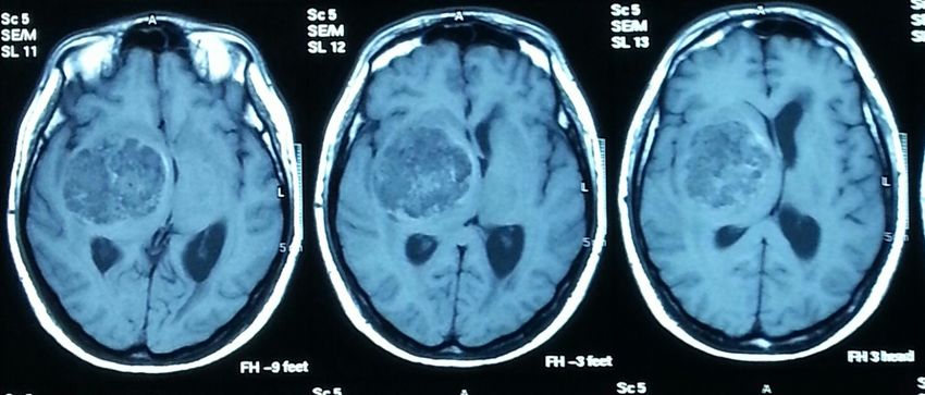
Computed Tomography (CT) scan of brain showed a large
heterogenous density mass in right temporoparietal region measuring
Correspondence to: Mohamed Saufi Awang, MBBS (Adelaide), M Surg
5.2 × 5.2 × 4.8cm with perilesional edema (Figure 1). The mass was (Neurosurgery/USM), Associate Professor, Department of Neurosurgery,
compressing the ipsilateral lateral and third ventricle with a midline International Islamic University Malaysia, Kuantan, Malaysia, Tel: 09-5706171,
shift of 0.9 cm. The contralateral left lateral ventricle and temporal horn Fax: 09-5146090, E-mail: saufiawang@iium.edu.my
of right lateral ventricle were dilated. Magnetic Resonance Angiograpy
Key words: choroid plexus carcinoma, basal ganglia, brain tumor
(MRI) brain revealed heterogenous intra axial right basal ganglia
mass (Figure 2). The mass was hypointense on T1, heterogeneously Received: April 05, 2017; Accepted: April 21, 2017; Published: April 24, 2017
Surg Rehabil, 2017 doi: 10.15761/SRJ.1000108 Volume 1(2): 1-4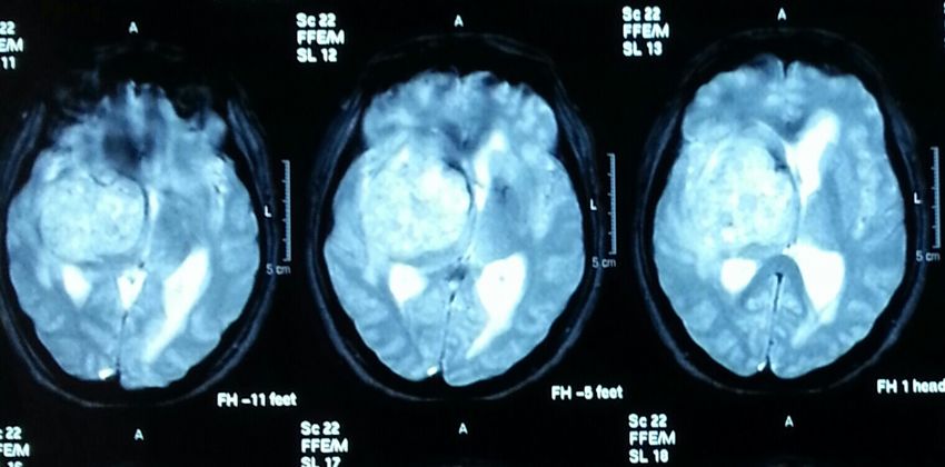
Azhani C (2017) Choroid plexus carcinoma: A case report and literature review

                                                                     Figure 1: Pre-operative CT brain (non contrast).

                         A

                         B

                                                  Figure 2: Pre-operative MRI. T1 weighted with Gado (a) and T2 weighted (b) images.

                                                                                               common than papilloma; estimated around 15-30% of choroid plexus
                                                                                               tumors [11]. Sampath, et al. conducted a 20 year retrospective reviewed
                                                                                               cases of CPT and found only 47 cases in total population, with 95%
                                                                                               out of 19 cases of adult CPT are choroid plexus papiloma [12]. Most
                                                                                               of the diagnosis of choroid plexus carcinoma is made in paediatrics
                                                                                               age group and rarely reported in adult. If present in adult, it should be
                                                                                               interpreted with caution and patient should be investigated thoroughly
                                                                                               to exclude metastatic tumors particularly from renal and thyroid
                                                                                               [1,11]. The median age of presentation is 3 years (ranging from 0 to 72
                                                                                               years old) with the youngest case recorded was an unusual congenital
                                                                                               choroid plexus carcinoma detected antenatally at 29 weeks in utero
                                                                                               [3,8]. In adult population, median age of CPT is 30 years and diagnosis
                                                                                               of choroid plexus carcinoma is rarely made [12]. Choroid plexus
Figure 3: Histologic characteristic of resected tumor. Solid growth of carcinoma with focal
                                                                                               papilloma is graded as WHO grade I tumor, whereas choroid plexus
papillary structures. H&E magnification 190x.                                                  carcinoma is WHO grade III.

Surg Rehabil, 2017             doi: 10.15761/SRJ.1000108                                                                                              Volume 1(2): 2-4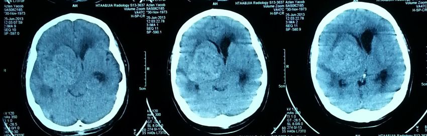
Azhani C (2017) Choroid plexus carcinoma: A case report and literature review

    The location of CPT are usually intraventricular; however                   metastatic papillary tumor is also making the diagnosis of choroid
extraventricular has been reported in several literatures [2,5,6,12].           plexus carcinoma more challenging. On immunohistochemistry,
Sampath et al also have found that CPT at ectopic site occurred only in         choroid plexus carcinoma will stain positive for cytokeratins and has
adults according to their review [12]. Sites that have been reported to be      variable expression for Vimentin, S100, transthyretin and glial fibrillary
ectopic include suprasellar region, foramen magnum, cerebellopontine            acidic protein.
angle and in the spinal canal in the absence of intracranial lesion. As
                                                                                    Owing to its low incidence, guideline in managing choroid plexus
choroid plexus tumors arise from epithelium of the choroid plexus,
                                                                                carcinoma is not well established. Best treatment option is still gross
possible plausible theory for occurrence of extraventricular tumors is
that it could either arise from dissemination through cerebrospinal             total resection, with Bettegowda, et al. defining it as reduction of
fluid or presence of small tuft of choroid plexus extending from                more than 75% in tumor size. Partial resection was defined as 25-
the Foramen of Luschka (in cerebellopontine angle choroid plexus                75% reduction in tumor size. Gross total resection is achieved in
carcinoma) [5]. Commonest site for adult is in fourth ventricle (63%),          40-75% in choroid plexus carcinoma as opposed to 95% in choroid
while in paediatric age group is lateral ventricle (72%) [12]. 12% of           plexus papilloma [11]. The difficulty resulting in partial resection is
patients with CPC estimated to have metastasis at presentation with             due to highly vascularized tumor in deep location making it prone to
the frequency is higher in supratentorial lesion [3].                           intraoperative hemorrhage. A review by Sampath et al. has seen that
                                                                                mean blood loss for total resection is 540 ml as compared to 890ml
    The presentations of symptoms vary and fall into 2 major                    with partial resection [12]. In contrast, they also found the incidence
categories which are intracranial hypertension and focal neurological           of tumor bed hematoma was higher in total resection, but extent of
deficit. Menon, et al. reviewed 25 cases of CPT and 72% of their                excision did not significantly correlate with tumor bed hematoma.
patients presented with increase intracranial pressure symptoms,                Other complications that have been more commonly reported post
whereas Sampath noted 90% of patients in both adult and paediatric              operatively includes subdural collection (32 to 43%), pneumocephalus
populations presented with hydrocephalus [9,11]. Most common focal
                                                                                (40%), focal deficit (36%) and persistent hydrocephalus requiring CSF
neurological deficit sustained is cranial nerve VI palsy attributed to
                                                                                diversion surgery more commonly in adult [9,12].
hydrocephalus. Other forms of neurodeficit include hemiparesis and
sensory dysfunction. In a reported case of cerebellopontine angle CPT,              Other therapeutic options have been used as adjunct to surgery
other cranial nerves will also involve. Most of the symptoms contributed        includes chemotherapy and radiotherapy particularly in patients with
by the mass effect and hydrocephalus (both communicating and                    residual tumor. These adjuvant treatments however are not suitable for
obstructive), but cases with primary bleeding from the tumor has                the young age group of less than 3 year old. Wolff et al have conducted
been reported and markedly in tumor at ectopic area [2]. Intratumoral           a meta analysis through literature review of 566 choroid plexus tumor
hemorrhage in a recurrent tumor has also been reported needing                  cases through 1966 till 1998 to determine the treatment modality.
evacuation [10].                                                                Their conclusions were, surgery significantly improve prognosis
    In investigating the tumor, both CT scan and MRI is needed, and             and radiotherapy significantly give better survival in choroid plexus
sometimes angiography will be useful especially if we are suspecting            carcinoma. Only 8 cases of 22 choroid plexus carcinoma were given
a highly vascularized tumor. In CT scan, 75% of tumors will show                chemotherapy and responded, thus the impact of this treatment
heterogenous density, and 10-25% will have calcifications [2]. MRI will         option could not be sufficiently analyzed to be statistically of value
usually show isointense on T1 image and heterogenous hyperintese                [3]. Nonetheless, Berrak, et al. conducted a meta analysis of 361
on T2 with enhancement post Gadolinium contrast. Angiography can                choroid plexus carcinoma and found out of those given chemotherapy,
sometimes identify tumor blush which suggest presence of blood supply           etoposide is the most effective agent.
to the tumor. In Menon et al review, 18 of 25 patients have tumor blush             Similar case has been reported by Lozier, et al. in a 68 year old
present. If feeding vessel is identified, endovascular embolization has
                                                                                lady with supratentorial extraventricular choroid plexus carcinoma.
been attempted previously but with low success rate in view of tortuous
                                                                                She successfully underwent gross total resection followed by adjuvant
tumor vessel and difficulty in cannulating the choroidal arteries. Further
                                                                                radiotherapy and Temozolamide. No recurrence or residual tumor
investigation will include searching for exclusion of primary tumor
                                                                                noted at 44 months follow up [5]. The difference with this patient was
in suspicion of metastatic papillary growth in adult diagnosed with
                                                                                that his Karnofsky performance scale was already poor prior to our
choroid plexus carcinoma. Extensive search of primary will include
                                                                                surgery thus rendered him poor prognosis.
doing CT scan of thorax, abdomen and pelvis and also mammography.
Most common primary resembling choroid plexus carcinoma includes                    The outcome of choroid plexus tumor depending on 3 factors
renal cell carcinoma, thyroid malignancies and esophageal cancer. Thus          which are; choroid plexus carcinoma histopathology, location of tumor
history and clinical examination should be directed towards excluding           and extent of resection [4,12,13]. However Wolff et al predicted that
these primaries as choroid plexus carcinoma is extremely rare in adult.         location of tumor has no prognostic relevance as opposed to Berrak,
Other investigations to be considered to exclude metastasis from                et al. who found survival is poorest in infratentorial tumor in choroid
choroid plexus carcinoma are MRI of spine to rule out leptomeningeal            plexus carcinoma. Mean survival documented for supratentorial tumor
spread and also a cerebrospinal fluid cytology.                                 was 26.9% at 10 years and none for infratentorial tumor. In case of
     The histopathology of choroid plexus papilloma and choroid plexus          a relapse after primary treatment of choroid plexus carcinoma, it is a
carcinoma are difficult to be distinguished. Differences are looked at the      poor prognostic factor for survival. 5 year survival of choroid plexus
appearances and histopathology characteristic [11]. Gross pathology             carcinoma is estimated to be 25-30% in patient with gross total resection
will usually show a friable papillary like or cauliflower like appearance.      [7,11]. Menon, et al. calculated the survival in subtotal resection for
Meanwhile, presence of increase in tumor necrosis, mitotic activity             choroid plexus carcinoma was 36 months from surgery and 58 months
and change in growth pattern are more likely to resemble choroid                for gross total resection [9]. Thus we should at best aim for gross total
plexus carcinoma than papilloma [7]. Likewise, the resemblance with             resection or multiple stage resection to prevent complications.

Surg Rehabil, 2017       doi: 10.15761/SRJ.1000108                                                                                      Volume 1(2): 3-4
Azhani C (2017) Choroid plexus carcinoma: A case report and literature review
Conclusion 6. Bekiesinska-Figatowska M, Madzik J, Biejat A, Maldyk J, Duczkowska A (2009)
Choroid plexus carcinoma of the spinal canal without cranial lesion. European Journal
Choroid plexus carcinoma is extremely rare in adult and its of Radiology Extra 72: 107–109.
frequency at ectopic sites such as in this case is only been reported 7. Gopal P, Parker JR, Debski R, Parker JC Jr (2008) Choroid plexus carcinoma. Arch
few in the literature. This case poses a great challenge to us in term of Pathol Lab Med 132: 1350-1354. [Crossref]
diagnosis and management. Total surgical resection was limited due to 8. Wilhelm M, Hirsch W, Merkenschlager A, Stepan H, Geyer C, Kiess W (2012) A rare
tumor vascularity and brain swelling. In the future, we hope a proper case of congenital choroid plexus carcinoma. Pediatr Hematol Oncol 29: 643-646.
guideline can be established. [Crossref]
9. Menon G, Nair SN, Baldawa SS, Rao RB, Krishnakumar KP, Gopalakrishnan CV
References (2010) Choroid plexus tumor: An institutional series of 25 patients. Neurol India 58:
1. Kishore S, Nei G, Meena H, Anuradha K, Ved Pathak P, Bansal KK (2012) Choroid 429-435. [Crossref]
plexus carcinoma in an adult. J Neurosci Rural Pract 3: 71-73. [Crossref] 10. Wyatt SS, Price RA, Holthouse D, Elsaleh H (2001) Choroid plexus carcinoma in an
2. Haroun RI, Li KW, Carson BS, Brem H (2000) Primary tumors of choroid plexus. adult. Australas Radiol 45: 369-371. [Crossref]
Contemporary neurosurgeryv22: 1-7. 11. Maimone G, Ganau M, Nicassio N, Paterniti S (2013) Paratrigonal choroid plexus
3. Wolff JE, Sajedi M, Brant R, Coppes MJ, Egeler RM (2002) Choroid plexus tumours. papilloma presenting with satellite multiple supra and infratentorial hemorrhages. Int J
Br J Cancer 87: 1086-1091. [Crossref] Surg Case Rep 4: 239-242. [Crossref]
4. Berrak SG, Liu DD, Wrede B, Wolff JE (2011) Which therapy works better in choroid 12. Sampath S, Nitin G, Yasha TC, Chandramouli BA, Devi BI, et al. (2008) Does choroid
plexus carcinomas?. 2011. J Neurooncol 103: 155-162. [Crossref] plexus tumour differ with age? Br J Neurosurg 22: 373-388. [Crossref]
5. Lozier AP, Arbaje YM, Scheithauer BW (2009) Scheithauer. Supratentoria, 13. Bettegowda C, Adogwa O, Mehta V, Chaichana KL, Weingart J, et al. (2012) Treatment
extraventricular choroid plexus carcinoma in an adult: Case report. Neurosurgery 65: of choroid plexus tumors: a 20-year single institutional experience. J Neurosurg Pediatr
816-817. [Crossref] 10: 398-405. [Crossref]
Copyright: ©2017 Azhani C. This is an open-access article distributed under the terms of the Creative Commons Attribution License, which permits unrestricted
use, distribution, and reproduction in any medium, provided the original author and source are credited.
Surg Rehabil, 2017 doi: 10.15761/SRJ.1000108 Volume 1(2): 4-4You can also read