Severe whipworm (Trichuris spp.) infection in the hamadryas baboon (Papio hamadryas) - Semantic Scholar
←
→
Page content transcription
If your browser does not render page correctly, please read the page content below
NOTE
Wildlife Science
Severe whipworm (Trichuris spp.) infection
in the hamadryas baboon (Papio hamadryas)
Kyung-Yeon EO1), Min-Goo SEO2), Hyun-Ho LEE1), Yeong-Mok JUNG1),
Dongmi KWAK3) and Oh-Deog KWON3)*
1)Conservation and Health Center, Seoul Zoo, Gwacheon, Gyonggido 13829, Korea
2)Animal and Plant Quarantine Agency, Gimcheon 39660, Korea
3)College of Veterinary Medicine, Kyungpook National University, Daegu 41566, Korea
ABSTRACT. A 3-year-old male hamadryas baboon (Papio hamadryas) at the Seoul Zoo, Korea,
J. Vet. Med. Sci. died without any previous symptoms. Necropsy revealed severe whipworm infection in the large
81(1): 53–56, 2019 intestine. The animal weighed 2.6 kg and had a blood clot at the anus. Numerous whipworms
were found attached to the intestinal wall, with their anterior ends embedded in the mucosa.
doi: 10.1292/jvms.17-0568 Fecal microscopy revealed typical barrel-shaped, brown eggs of Trichuris spp., with hyaline polar
plugs at each end. Histopathological examination revealed the thin anterior part of Trichuris
spp. embedded in the mucosal layer and the thick posterior part at the mucosal surface or
Received: 20 October 2017
hanging freely in the intestinal lumen. This case emphasizes the importance of parasitic infection
Accepted: 12 April 2018 management in zoo animals.
Published online in J-STAGE:
KEY WORDS: hamadryas baboon, Papio hamadryas, trichuriasis, zoo
21 November 2018
Trichuris spp. are general parasitic helminths in non-human primates. The prevalence of whipworms in non-human primates
is usually high, and the common Trichuris sp. that infects non-human primates is Trichuris trichiura, which is also known to
parasitize humans [1]. The species of non-human primates that can be infected include macaques, African green monkeys,
baboons, squirrel monkeys and woolly monkeys. Although light infection is asymptomatic, severe infection produces clinical signs
such as severe enteritis, anorexia, gray mucoid diarrhea and sometimes death [2]. Globally, T. trichiura is still one of the most
important soil-transmitted helminths, in addition to Ascaris lumbricoides and hookworms [18].
Hamadryas baboon (Papio hamadryas), classified as a least concern species by the International Union for Conservation of
Nature Red List, is widespread and abundant, and has no chief range-wide threats believed to result in any important decline in
numbers [19]. This species is endemic to northeast Africa and is mainly found in Ethiopia, Sudan, northern Somalia, south-west
Arabian Peninsula and Egypt [18]. In January 2016, a 3-year-old male hamadryas baboon weighing 2.6 kg was found dead without
any previous symptoms at the Seoul Zoo, Korea. This animal was fed twice per day with a commercial primate biscuit (ZuPreem
Primate Dry Biscuit no. 6985, Shawnee Mission, KS, U.S.A.), lettuce, kale, celery, sweet potato, Chinese cabbage, carrot, banana
and apple. It was exhibited in a 422 m2 outdoor enclosure and 20 m2 indoor room as part of a troop.
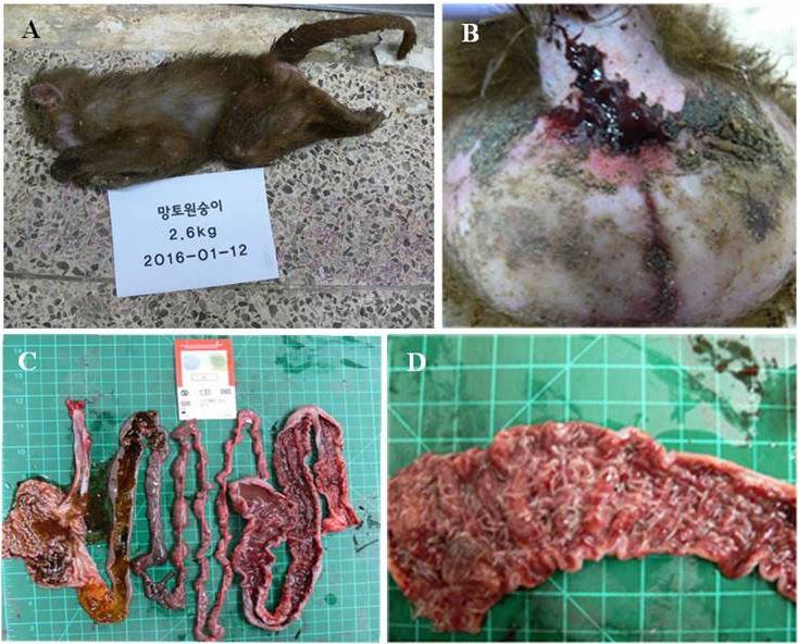
External observation revealed loose body hair and a blood clot at the anus (Fig. 1A, 1B). Necropsy revealed severe whipworm
infection in the large intestine. There was no food in the stomach and small intestine. Numerous whipworms were found attached
to the intestinal wall, with their anterior ends embedded in the mucosal layer. Hemorrhage and enteritis with a thickened and
ulcerative intestinal wall was observed throughout the tract, including the cecum and rectum (Fig. 1C, 1D). Although bacterial
cultures, protozoa detection tests and viral tests were needed to clarify the cause of enteritis in this case, we did not conduct these
tests because the lesion caused by Trichuris infection was very prominent and severe in the large intestinal wall at necropsy.
For histopathological examination, a piece of the colon containing the whipworms was collected and stored in 10% neutral
buffered formalin. Many adult whipworms were found embedded in the colon mucosa, with their thick posterior parts on the
mucosal surface (Fig. 2A). Five-micrometer-thick sections of the paraffin-embedded tissue were cut using a microtome and affixed
to glass slides. The sections were dewaxed and hydrated by passing them through a xylene-ethanol series. The slides were stained
with hematoxylin and eosin for light microscopic examination [6]. The mucosa in the vicinity of the embedded worms was friable
and edematous because of inflammation. The stained sections showed the embedded anterior parts of the whipworms surrounded
by leucocytes and fibroblasts (Fig. 2B, 2D). Some free-hanging segments of whipworms in the intestinal lumen and slender
anterior parts of the Trichuris spp. embedded in the mucosa were also observed. The uterus of the whipworms was filled with
smooth-walled eggs with unstained bipolar plugs (inset in Fig. 2B). Microscopic examination of the direct smear from the fecal
*Correspondence to: Kwon, O.D.: odkwon@knu.ac.kr
©2019 The Japanese Society of Veterinary Science
This is an open-access article distributed under the terms of the Creative Commons Attribution Non-Commercial No Derivatives (by-nc-nd)
License. (CC-BY-NC-ND 4.0: https://creativecommons.org/licenses/by-nc-nd/4.0/)
53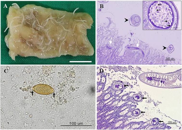
K.-Y. EO ET AL.

Fig. 1. External appearance and gross findings at necropsy of a 3-year-old male hamadryas baboon exhibited at a zoo. (A) Loose body hair and
   light emaciation are observed. (B) A blood clot is seen at the anus. (C) Thickening of the intestinal wall is observed from the cecum to the
   rectum. (D) Numerous adult whipworms are firmly attached to the intestinal wall of the colon, with their anterior ends embedded in the mucosa.

sample revealed barrel-shaped, brown eggs with hyaline polar plugs at each end (Fig. 2C).
   Parasitic infection and disease are the main causes of concern as well as of loss of animal life at zoos. Other infections are
usually due to contact with agents such as bacteria, fungi, rickettsiae or viruses. Biohazards should be considered not only among
the various animal populations but also among humans, especially those coming in contact with non-human primates [11].
   In such cases of infection resulting in gastrointestinal disease, both helminth and protozoan parasites are general causes.
Effective parasite treatment regimens, in particular, have enhanced the ability to maintain non-human primates in naturalistic
exhibits [2]. Nevertheless, from the perspective of zoonosis, a previous study showed that the fecal samples of four of 54 animal
caretakers in charge of non-human primates at five zoos in Belgium tested positive for T. trichiura eggs under microscopic
examination [13]. The design of exhibits and off-exhibit holding areas should prevent disease transmission between human and
non-human primates, such as through the use of glass or other physical separations. Moreover, the management and husbandry
protocols should be improved to ensure the health and safety of both human and non-human primates [2].
   Various pathogens, such as Strongyloides spp., Enterobius vermicularis, T. trichiura, Oesophagostomum sp., Entamoeba
histolytica, Balantidium coli, Giardia lamblia, Shigella flexneri, Sh. sonnei, Salmonella enteritidis, Sa. typhimurium, adenovirus
and hepatitis virus can cause gastrointestinal diseases in non-human primates [10]. Cases of fatal Trichuris infection in captive
baboons (Pa. hamadryas) were reported in Australia and Nigeria [3, 9]. A black-and-white colobus monkey (Colobus guereza
kikuyuensis) at a zoo in Indiana, U.S.A., died of severe Trichuris infection. At necropsy, many Trichuris worms were seen
protruding from necrotic nodules, having diameters ranging from 0.5 to 2 cm, found in the stomach. However, nematodes were
not found in other portions of the gastrointestinal tract [14]. Trichuris infection has also been reported in other wild animals. For
instance, Trichuris spp. were detected in the fecal samples (19.1%, 22/115) of Arabian sacred baboons (Pa. hamadryas arabicus)
living in human-populated residential and non-residential areas in Asir province, Saudi Arabia, in 1988 [16]. They were also
detected in the fecal samples of free-ranging individuals of the three colobus monkey species, including the endangered red colobus
(37.8%, 607/1,608, Piliocolobus tephrosceles), the eastern black-and-white colobus (79.0%, 376/476, C. guereza) and the Angolan
black-and-white colobus (100%, 19/19, C. angolensis), in Uganda from 1997 to 2003 [7]. In addition, they were detected in the
fecal samples (72.9%, 62/85) of free-ranging baboons (Pa. cynocephalus and Pa. anubis) in Kenya between 1993 and 1995 [8].
Further, T. trichiura was detected in the fecal samples of captive and wild-trapped non-human primates such as olive baboons
(71.9%, 120/167, Pa. cynocephalus anubis), grivets (50%, 25/50, Cercopithecus aethiops) and blue monkeys (56.1%, 55/98, Ce.
mitis) in Kenya in 1998 [15].
   Trichuris infection has also been detected in zoo animals. For example, Trichuris spp. were detected in the fecal samples of
baboons (100%, 6/6, Pa. cynocephalus), ring-tailed lemurs (100%, 6/6, Lemur catta) and Bactrian camels (100%, 6/6, Camelus

doi: 10.1292/jvms.17-0568                                                                                                                     54
WHIPWORM INFECTION IN HAMADRYAS BABOON

Fig. 2. Histopathological study of the colon after staining with hematoxylin and eosin. (A) A part of the dissected colon after formalin fixation.
   Whipworms are found embedded in the intestinal mucosa. The bar represents 1 cm. (B) Microscopic observation of the intestinal tissue
   embedded with whipworms. Free-hanging segments of worms in the intestinal lumen (arrowheads) and the slender anterior parts of Trichuris
   spp. embedded in the colon mucosa (white arrows). Inset: Note the eggs within the uterus of the whipworms, showing smooth walls and bipolar
   plugs (black arrows). (C) Fecal microscopy shows a barrel-shaped egg with hyaline plugs at each end (arrows). (D) The anterior parts (arrows)
   of the whipworms invade into the large intestinal wall.

bactrianus) at two Italian zoological gardens in 2007 [5]. Further, they were detected in the fecal samples of baboons (75.0%, 3/4)
and peacocks (100%, 2/2) at the zoological garden in Nekede, Nigeria, in 2010 [17]. They were also detected in the fecal samples
of Celebes crested macaques (20.0%, 2/10, Macaca nigra), hamadryas baboons (15.7%, 16/102, Pa. hamadryas), Hamlyn’s
monkey (2.9%, 1/35, Ce. hamlyni) and black-crested gibbon (33.3%, 1/3, Hylobates concolor) at four zoological gardens in
Belgium between 2004 and 2006 [12]. Trichuris spp. were also the cause of infection in three dromedary camels (Ca. dromedarius)
at the Seoul Zoo, Korea, in 2013 [4].
   We monitor gastrointestinal parasitic infection in hamadryas baboon troops twice a year by using a typical microscopic fecal
test. On the basis of the findings of the fecal test, we prescribe anthelmintics to treat parasitic infection. However, in the case
of baboons, individual administration is difficult because of the large troop size, and oral administration of drugs is challenging
because of the baboon’s ability to detect medicated food or drink. Hence, it is uncertain whether the anthelmintics were consumed
by infected animals. Therefore, to eradicate whipworm infection in the whole troop, long-term monitoring, regular fecal
examination and individual drug administration are important and necessary. This case of severe whipworm infection in a captive
hamadryas baboon serves as an example of pathological trichurid infection and emphasizes the importance of parasitic infection
management in zoo animals.

REFERENCES

 1. Abee, C. R., Mansfield, K., Tardif, S. D. and Morris, T. 2012. Parasitic diseases of nonhuman primates. pp. 243–244. In: Nonhuman Primates in
    Biomedical Research: Diseases, 2nd ed., Elsevier Academic, London.
 2. Calle, P. P. and Joslin, J. O. 2014. New world and old world monkeys. pp. 301–335. In: Zoo and Wild Animal Medicine, 8th ed. (Fowler, M. E. and
    Miller, R. E. eds.), Elsevier Saunders, St. Louis.
 3. Emikpe, B. O., Ayoade, G. O., Ohore, O. G., Olaniyan, O. O. and Akusu, M. O. 2002. Fatal trichuriosis in acaptive baboon (Papio anubis) in Ibadan
    Nigeria: A case report. Trop. Vet. 20: 36–39.
 4. Eo, K. Y., Kwak, D. and Kwon, O. D. 2014. Severe whipworm (Trichuris spp.) infection in the dromedary (Camelus dromedarius). J. Zoo Wildl.
    Med. 45: 190–192. [Medline] [CrossRef]
 5. Fagiolini, M., Lia, R. P., Laricchiuta, P., Cavicchio, P., Mannella, R., Cafarchia, C., Otranto, D., Finotello, R. and Perrucci, S. 2010. Gastrointestinal
    parasites in mammals of two Italian zoological gardens. J. Zoo Wildl. Med. 41: 662–670. [Medline] [CrossRef]
 6. Fischer, A. H., Jacobson, K. A., Rose, J. and Zeller, R. 2008. Hematoxylin and eosin staining of tissue and cell sections. CSH Protoc 2008: pdb.
    prot4986. [Medline]

doi: 10.1292/jvms.17-0568                                                                                                                                 55
K.-Y. EO ET AL.
7. Gillespie, T. R., Greiner, E. C. and Chapman, C. A. 2005. Gastrointestinal parasites of the colobus monkeys of Uganda. J. Parasitol. 91: 569–573.
[Medline] [CrossRef]
8. Hahn, N. E., Proulx, D., Muruthi, P. M., Alberts, S. and Altmann, J. 2004. Gastrointestinal parasites in free ranging Kenyan baboons (Papio
cynocephalus and P. anubis). Int. J. Primatol. 24: 271–279. [CrossRef]
9. Hennessy, A., Phippard, A. F., Harewood, W. J., Horam, C. J. and Horvath, J. S. 1994. Helminthic infestation complicated by intussusception in
baboons (Papio hamadryas). Lab. Anim. 28: 270–273. [Medline] [CrossRef]
10. Joslin, J. O. 2003. Other primates excluding great apes. pp. 346–381. In: Zoo and Wild Animal Medicine, 5th ed. (Fowler, M. E. and Miller, R. E.
eds.), Elsevier Saunders, St. Louis.
11. Kalter, S. S. 1989. Infectious diseases of nonhuman primates in a zoo setting. Zoo Biol. 8: 61–76. [CrossRef]
12. Levecke, B., Dorny, P., Geurden, T., Vercammen, F. and Vercruysse, J. 2007. Gastrointestinal protozoa in non-human primates of four zoological
gardens in Belgium. Vet. Parasitol. 148: 236–246. [Medline] [CrossRef]
13. Levecke, B., Dorny, P., Vercammen, F., Visser, L. G., Van Esbroeck, M., Vercruysse, J. and Verweij, J. J. 2015. Transmission of Entamoeba nuttalli
and Trichuris trichiura from nonhuman primates to humans. Emerg. Infect. Dis. 21: 1871–1872. [Medline] [CrossRef]
14. Loomis, M. R. and Wright, J. F. 1986. Gastric trichuriasis in a black and white colobus monkey. J. Am. Vet. Med. Assoc. 189: 1214–1215. [Medline]
15. Munene, E., Otsyula, M., Mbaabu, D. A., Mutahi, W. T., Muriuki, S. M. and Muchemi, G. M. 1998. Helminth and protozoan gastrointestinal tract
parasites in captive and wild-trapped African non-human primates. Vet. Parasitol. 78: 195–201. [Medline] [CrossRef]
16. Nasher, A. K. 1988. Zoonotic parasite infections of the Arabian sacred baboon Papio hamadryas arabicus Thomas in Asir Province, Saudi Arabia.
Ann. Parasitol. Hum. Comp. 63: 448–454. [Medline] [CrossRef]
17. Opara, M. N., Osuji, C. T. and Opara, J. A. 2010. Gastrointestinal parasitism in captive animals at the zoological garden, Nekede Owerri, Southeast
Nigeria. Rep. Opinion. 2: 21–28.
18. Pullan, R. L., Smith, J. L., Jasrasaria, R. and Brooker, S. J. 2014. Global numbers of infection and disease burden of soil transmitted helminth
infections in 2010. Parasit. Vectors 7: 37. [Medline] [CrossRef]
19. The IUCN Red List of Threatened Species. Papio hamadryas. http://iucnredlist.org/details/summary/16019/0 [accessed on June 7, 2017].
doi: 10.1292/jvms.17-0568 56You can also read