Pigeon Fever (Corynebacterium pseudotuberculosis infection)
←
→
Page content transcription
If your browser does not render page correctly, please read the page content below
Pigeon Fever (Corynebacterium pseudotuberculosis infection)

Definition
       Corynebacterium pseudotuberculosis is a gram-positive bacteria with worldwide
       distribution. In North America, cases have been reported throughout the United States.
       Infection has been reported in equids, sheep, goats, cattle, buffalo, camelids, and rarely
       humans. Biotypes isolated from small ruminants and camelids are nitrate negative, while
       those from horses are nitrate positive. Natural cross-species transmission does not occur
       between sheep and horses, however cattle can have infection from either biotype.

Clinical Signs
       Three forms have been described in horses: ulcerative lymphangitis or limb infection,
       external abscesses and internal infection. Ulcerative lymphangitis and internal infection
       must be treated more aggressively with antimicrobial therapy, while use of
       antimimicrobials for external abscesses is often unnecessary.

       Ulcerative lymphangitis is the least common form seen in North America, although this
       form of disease has been reported worldwide. Ulcerative lymphangitis manifests as a
       severe limb swelling and cellulitis, with multiple draining tracts following lymphatics.
       Most commonly one or both hind limbs are affected. Horses often develop a severe
       lameness, fever, lethargy and anorexia. Aggressive medical therapy (antimicrobial and
       anti-inflammatory) is necessary or the disease often becomes chronic, resulting in limb
       edema, prolonged or recurrent infection, lameness, weakness, and weight loss.

       Ulcerative lymphangitis (Photo by Sharon Spier, DVM, Ph.D, University of California, Davis)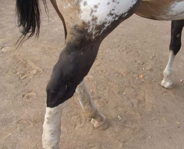
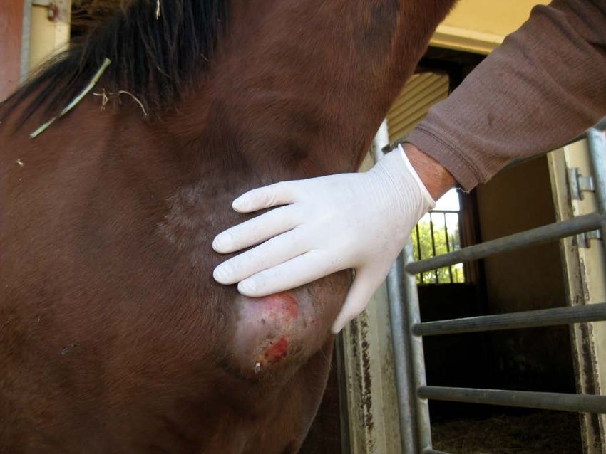
External Abscesses are the most common manifestation, and may occur anywhere on the
body, but most frequently develop in the pectoral region (swelling resembles a pigeon’s
breast) and along the ventral midline of the abdomen. Abscesses contain tan, odor-free
purulent exudate and are usually well encapsulated. Additional sites for abscess
formation are the prepuce, mammary gland, triceps, axilla, limbs, and head. Septic joints
and osteomyelitis have been reported. Horses may have a single or multiple abscesses
involving different regions of the body. Horses with external abscesses do not usually
develop signs of systemic illness, however one-quarter will develop fever. If signs of
systemic illness are present, further diagnostics to rule out internal infection are
warranted, and antimicrobial therapy should be considered. While there is considerable
variation in severity among horses, most straight – forward cases can be treated with
lancing and draining the abscesses when mature. The case fatality for horses with
external abscesses is very low (0.8%).

Typical pectoral abscess with flies attracted to exudates
(Photo by Sharon Spier, DVM, Ph.D, University of California, Davis)

Internal infection occurs in approximately 8% of affected horses, which is associated
with a high case fatality rate (30 to 40%). Diagnosis can be challenging, and long-term
antimicrobial therapy is imperative for successful outcome. In a retrospective study, the
organs most commonly involved were liver, kidney, spleen and lungs. Abortion due to
placentitis or fetal infection has been reported.
Transmission
      The portal of entry of this soil-dwelling organism is thought to be through abrasions or
      wounds in the skin or mucous membranes. Many insects have been incriminated as
      vectors for the transmission of the disease to horses, and studies have shown that
      Haematobia irritans, Musca domestica, Stomoxys calcitrans can act as mechanical
      vectors of this disease. The regional location of abscesses suggests that ventral midline
      dermatitis is a predisposing cause of infection. Temporal and special analysis indicated
      an incubation period of 3 to 4 weeks. Within a geographic area, the disease appeared to
      be transmitted between 7 and 56 days throughout a 4.3 to 6.5 km distance, strongly
      suggesting that the disease could be transmitted through horse-to-horse contact or from
      infected to susceptible horses via insects, other vectors, or contaminated soil. The
      organism has been shown to survive for up to 2 months in hay and shavings, and more
      than 8 months in soil samples at environmental temperatures. The incidence of disease
      fluctuates considerably from year to year presumably due to herd immunity and
      environmental factors such rainfall and temperature. Disease incidence is seasonal, with
      highest number of cases occurring during the dry months of the year, which is summer
      and fall in the Southwestern US, although cases may be seen all year. Horses with
      internal infection are more frequently seen one to two months following the peak number
      of cases with external abscesses.

Diagnostic Sampling, Testing and Handling
      Bacterial culture of aspirates or exudate is used to confirm diagnosis and the organism
      survives for prolonged periods. Corynebacterium pseudotuberculosis grows well at 36°C
      on blood agar in 24 to 48 hours, and it forms small, pinpoint in diameter, whitish, opaque
      colonies that are surrounded by a weak zone of hemolysis. Biotypes isolated from small
      ruminants and camelids are nitrate negative, while those from horses are nitrate positive.
      Corynebacterium pseudotuberculosis produces various extracellular exotoxins, which
      play a role in virulence; the most studied is phospholipase D (PLD). The bacterial
      phospholipase D is similar to the PLD of the brown recluse spider, which explains the
      presence of pain and edema at the site of infection. The synergistic activity of C.
      pseudotuberculosis exotoxins with the exotoxins of Rhodococcus equi in lysing red blood
      cells in agar forms the basis for the synergistic hemolysis inhibition (SHI) test. The SHI
      test is a used to detect IgG antibody to C. pseudotuberculosis in horses with internal
      infections where external abscesses are not present.

       Clinical pathologic abnormalities that may be observed include anemia of chronic
       disease, leukocytosis with neutrophilia, hyperfibrinogenemia, and hyperproteinemia.
       These hematological parameters can occur with either internal or external abscesses but
       are more consistently observed with internal abscesses.

       A diagnosis of internal infection is based on clinical signs, clinicopathologic data,
       serology, diagnostic imaging and bacterial culture. The most common clinical signs are
       concurrent external abscesses, decreased appetite, fever, lethargy, weight loss, and signs
       of respiratory disease or abdominal pain. Other signs observed in horses with internal
       abscesses include ventral edema, ventral dermatitis, ataxia, hematuria (due to renal
       abscesses), and uncommonly, abortion.
Serologic testing using the Synergistic Hemolysis Inhibition (SHI) test can be useful in
aiding the diagnosis of internal abscesses and is available through the California Animal
Health and Food Safety Laboratory System in Davis, California, and the Colorado State
University Veterinary Diagnostic Laboratories in Fort Collins, Colorado. Serology is
generally not helpful for diagnosis of external abscesses and may be negative early in the
course of disease and even the time of abscess drainage. Positive SHI titers must be
interpreted carefully and combined with clinical signs to distinguish active infection from
exposure or convalescence. Both published and unpublished data from the University of
California suggests a reciprocal titer of ≥256 is indicative of active infection. Horses
with internal abscesses generally have SHI titers ≥512. Titers ≤ 16 are considered
negative, while titers between 16 and 128 are considered suspicious or indicative of
exposure. These are rough guidelines, however, as there is considerable overlap in results
from horses with active disease, exposure and recovery from infection. The SHI test is
most accurate for diagnosis of internal infection in the absence of external abscesses.
The SHI test should not be used alone to diagnose internal infection without other
supportive diagnostics.
Abdominal ultrasonography is the most useful tool for identifying affected internal
organs and also for revealing the nature and extent of involvement. Abdominal
ultrasonography also facilitated transcutaneous liver and kidney biopsy procedures and
aspiration of abscess fluid for definitive diagnosis. Ultrasonography should be used in
conjunction with hematologic and serum biochemical analyses to monitor response to
treatment and may be the only available modality to monitor horses in which there is no
clinicopathologic evidence of organ disease.
Environmental Persistence
The organism has been shown to survive for up to 2 months in hay and shavings, and
more than 8 months in soil samples at environmental temperatures. In experimental
studies, the presence of manure favored survival and replication of bacteria in soil.
Specific Control and Treatment Measures
Biosecurity Measures
Implementation of biosecurity practices to limit the spread of Corynebacterium
pseudotuberculosis are aimed at reducing environmental contamination and spread via
insects or fomites. The bacterium is endemic in many regions of the world and survives
for months in soil, particularly when contaminated with manure.
• Wearing of disposable examination gloves when working with affected horses
followed by hand washing is indicated.
• Isolation of affected horses from naive herd mates
• Protecting horses from insect exposure by regular application of insect repellants
to the horse including the ventral midline (prevention of ventral midline
dermatitis).
• Meticulous wound care (topical fly repellants, antimicrobial ointments and
bandages) to prevent infection from a contaminated environmentVaccination There is currently no licensed commercially available vaccine in the United States for control of Corynebacterium pseudotuberculosis in horses. Use of autogenous bacterin - toxoids designed for horses demonstrated increased SHI titers following 2 injections, however the protection remains to be established. A commercial bacterin - toxoid is clearly needed to protect horses as the disease becomes endemic in more geographic regions. Treatment The treatment regimen for external abscesses must be individualized for each horse depending on the severity of disease, including the presence of systemic illness such as fever or anorexia, the extent of soft tissue inflammation, the maturity of the abscess and the ability to successfully establish drainage of purulent exudate. Establishing drainage is the most important treatment and ultimately leads to faster resolution and return to athletic performance. The proximity of the fibrous abscess capsule to the skin varies, often being
therapy for internal infection is 4-6 weeks, and is best determined by repeat abdominal
       ultrasound and clinicopathologic results.

       Horses with ulcerative lymphangitis or cellulitis should be treated early and aggressively
       with antimicrobials or residual lameness or limb swelling may occur. Typically
       intravenous antimicrobials (ceftiofur or penicillin G) alone or in combination with
       rifampin (orally) are used until lameness and swelling improves, and then therapy with
       orally administered antimicrobials such as trimethoprim sulfamethoxazole or rifampin are
       continued to prevent relapse. The time to resolution reported in one study was
       approximately 35 days. Physical therapy, including hydrotherapy, hand walking, and leg
       wraps, as well as NSAIDs are recommended.

Biosecurity Management for Receipt of Animals
      Once horses are recovered and there is no drainage from abscesses no precautions should
      be needed to reduce the risk these horses pose for spread of infection. There is no
      practical way at this time for eliminating the bacteria from soil.

Zoonotic Potential
      There exist few reports of human illness through working with infected sheep, mostly in
      Australian sheep shearers who had open wounds on their hands and developed axillary
      lymphadenitis. One veterinary student from California developed pneumonia following
      exposure to an infected horse, presumably from inhalation of the bacteria from a
      contaminated environment.

                                       © Copyright AAEP 2013
You can also read