ULTRASONOGRAPHY OF THE LARYNX: NOVEL USE DURING THE SARS COV 2 PANDEMIC (REVIEW)
←
→
Page content transcription
If your browser does not render page correctly, please read the page content below
EXPERIMENTAL AND THERAPEUTIC MEDICINE 21: 273, 2021

                 Ultrasonography of the larynx: Novel use during
                       the SARS‑CoV‑2 pandemic (Review)
              ROMICA CERGAN1, MIHAI DUMITRU1, DANIELA VRINCEANU2, ADRIANA NEAGOS3,
                       IONUT ISAIA JEICAN4 and RADU CONSTANTIN CIULUVICA1

    1
     Department of Anatomy, ‘Carol Davila’ University of Medicine and Pharmacy, 050474 Bucharest; 2ENT Department,
           Bucharest Emergency University Hospital, 010271 Bucharest; 3ENT Department, ‘George Emil Palade’
             University of Medicine, Pharmacy, Science and Technology of Targu Mures, 540139 Târgu Mureș;
         4
          Department of Anatomy, ‘Iuliu Hatieganu’ University of Medicine and Pharmacy, 400012 Cluj, Romania

                                    Received October 12, 2020; Accepted November 10, 2020

                                                   DOI: 10.3892/etm.2021.9704

Abstract. Few articles have been published on the subject of        1. Introduction
laryngeal ultrasonography. However, considering the increased
power and accuracy of ultrasound technology, this imaging           If one would query international databases using search words
modality should be reevaluated. The present review aimed            such as larynx and ultrasound, they would find 17 free full text
to increase the awareness of fellow specialists regarding the       articles on the subject of ultrasonography of the larynx. One
use of this imaging tool in healthcare units that do not benefit    of the first articles on this subject was published in 1987 by
from onsite ear, nose and throat (ENT) service. We illustrate       Raghavendra et al (1). They analyzed 41 healthy volunteers,
the ultrasonographic examination protocol for the larynx            concluding that sonography may prove to be a potentially
along with the relevant anatomic landmarks. We review cases         useful technique for the examination of the vocal cords. An
with laryngeal tumoral pathology that underwent ultrasono‑          analysis of 229 healthy volunteers by Hu et al (2) revealed that
graphic examination for improved management. All findings           high frequency sonography can quantitatively measure both
were confirmed through computerized tomography (CT) and             true and false vocal cords with good reliability and reproduc‑
endoscopy performed by the ENT specialist. The ultrasound           ibility. A detailed anatomical study of the upper airway was
of the larynx has potential utility in diagnosis (e.g., laryngeal   performed by Singh et al (3), with potential utility in diag‑
abnormalities, speech and swallowing abnormalities, iden‑           nosis (e.g., laryngeal abnormalities, speech and swallowing
tification of endotracheal tube placement), treatment (e.g.,        abnormalities, identification of endotracheal tube placement),
guidance of percutaneous tracheostomy and cricothyrotomy)           treatment [e.g., guidance of percutaneous tracheostomy and
and prognosis (e.g., prediction of postextubation stridor and       cricothyrotomy (4)], and prognosis [e.g., prediction of postex‑
difficult intubation). This imaging modality could be useful in     tubation stridor (5) and difficult intubation]. We are still at
the current SARS‑CoV‑2 pandemics in reducing the exposure           the level of using ultrasound for analyzing healthy volunteers
to invasive maneuvers producing aerosol, such as endoscopy.         in order to ascertain whether there are gender differences in
                                                                    vocal cord size and that there is no correlation between vocal
                                                                    cord measurements and individual body mass index (BMI) (6).
Contents                                                            In a preliminary bedside study, sonography was used for
                                                                    diagnosing acute epiglottitis in the emergency department (7).
1. Introduction                                                     Ultrasound examination proved especially useful in managing
2. Aims of the ultrasonography of the larynx during the             post intubation larynx traumas (8). Moreover, real‑time
   SARS‑CoV‑2 pandemic                                              high‑resolution ultrasonography was used to assess the status
3. Anatomical basis                                                 of thyroglossal duct cysts by the ENT surgeon firsthand (9).
4. Ultrasonographic findings in laryngeal pathology                 Finally, the utility of sonography for evaluation of clinical T1
5. Conclusions                                                      and T2 glottic carcinoma has been proven (10).

                                                                    2. Aims of the ultrasonography of the larynx during the
                                                                    SARS‑CoV‑2 pandemic
Correspondence to: Dr Daniela Vrinceanu, ENT Department,
Bucharest Emergency University Hospital, Aleea Lunguletu 2,         The ultrasonography of the larynx is a diagnostic imaging
010271 Bucharest, Romania                                           modality that is rather neglected due to the lack of experi‑
E‑mail: vrinceanudana@yahoo.com                                     ence and the proximity of ENT departments that can perform
                                                                    endoscopic diagnosis of laryngeal pathology. However, in
Key words: ultrasound, larynx, ENT, SARS‑CoV‑2, COVID‑19            an emergency setting, a simple visualization of the larynx
                                                                    could be implemented as a quick extension to any FAST‑like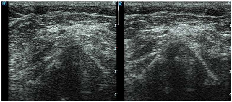
2                                              CERGAN et al: ULTRASONOGRAPHY OF THE LARYNX

Figure 1. Longitudinal midline view of the larynx. H, hyoid bone; E, epiglottis; T, thyroid cartilage; C, cricoid cartilage; G, thyroid gland isthmus; TRAH,
trachea.

                 Figure 2. Transverse view at the level of vestibular folds. MPL, anterior muscles of the neck; T, thyroid; V, vestibular folds.

protocol. We hope to increase the awareness of the scientific                     cartilage (C), thyroid gland isthmus (G) and trachea (TRAH)
community regarding this imaging tool that in the future,                         (Fig. 1).
after a thorough validation and standardization, could lower                          Fig. 2 illustrates the vestibular folds in transverse view:
the associated costs of other investigations. Furthermore,                        MPL, anterior muscles of the neck; T, thyroid cartilage and V,
current guidelines encourage reducing the use of aerosol                          vestibular folds.
producing procedures, such as laryngeal endoscopy during the                          The vocal cords are visualized in Fig. 3 in transverse view:
SARS‑CoV‑2 pandemic. Laryngeal ultrasound could lower                             MPL, anterior muscles of the neck; T, thyroid cartilage; CV,
the risk of ENT specialists in contracting the COVID‑19 viral                     vocal cords and ARI, arytenoid cartilage.
infection.
                                                                                  4. Ultrasonographic findings in laryngeal pathology
3. Anatomical basis
                                                                                  There are many sonography signs and anatomy landmarks
We reviewed cases with laryngeal pathology that received                          that need to be explained, illustrated, and understood by clini‑
ultrasound examination. All cases had ultrasonographic find‑                      cians before relying on ultrasound diagnosis at the level of the
ings confirmed by CT and underwent surgery with subsequent                        larynx.
pathology diagnosis. All images presented in this review                              Any imaging modality should be valuable in visualizing
come from patients admitted to a tertiary ENT department.                         the extent of the tumor. In a 50‑year‑old male patient presenting
All patients signed an informed consent form in compliance                        with breathing difficulty and impaired swallowing, ENT exam
with the Declaration of Helsinki and current Good Clinical                        revealed a tumor comprising the epiglottis and extending into
Practice.                                                                         the larynx. The CT showed that the tumor extended from the
    We used a Sonoscape S2 machine with a linear probe in                         epiglottis downwards to the level of the cricoid cartilage. The
order to examine the patients with laryngeal pathology. The                       ultrasonographic exam confirmed the complete neoplastic
patient was examined in a horizontal position with the head in                    change in the content of the larynx. All these findings were
hyperextension. The first step included a longitudinal midline                    confirmed through total laryngectomy (Figs. 4 and 5).
view of the larynx and trachea with direct inspection of the                          Fig. 6 comprises serial transverse images of a 70‑year‑old
hyoid bone (H), epiglottis (E), thyroid cartilage (T), cricoid                    patient, presenting a pharyngolaryngeal tumor invading the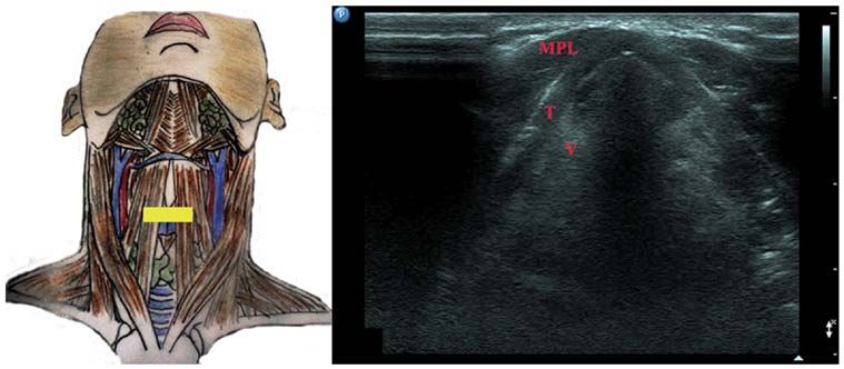
EXPERIMENTAL AND THERAPEUTIC MEDICINE 21: 273, 2021 3
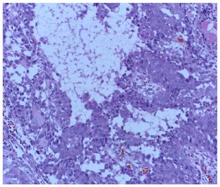
Figure 3. Ultrasonographic view of the vocal cords. MPL, anterior muscles of the neck; T, thyroid cartilage; CV, vocal cords; ARI, arytenoid cartilage.
Figure 4. Endolaryngeal tumor computerized tomography (CT) and ultra‑
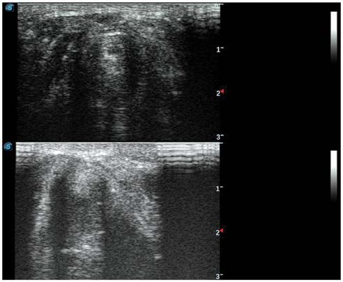
Figure 6. Upper view of the tumor at the base of the tongue and epiglottis,
sound (US) view.
middle view of the tumor at the level of the vestibular folds and lower view of
the tumor invading the vocal cord.
Figure 5. Resection piece and microscopy pathology result, squamous carci‑ Figure 7. Microscopy pathology result, acantholytic squamous carcinoma,
noma, in the same case as shown in Fig. 4. H&E; magnification, x100. H&E, from the same case as shown in Fig. 6. H&E magnification, x100. H&E,
hematoxylin and eosin. hematoxylin and eosin.
base of the tongue, the epiglottis and extending downwards of the left vocal cord. The pathology result confirmed an acan‑
through the vestibular folds until the level of the anterior part tholytic squamous laryngeal carcinoma (Fig. 7).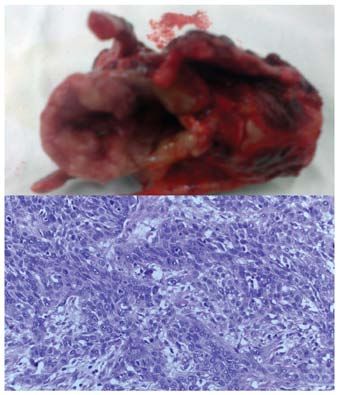
4                                       CERGAN et al: ULTRASONOGRAPHY OF THE LARYNX

                                        Figure 8. Vocal fold paresis confirmed through ultrasound (US).

    Another situation is that of an external mass effect. A              Funding
66‑year‑old woman with a history of thyroid pathology that
neglected endocrinology treatment presented with a giant                 No funding was received.
tumor comprising the left thyroid lobe and extending to adja‑
cent structures. CT raised the suspicion of vocal fold paresis.          Availability of data and material
This was confirmed through ultrasonography and endoscopy
with subsequent biopsy (Fig. 8). The patient was referred to             All data generated or analyzed during this study are included
oncologic palliative care.                                               in this published article.
    Moreover, laryngeal ultrasound has a promising use in
trauma departments enabling quick analysis of the motility of            Authors' contributions
the vocal cords (11).
    SARS‑CoV‑2 pandemic changed current diagnosis and                    RC and MD contributed substantially to the conception and
treatment protocols worldwide, minimizing the use of aerosol             design of the study, the acquisition, analysis and interpretation
producing maneuvers, such as indirect or direct flexible laryn‑          of the data, and were involved in the drafting of the manu‑
goscopy, at least until the development of a safe vaccine (12,13).       script. DV and AN contributed substantially to the acquisition,
                                                                         analysis and interpretation of the data and were involved in the
5. Conclusions                                                           drafting of the manuscript. RCC and IIJ contributed substan‑
                                                                         tially to the acquisition of the data and were involved in the
The ultrasonographic imaging of the larynx is a rather                   critical revisions of the manuscript for important intellectual
neglected imaging tool, mainly due to CT and magnetic                    content. All authors agreed to be accountable for all aspects of
resonance imaging (MRI) being accessible. However, ultra‑                the work in ensuring that questions related to the accuracy or
sonography is valuable in emergency room settings as an                  integrity of any part of the work are appropriately investigated
extension of FAST protocols in neck trauma. This review                  and resolved. All authors read and approved the final version
confirmed the fact that ultrasound enables indirect examina‑             of the manuscript.
tion of the vocal cords even by doctors of other specialties
other than ENT surgeons. Moreover, ultrasound was used in                Ethics approval and consent to participate
cases where further information was required for the manage‑
ment of the case, minimizing the costs associated with other             The information concerning the patients followed the inter‑
advanced imaging modalities. Needless to mention the fact                national regulations in accordance with the Declaration of
that ultrasonography is fast, nonradiating and permits serial            Helsinki.
real‑time examination of the patient. Further studies are
needed in order to gather data concerning the sensitivity and            Patient consent for publication
specificity of this imaging modality. New guidelines issued
during the SARS‑CoV‑2 pandemic recommend reducing the                    Patient informed consent for publication of the data/images
use of aerosol producing procedures, such as video endoscopy.            associated with the manuscript was obtained. The authors
Laryngeal ultrasound could be used to prevent the exposure of            followed the international and national regulations in accor‑
ENT specialists to COVID‑19.                                             dance with the Declaration of Helsinki and all identifying
                                                                         information was removed.
Acknowledgements
                                                                         Competing interests
Professional editing, linguistic and technical assistance
performed by Irina Radu, Individual Service Provider.                    The authors declare that they have no competing interests.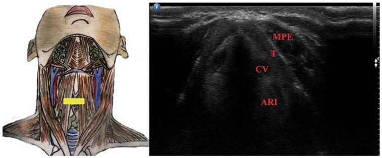
EXPERIMENTAL AND THERAPEUTIC MEDICINE 21: 273, 2021                                                  5

References                                                              9. Vrinceanu D, Dumitru M, Cergan R, Anghel AG, Patrascu ET,
                                                                           Sarafoleanu CG and Costache A: Correlations between ultra‑
                                                                           sonography performed by the ENT specialist and pathologic
1. Raghavendra BN, Horii SC, Reede DL, Rumancik WM,                        findings in the management of three cases with thyroglossal duct
   Persky WM and Bergeron T: Sonographic anatomy of the larynx,            cyst. Med Ultrason 20: 524‑526, 2018.
   with particular reference to the vocal cords. J Ultrasound Med 6:   10. Costache A, Dumitru M, Anghel I, Cergan R, Anghel AG and
   225‑230, 1987.                                                          Sarafoleanu C: Ultrasonographic anatomy of head and neck‑a
2. Hu Q, Zhu SY, Luo F, Gao Y and Yang XY: High‑frequency sono‑            pictorial for the ENT specialist. Med Ultrason 17: 104‑108, 2015.
   graphic measurements of true and false vocal cords. J Ultrasound    11. Anghel AG, Anghel I, Dumitru M and Soreanu CC: Respiratory
   Med 29: 1023‑1030, 2010.                                                and phonatory impairment due to iatrogenic vocal fold paralysis
3. Singh M, Chin KJ, Chan VW, Wong DT, Prasad GA and Yu E:                 and paresis. A retrospective study of 188 patients. Rom J Leg
   Use of sonography for airway assessment: An observational               Med 20: 287‑290, 2012.
   study. J Ultrasound Med 29: 79‑85, 2010.                            12. Docea AO, Tsatsakis A, Albulescu D, Cristea O, Zlatian O,
4. Suzuki A, Iida T, Kunisawa T, Henderson JJ, Fujita S and                Vinceti M, Moschos SA, Tsoukalas D, Goumenou M,
   Iwasaki H: Ultrasound‑guided cannula cricothyroidotomy.                 Drakoulis N, et al: A new threat from an old enemy: Re‑emergence
   Anesthesiology 117: 1128, 2012.                                         of coronavirus (review). Int J Mol Med 45: 1631‑1643, 2020.
5. Ding LW, Wang HC, Wu HD, Chang CJ and Yang PC: Laryngeal            13. C a l i n a D, D o c e a AO, P e t r a k i s D, Ego r ov A M,
   ultrasound: A useful method in predicting post‑extubation               Ishmukhametov AA, Gabibov AG, Shtilman MI, Kostoff R,
   stridor. A pilot study. Eur Respir J 27: 384‑389, 2006.                 Carvalho F, Vinceti M, et al: Towards effective COVID‑19
6. Sólyom R, Csiszér I and Neagos A: Tonsillar hypertrophy impli‑          vaccines: Updates, perspectives and challenges (review). Int J
   cations in sleep disorders in adults and children. Rom J Morphol        Mol Med 46: 3‑16, 2020.
   Embryol 55 (2 Suppl): S603‑S606, 2014.
7. Ko DR, Chung YE, Parl I, Lee HJ, Park JW, You JS, Chung TN,
   Park YS, Chung SP and Kim S: Use of bedside sonography for
   diagnosing acute epiglottitis in the emergency department: A
   preliminary study. J Ultrasound Med 31: 19‑22, 2012.                                 This work is licensed under a Creative Commons
8. Costache A, Dumitru M, Tweedie D, Sarafoleanu C and Anghel I:                        Attribution-NonCommercial-NoDerivatives 4.0
   Adult cervical lymphangioma‑ultrasonography, surgical removal,                       International (CC BY-NC-ND 4.0) License.
   and pathology results. Case report. Med Ultrason 17: 411‑413,
   2015.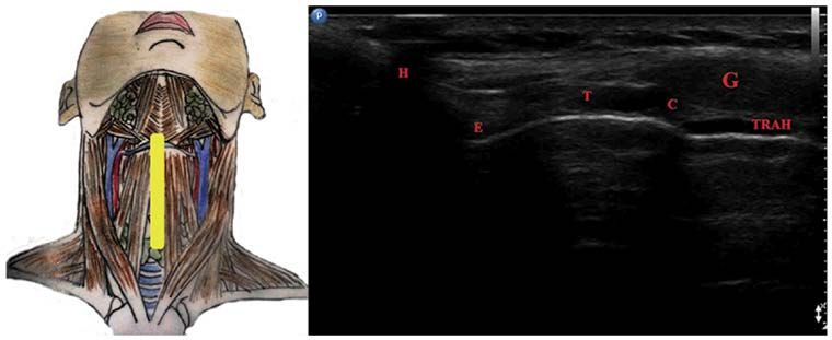
You can also read