Ultrasound Examination of Gynecomastia Case in Male Patient: Case Report
←
→
Page content transcription
If your browser does not render page correctly, please read the page content below
Global Journal of Health Science; Vol. 10, No. 4; 2018
                                                                                   ISSN 1916-9736 E-ISSN 1916-9744
                                                                Published by Canadian Center of Science and Education

    Ultrasound Examination of Gynecomastia Case in Male Patient: Case
                                Report
Puji Supriyono1, Gando Sari1, Nursama Heru Apriantoro1, Win Priantoro1, Samsun1, Mayarani1, Eka Putra Syarif1,
                              Sri Rebekka2, Sri Kartini3 & Humala Prika Aditama4
1
    Department of Radiodiagnostic and Radiotherapy, Politeknik Kesehatan Kemenkes Jakarta II, Jakarta, Indonesia
2
    Radiology Unit, District General Hospital of Cengkareng, Jakarta Barat, Jakarta, Indonesia
3
    Radiology Unit, Dr Mintohardjo Navy Hospital Jakarta, Indonesia
4
    General Practitioner, Puri Indah, Pondok Indah Hospital, Jakarta, Indonesia
Correspondence: Puji Supriono, Department of Radiodiagnostic and Radiotherapy, Politeknik Kesehatan
Kemenkes Jakarta II, Hang Jebat III/F-3, Jakarta 12120, Indonesia. Tel/Fax: 62-21-720-6239. E-mail:
puji_supriyono@yahoo.com

Received: January 21, 2018       Accepted: February14, 2018      Online Published: February 26, 2018
doi:10.5539/gjhs.v10n4p7              URL: https://doi.org/10.5539/gjhs.v10n4p7

Abstract
Gynecomastia is hypertrophy abnormality of male’s breast gland soft tissue. In that case, the mammography is
difficult to show the malignant masses. The ultrasonography is often useful and really important examination to
confirm the diagnosis by showing retroareolar hypoechoic mass and or elevation of hyperechoic on parenchyma
gland. Gynecomastia is very rare cases in older man. This case report shows 67 years old patient on compos mentis
state, who has left breast enlargement.
Keywords: gynecomastia, male’s breast, benign, ultrasonography
1. Introduction
Gynecomastia is defined as an enlargement of the male breast. It is often benign and can be the source of
significant embarrassment and psychological distress (Lemaine, 2013). It is an abnormal and excessive growth of
male’s breast tissue, commonly occurs in adults and middle age groups in general as a result of medications,
nutritional supplements, and anabolic steroid consumption, usually athletes and bodybuilders (Stavros, 2004). In
some cases, there were due to physiological, pathological and pharmacological factors (Oates, 2004).
Physiologically, gynecomastia is caused by an imbalance between free estrogen and free androgen actions in the
breast tissue. (Mathur & Braunstein, 1997; Braunstein, 2007). It can occur in neonates, at puberty, and with obesity
and aging. (Niewoehner & Schorer, 2008; Johnson & Murad, 2009). The basic mechanism of gynaecomastia
caused by lowering of androgen production, elevation of estrogen production and elevation of estrogen precursor
availability for estradiol conversion, therefore there are changes in estrogen and androgen ratio. Transition time
from puberty to post-puberty is followed by elevation of testosterone by 30 times and estrogen by 3 times.
The main causes of gynecomastia were reported by (Cooper et al., 1994; Appelbaum et al., 1999; Charlot et al.,
2013). That is Physiological (senescence, puberty), Endocrine (Klinefelter syndrome, hypogonadism,
hypothyroidism), systemic disease (cirrhosis, chronic renal failure), tumors (testicular tumors, adrenal carcinoma,
pituitary adenoma, hepatocellular carcinoma), Toxicity Medication (alcohol, cannabis, digoxin, cimetidine,
thiazide diuretics, omeprazole, tricyclic antidepressants, spironolactone, diazepam, anabolic-androgen steroids,
exogenous oestrogens), and idiopathic.
The primary modality of mammography is usually to diagnosis and classification of the breast (Chen & Slanetz,
2014). However. it is not necessary to confirm gynecomastia through a mammogram (Tan et al., 2016).
Furthermore, ultrasonography technique is appropriate to diagnose gynecomastia case to produce visualization
image of breast tissue. (Berg & Yang, 2014)
2. Case Report
67years old male patient was referred from surgeon’s outpatient clinic to Radiology Unit for ultrasound
examination of the breast. The physical feature of left breast is bigger than right breast, not symmetrical and
                                                          7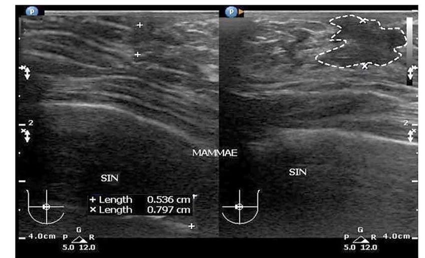
gjhs.ccsenet.org                             Global Journal of Health Science                      Vol. 10, No. 4; 2018

uncomfortable feeling, slight pressure pain, no fever, no history of trauma, no history of similar illness in the
family, no significant weight loss. The patient has suffered this condition since one year ago. Body Mass Index of
31.22, the body temperature of 37 oC, heart rate of 64 bpm, Blood pressure of 125 per 80 mmHg. On the previous
examination, Thorax examination, laboratory routine, showed that there are no abnormalities in the chest.
The Ultrasound technique to show breast tissue until surface layer, cutis, and subcutis. Ultrasound machine used on
the right hand was Phillips EPIQ 5G, which was calibrated until November 2019, A linear matrix probe transducer
type L12-3 was used from Phillips with the frequency of 7 MHz to 12 MHz, it was intended to visualize superficial
mammary tissue, cutis, and subcutis layer.
Patient examined on supine position or abnormal part is located higher and hands on the same side above the head.
Scanning technique on breast examination is different with common examination, the scanning was done with
transversal and longitudinal projection on breast, on both breast, one by one, scanning was started from right axilla
region to the left axilla region, next right breast on transversal projection by moving the transducer away from
nipple on 12 o’clock, 3 o’clock, 6 o’clock, and 9 o’clock moved circularly with nipple as the axis, and then
continue to left breast with measurement of hypoechoic lesion. (Berg & Yang, 2014). The Image ultrasound of
breast examination was an interpretation based on BI-RADS (Breast Imaging Reporting and Data System
Standard) guidelines as shown Table 1.

Table 1. Classification of Ultrasoundbreast examination based on BI-RADS (ACR, 2014)
BI-RAD* Catagory            Reporting
0                           Need additional information. And/or prior mammogram for comparison
1                           Negative
2                           Benign Finding
3                           Probably benign finding. Short interval follow up suggested
4                           Suspicious abnormality-biopsy should be considered
5                           Highly suggestive of malignancy-Appropriate action should be taken
6                           Known biopsy-proven malignancy appropriate action should be taken
*Breast Imaging Reporting and Data System.

3. Results and Discussion
For gynecomastia cases in male patients, a mammography is not necessary for confirming a diagnosis. However,
ultrasound examination is the most common and important to diagnosis of true gynecomastia, which has a specific
presentation for detecting nodular diseases or malignant lesions (Muñoz et al., 2010; Draghi et al., 2011; Foo et al.,
2017). In fact, more than of 95% of the male breast lesions were benign, as other authors reported (Chen et al.,
2006; Muñoz et al., 2010). For optimal results of gynecomastia cases, it needs cooperation from the patient to be in
supine position. To describe mammary soft tissue, therefore visualizing soft tissue of cutis, subcutis,
fibro-glandular and muscular tissue with a transversal and longitudinal view, it should be done in transversal and
longitudinal scanning. Scanning starts from right axilla region and then left axilla region, its purpose is to look for
lesions in the axilla region so as to visualize cuticle tissue, subcutis, and muscle and lymph node lesions as an
indicator of spread through lymph nodes (Rasad & Ekayuda, 2005). To look for a benign lesion in gynecomastia
cases around the breast, it was done on the transversal projection by moving the transducer away from nipple as
clock direction on 12 o’clock, 3 o’clock, 6 o’clock and 9 o’clock. Scanning results of left breast in gynecomastia
cases have been shown in Figure 1, and Figure 2 the scanning result appears bordered inhomogenous lesion in the
left axilla. A result of left breast with Doppler Ultrasound Scanning has been seen in Figure 3.

                                                            8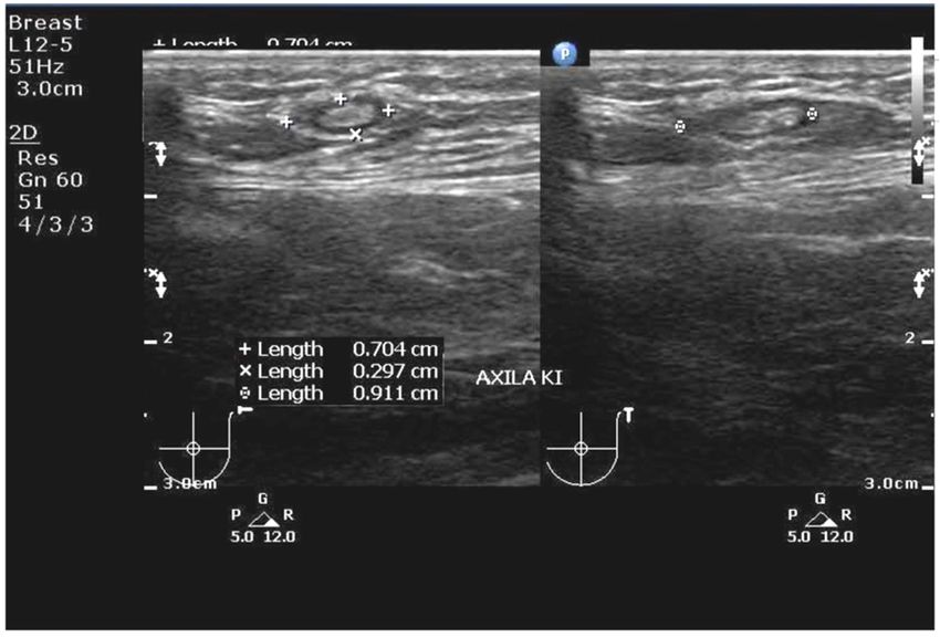
gjhs.ccsenet.org                          Global Journal of Health Science                      Vol. 10, No. 4; 2018

  Figure1. Ultrasound image of Left Breast Scanning: It shown irregular edge hypoechoic lesions in left breast
                                                 retroareola

   Figure 2. Ultrasound image of left axilla scanning: it shows irregular edge hypoechoic lesions in left axilla
                                                  (lymphnode)

                                                         9
gjhs.ccsenet.org Global Journal of Health Science Vol. 10, No. 4; 2018
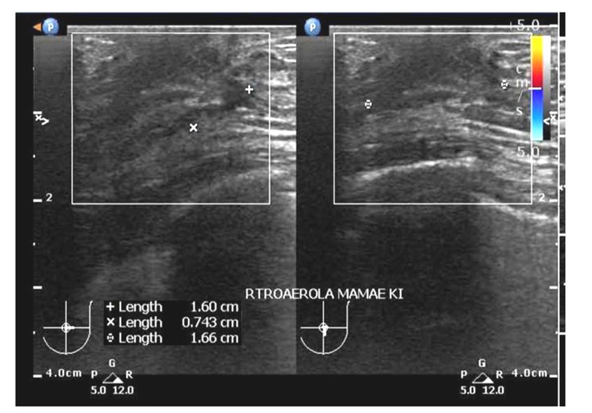
Figure 3. Result of left breast with Doppler Ultrasound Scanning: It inhomogeneous lesions are firmly defined in
left axilla region. There is no blood vessels in lession area
For ultrasound evaluation in gynecomastia case on the left breast. Figure 1 shows cutis and subcutis are not
thickening, this case demonstrates that there is hypoechoic lesion with an irregular edge at retro areola breast with
the size of 0.536 x 0.797 cm2. In Figure 2, the result of left axilla scanning of gynecomastia case, it shows irregular
edge hypoechoic lesions in left axilla (lymph node) with the size of 0.704 x 0.297 x 0.911 cm3. The result of left
breast with Doppler ultrasound scanning as shown in Figure 3, it appears inhomogeneous lesions with no vascular
flow in the lesion, it's signed benign mass at retro areola left breast with the size of 1.60 x 0.743 x 1.66 cm3. Based
on the evaluation of the ultrasound scanning images and physical examination of a patient, it was found a benign
lesion classified as BI-RAD 4 category, that means biopsy technique should be used as the highest standard (gold
standard) in the next procedure,it was obtain to detect and identify benign proliferative lesion of the male breast. A
biopsy is mandatory in these cases to exclude malignancy. (Foo et al., 2017)
4. Conclusion
Ultrasound is the right examination to the benign diagnosis. In the case was shown lesion on retro areola left breast
in accordance BI-RADS 4 with lymphadenopathy in the left axilla. Base on the findings results, biopsy results
should be used to establish a diagnosis and to make sure benign lesion of the male breast.
5. Suggestion
From the above results, a breast ultrasound examination in the case of gynecomastia is recommended to use
adequate equipment with Doppler ability, to uphold the diagnosis and to ensure a benign or malignancy by using
biopsy action.
Acknowledgements
We would like to thank the Director of Radiodiagnostic and Radiotherapy Department of Politeknik Kesehatan
Kementerian Kesehatan Jakarta II, Ministry of Health for his contributions during the study. We are sincerely
grateful to the Directors, Sonographers and staffs of District General Hospital of Cengkareng Jakarta for their
valuable contributions, facilities, assistance and constructive suggestions during our study.
Competing Interests Statement
The authors declare that they have no competing or potential conflicts of interest.
References
ACR.The American College of Radiologi. (2014). ACR BI-RADS Atlas Fifth Edition. Quick Reference. Retrieved
from https://www.acr.org/-/media/ACR/Files/RADS/BI-RADS/ BIRADS-Reference-Card.pdf
10gjhs.ccsenet.org Global Journal of Health Science Vol. 10, No. 4; 2018
Appelbaum, A. H., Evans, G. F., Levy, K. R., Amirkhan, R. H., & Schumpert, T. D. (1999). Mammographic
appearances of male breast disease. RSNA Radiographics, 19 (3), 559-568.
https://doi.org/10.1148/radiographics.19.3.g99ma01559
Berg, W. A., & Yang, W. T. (2014). Diagnostic Imaging: Breast (2nd ed.). Manitoba: Amirsys
Braunstein, G. D. (2007). Clinical Practice, Gynecomastia. New England Journal of Medicine, 357(12),
1229-1237. https://doi.org/10.1056/NEJMcp070677
Charlot, M., Béatrix, O., Chateaua, F., Dubuissonb, J., Golfier, F., Valettea, P. J., & Rétya, F. (2013).Breast
imaging Pathologies of the male breast. Diagnostic and Interventional Imaging, 94, 26-37.
https://doi.org/10.1016/j.diii.2012.10.011
Chen, L., Chantra, P. K., Larsen, L. H., Barton, P., Rohitopakarn, M., Zhu, E. Q., & Bassett, L. W. (2006) Imaging
characteristics of malignant lesions of the male breast. Radiographics, 26, 993-1006.
https://doi.org/10.1148/rg.264055116
Chen, P. H., & Slanetz, P. J. (2014). Incremental clinical value of ultrasound in men with mammographically
confirmed gynecomastia. Eur J Radiol., 83, 123-129. https://doi.org/10.1016/j.ejrad.2013.09.021.
Cooper, R. A., Gunter, B. A., & Ramamurthy, L. (1994). Mammography in men. Radiology, 191, 651-656.
https://doi.org/10.1148/radiology.191.3.8037795
Draghi, F., Tarantino, C. C., Madonia, L., & Ferrozzi, G. (2011).Ultrasonography of the male breast. J.
Ultrasound., 4(3), 122-129. https://doi.org/10.1016/j.jus.2011.06.004
Foo, E. T., Lee, A. Y., Ray, K. M., Woodard, G. A., Freimanis, R. I., & Joe, B. N. (2017). Value of diagnostic
imaging for the symptomatic male breast: Can we avoid unnecessary biopsies? Clin Imaging, 45, 86-91.
https://doi.org/10.1016/j.clinimag.2017.06.004
Johnson, R. E., &Murad, M. H. (2009). Gynecomastia: Pathophysiology, Evaluation, and Management. Mayo
Clin Proc., 84(11), 1010-1015. https://doi.org/10.1016/S0025-6196(11)60671-X
Lemaine, V., Cayci, C., Simmons, P. S., & Petty, P. (2013). Gynecomastia in Adolescent Males. Semin Plast Surg.,
27(1), 56-61. https://doi.org/10.1055/s-0033-1347166
Mathur, R., & Braunstein, G. D. (1997).Gynecomastia: pathomechanisms and treatment strategies. Hormone
Research, 48(3), 95-102. PMID: 11546925. https://doi.org/10.1159/000185497
Mu-oz, C. R., Álvarez, B. M., Mu-oz, G. E., Raya, P. J. L., & Martínez, P. M. (2010) Mammography and
ultrasound in the evaluation of male breast disease. European Radiology, 20(12), 2797-2805.
https://doi.org/10.1007/s00330-010-1867-7
Niewoehner, C. B., & Schorer, A. E. (2008). Gynaecomastia and breast cancer in men. British Medical Journal.
336(7646), 709-713. https://doi.org/10.1136/bmj.39511.493391.BE
Oates, S. D. (2004). Gynecomastia. In: S. E. Singletary, G. L. Robb, & Hortobagyi, Advanced Therapy of Breast
Disease. GN.London : BC Decter Inc.
Rasad, S., & Ekayuda, I. (2005). Radiologi Diagnostik (2nd.). Jakarta: Faculty of Medicine of Universitas
Indonesia.
Stavros, A. T. (2004). Breast Ultrasound. Philadelphia : Lippincott Williams & Wilkins
Tan, W., DePeri, E., Feigel, D., Perez, E., &Yasrebi, M. (2016). Use Of Mammograms In Men Is Rising, But
Researchers Find Little Reason To Administer. Mayo Clinic. Retrieved from
https://www.sciencedaily.com/releases/2006/12/061216104542.htm
Copyrights
Copyright for this article is retained by the author(s), with first publication rights granted to the journal.
This is an open-access article distributed under the terms and conditions of the Creative Commons Attribution
license (http://creativecommons.org/licenses/by/4.0/).
11You can also read