THERAPEUTIC CHALLENGE - Treatment of upper limb arterial occlusion caused by a cervical rib
←
→
Page content transcription
If your browser does not render page correctly, please read the page content below
ISSN 1677-7301 (Online)
THERAPEUTIC CHALLENGE
Treatment of upper limb arterial occlusion caused
by a cervical rib
Tratamento de oclusão arterial em membro superior causada por costela cervical
Vanessa Aline Miranda Vieira Milagres1 , Roberto Lucas de Sena Avellar1, Ana Paula Pires Silva1 ,
Pedro José Pires Neto1, Daniel Mendes Pinto1
Abstract
The cervical rib syndrome occurs when the interscalene triangle is occupied by a cervical rib, displacing the brachial
plexus and the subclavian artery forward, which can cause pain and muscle spasms. The objective of this study is to
discuss diagnosis of the cervical rib syndrome and treatment possibilities. This therapeutic challenge describes clinical
and surgical management of a 37-year-old female patient with upper limb arterial occlusion caused by a cervical rib.
Keywords: cervical rib; thoracic outlet syndrome; subclavian artery; subclavian vein; brachial plexus.
Resumo
A síndrome da costela cervical ocorre quando o triângulo intercostoescalênico é ocupado por uma costela cervical,
deslocando o plexo braquial e a artéria subclávia anteriormente, o que pode gerar dor e espasmo muscular. O objetivo
deste estudo é discutir sobre o diagnóstico da síndrome da costela cervical e as possibilidades de tratamento. Este
desafio terapêutico descreve a condução clínica e cirúrgica de uma paciente de 37 anos com obstrução arterial em
membro superior causada por costela cervical.
Palavras-chave: costela cervical; síndrome do desfiladeiro torácico; artéria subclávia; veia subclávia; plexo braquial.
How to cite: Milagres VAMV, Avellar RLS, Silva APP, Pires Neto PJ, Pinto DM. Treatment of upper limb arterial occlusion
caused by a cervical rib. J Vasc Bras. 2021;20:e20200193. https://doi.org/10.1590/1677-5449.200193
1
Hospital Felício Rocho, Belo Horizonte, MG, Brasil.
Financial support: None.
Conflicts of interest: No conflicts of interest declared concerning the publication of this article.
Submitted: September 08, 2020. Accepted: March 08, 2021.
The study was carried out at Hospital Felício Rocho, Belo Horizonte, MG, Brazil.
Copyright© 2021 The authors. This is an Open Access article distributed under the terms of the Creative Commons Attribution License, which
permits unrestricted use, distribution, and reproduction in any medium, provided the original work is properly cited.
Milagres et al. J Vasc Bras. 2021;20:e20200193. https://doi.org/10.1590/1677-5449.200193 1/5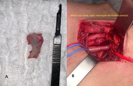
Arterial occlusion caused by a cervical rib

INTRODUCTION                                                               PART II – WHAT WAS DONE

    A cervical rib is an anatomic variant that is present                     The patient was admitted and given pain control
in 1% of the population.1 It originates from the                           and anticoagulation with enoxaparin at 1 mg/kg every
transverse process of the seventh cervical vertebra.1                      12 h. Angiotomography of the thoracic aorta and
The majority of patients are asymptomatic and just                         the right upper limb identified an accessory cervical
                                                                           rib (C7) on the right, joining to the first ipsilateral
10% manifest symptoms.1 When present, symptoms
                                                                           rib anteriorly. The subclavian artery was patent, but
are caused by compression of neurovascular structures                      subjected to considerable compression between the
in the thoracic outlet region, constituting the cervical                   accessory rib described above and the ipsilateral
rib syndrome, which is one type of thoracic outlet                         clavicle, increasing notably during abduction of the
syndrome (TOS). A cervical rib is present in almost                        right upper limb (Figure 1). Other patent segments of
30% of cases of TOS.2                                                      the right subclavian artery did not exhibit evidence
    Surgical treatment of the cervical rib syndrome                        of compression. Having established a diagnosis of
can be performed via conventional access routes,                           TOS, the patient was discharged on warfarin and
such as the supraclavicular, posterior, transaxillary,                     acetylsalicylic acid, with an international normalized
                                                                           ratio result of 2.39, and was instructed to attend
or combined approaches.3,4 More recently described
                                                                           outpatients follow-up and schedule cervical rib
approaches include video-assisted surgery and
                                                                           resection surgery.
transthoracic with robotic assistance.5,6                                     After 1 month on therapeutic anticoagulation, in
    The objective of this study is to discuss diagnosis of                 October 2019, the patient was admitted for elective
cervical rib syndrome and the treatment possibilities.                     surgical treatment of TOS. Access was obtained via
We present the case of a 37-year-old female patient with                   a supraclavicular incision, as illustrated in Figure 2,
an upper limb arterial obstruction caused by a cervical                    with identification of the cervical rib, the subclavian
rib and discuss its clinical and surgical management.                      artery, and the brachial plexus (Figure 3).
The protocol was approved by the Ethics Committee                             The patient underwent resection of the right cervical
at our institution (CAAE 35649620.2.0000.5125,                             rib (Figure 4), with dissection and release of adhesions
                                                                           to the right subclavian artery and exploration of the
approval ruling no. 4.303.586).
                                                                           brachial plexus. The surgical operation was well-
                                                                           tolerated and was conducted with no intraoperative
PART I – CLINICAL SITUATION                                                complications. Perioperative chest X-ray did not show
                                                                           pneumothorax. During the postoperative period, the
   A 37-year-old, previously healthy, female patient                       patient recovered well, with improvement in pain,
was admitted to an Urgent Care Center in September                         conservation of upper limb sensitivity and motricity,
2019 with pain, pallor, and paresthesia in the right                       and full and symmetrical radial pulses. She was
upper limb. She reported onset of symptoms 4 months                        discharged on the first postoperative day on 100 mg
previously, with deterioration over the last 2 weeks.                      of acetylsalicylic acid per day. At 6 months’ follow-
Physical examination of the right upper limb found                         up, she was asymptomatic and had good perfusion
                                                                           of the right upper limb.
distal pallor, a palpable pulse in the right supraclavicular
region, weak brachial pulse, and absent radial and
ulnar pulses. There was a palpable cervical rib on the
right. Arterial duplex ultrasound of the right upper
limb revealed thrombi with a chronic appearance
in the radial and ulnar arteries, with occlusion.
Radiography of the cervical spine and thorax showed
an articulated cervical rib on the right. This situation
raised a number of treatment options:

  1- Systemic anticoagulation with heparin;
  2- Catheter-guided thrombolysis;
                                                                           Figure 1. Angiotomography showing the cervical rib on the
  3- Surgery to resect the cervical rib.                                   right and compression of the subclavian artery.

Milagres et al. J Vasc Bras. 2021;20:e20200193. https://doi.org/10.1590/1677-5449.200193                                                 2/5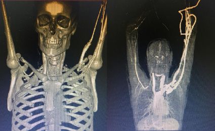
Arterial occlusion caused by a cervical rib
Thoracic outlet syndrome encompasses symptoms
caused by compression of neurovascular structures
in the region of the thoracic outlet.5 Cervical rib
syndrome occurs when the interscalene triangle is
occupied by a cervical rib, displacing the brachial
plexus and subclavian artery forward, causing pain
and muscle spasms.1
Treatment of the majority of patients with TOS is
clinical, involving analgesics, anti-inflammatories,
benzodiazepines, and postural changes.1 In the case
presented here, clinical treatment was initiated with
Figure 2. “Necklace” incision, 2 cm superior of the clavicle.
analgesics and therapeutic anticoagulation because the
patient had presented with arterial occlusion. There
are formal indications for surgery in 15% of cases and
the majority of operations to treat TOS are conducted
in patients with neurogenic compression.1 Presence
of cervical rib, symptomatic bone abnormalities,
and vascular complications such as aneurysms and
thromboses, are indications for mandatory surgery.1,2
According to Daniels et al.,3 when thrombus is
present, catheter-guided thrombolysis is the initial
treatment of choice. In this case, we decided to
initiate treatment with anticoagulation because the
patient had symptoms of chronic ischemia. She
exhibited satisfactory progress, without needing
thrombolysis. Once the thrombus has been resolved,
treatment should proceed with surgical release of
thoracic outlet compression. Surgical treatment of
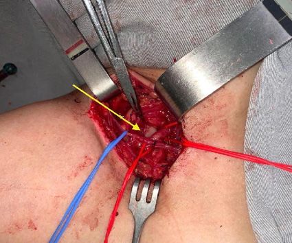
Figure 3. The yellow arrow indicates the cervical rib. Repair of cervical rib syndrome consists of resection, which
the subclavian artery with a red vessel loop. can be accomplished via supraclavicular, posterior,
transaxillary, or combined approaches, and via more
recently described techniques such as video-assisted
and transthoracic surgery with robotic assistance.4-6
Resection of the cervical rib and/or first rib via
the supraclavicular approach provides access to the
subclavian artery, which is of relevance in patients
with aneurysms and/or thrombosis caused prolonged
compression of the artery by the cervical rib, as was
the case of the patient described in this report. The
degree of integrity of the artery will determine whether
repair or resection are needed.3 If the artery is only
compressed, relieving the compression is a sufficient
Figure 4. (A) Cervical rib; (B) Subclavian artery after resection treatment.3 If there is greater arterial compromise
of the cervical rib. or aneurysmal degeneration, a bypass is generally
performed.3 In the case presented, release of adhesions
from the right subclavian artery proved effective.
DISCUSSION The transaxillary surgical approach is a safe technique
This study draws attention to arterial occlusion in involving reduced manipulation of the brachial plexus,
young patients, which can be caused by mechanical which can achieve a lower incidence of perioperative
compression. About 50% of patients with cervical rib complications related to nerve damage.4,7 It enables
syndrome present with arterial compression.2 Treatment safe resection of cervical ribs and/or first ribs and
via a cervical approach is appropriate in this situation, is the approach most often used when concomitant
because it enables safe access to the brachial plexus, resections are performed.4 It also produces better
the subclavian artery, and the cervical rib. esthetic results than the supraclavicular approach.4
Milagres et al. J Vasc Bras. 2021;20:e20200193. https://doi.org/10.1590/1677-5449.200193 3/5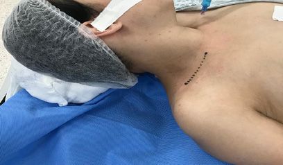
Arterial occlusion caused by a cervical rib

The disadvantage of this technique is that it does                         CONCLUSIONS
not offer adequate access to the subclavian artery.
There is also a higher incidence of pneumothorax,                             Cervical rib syndrome is rare, but has great potential
probably due to the proximity of the pleura to the                         to become severe, causing significant morbidity if not
area dissected in the transaxillary approach.4 The                         treated adequately. We should remember mechanical
combined approach should be used in cases in which                         compression as a possible cause of cases of arterial
a transaxillary approach does not provide an adequate                      occlusion in young patients.
view for resection of the cervical rib.4                                      Resection of the cervical rib via a supraclavicular
    Video-assisted surgery for resection of cervical                       approach is a safe treatment that offers satisfactory
ribs and/or first ribs offers better surgical access and                   access to the subclavian artery, good clinical results,
enables the surgical team to clearly identify anatomic                     and a favorable impact on the recovery of patients with
structures. It also allows for safer dissection and reduces                cervical rib syndrome. In these cases, anticoagulants
the number of complications.6 One disadvantage of                          are indicated for initial treatment of thrombosis and
the video-assisted approach is difficulty in accessing                     anticoagulation is generally unnecessary during the
the superior portions of the scalene muscles; only the                     postoperative period.
inferior 2 cm can be resected.6 Video-assisted surgery
is more expensive than conventional surgery, but                           REFERENCES
is less expensive than robotically-assisted surgery.                       1. Ciorlin E, Araújo JD, Araújo JD Fo. Síndromes compressivas
    Transthoracic robotically-assisted resection of the                       neurovasculares cervicotoracoaxilares (Síndrome do desfiladeiro).
cervical rib is a minimally invasive technique that                           In: Brito CJ, Silva RM, Araújo, EL. Cirurgia vascular: cirurgia
                                                                              endovascular, angiologia. 4. ed. Rio de Janeiro: Thieme Revinter
offers adequate visualization of the neurovascular
                                                                              Publicações; 2020. p. 733-747.
and musculoskeletal structures. The improved view
                                                                           2. Henry BM, Vikse J, Sanna B, et al. Cervical rib prevalence and
improves safety and enables complete surgical                                 its association with thoracic outlet syndrome: a meta-analysis
decompression. It also yields better esthetic results,                        of 141 studies with surgical considerations. World Neurosurg.
since just three small surgical incisions are made,                           2018;110:e965-78. http://dx.doi.org/10.1016/j.wneu.2017.11.148.
with the largest, at 15 mm, at the level of the armpit.5                      PMid:29203316.
However, this is a new technique with few cases                            3. Daniels B, Michaud L, Sease F Jr, Cassas KJ, Gray BH. Arterial thoracic
reported and higher costs.                                                    outlet syndrome. Curr Sports Med Rep. 2014;13(2):75-80. http://
                                                                              dx.doi.org/10.1249/JSR.0000000000000034. PMid:24614419.
    Use of anticoagulants for initial treatment of
                                                                           4. Jayaraj A, Duncan AA, Kalra M, Bower TC, Gloviczki P. Outcomes
thrombosis is well-defined in treatment of TOS. Use                           of transaxillary approach to cervical and first-rib resection for
of anticoagulants after resection of the first rib is                         neurogenic thoracic outlet syndrome. Ann Vasc Surg. 2018;51:147-
controversial. Fairman et al.8 recommend preoperative                         9. http://dx.doi.org/10.1016/j.avsg.2018.02.029. PMid:29772332.
thrombolysis, with the advantage of potential elimination                  5. Wybaillie E, Maene L, Cooreman F, Beelen R. Robotically assisted
of the risk of postoperative anticoagulation to treat                         transthoracic cervical rib resection. Ann Thorac Surg. 2018;106(5):e253-5.
Paget-Schroetter syndrome, which is the venous                                http://dx.doi.org/10.1016/j.athoracsur.2018.04.016. PMid:29752917.
vascular form of TOS. Gelabert et al.9 describe use of                     6. Chan YC, Gelabert HA. Hight-definition video-assisted transaxillary
                                                                              first rib resection for thoracic outlet syndrome. J Vasc Surg.
anticoagulation with warfarin during the postoperative
                                                                              2013;57(4):1155-8. http://dx.doi.org/10.1016/j.jvs.2012.10.089.
period and recommend against use of heparin because                           PMid:23357519.
of the higher risk of bleeding. In the case reported,                      7. Gelabert HA, Rigberg DA, O’Connell JB, Jabori S, Jimenez JC, Farley
the patient was treated with warfarin preoperatively,                         S. Transaxillary decompression of thoracic outlet syndrome patients
with good response in terms of reduction of symptoms                          presenting with cervical ribs. J Vasc Surg. 2018;68(4):1143-9. http://
and no need for thrombolysis. Anticoagulation was                             dx.doi.org/10.1016/j.jvs.2018.01.057. PMid:29705086.
not used during the postoperative period. The most                         8. Fairman AS, Fairman RM, Foley PJ, Etkin Y, Jackson OA, Jackson BM.
recent studies demonstrate that patients are often                            Is routine postoperative anticoagulation necessary in all patients
                                                                              after first rib resection for paget-schroetter syndrome? Ann Vasc
discharged on aspirin alone during the postoperative                          Surg. 2020;69:217-23. http://dx.doi.org/10.1016/j.avsg.2020.05.042.
period, with no need for anticoagulation.8                                    PMid:32497616.
    Immediate treatment of cervical rib syndrome                           9. Gelabert HA, Jimenez JC, Davis GR, Derubertis BG, O’Connell
is important to prevent long-term complications of                            JB, Rigberg DA. Early postoperative hemorrhage after first rib
neural and/or vascular compression.10 Asymptomatic                            resection for vascular thoracic outlet syndrome. Ann Vasc Surg.
                                                                              2011;25(5):624-9. http://dx.doi.org/10.1016/j.avsg.2011.02.023.
patients in whom a cervical rib is found as an incidental
                                                                              PMid:21724102.
diagnosis should be given guidance on the symptoms
                                                                           10. Morel J, Pirvu A, Elie A, Gallet N, Magne JL, Spear R. Functional
of neurovascular compression, so that they can                                 results of cervical rib resection for thoracic outlet syndrome:
seek appropriate treatment rapidly in the event that                           impact on professional activity. Ann Vasc Surg. 2019;56:233-9.
symptoms emerge.2                                                              http://dx.doi.org/10.1016/j.avsg.2018.09.007. PMid:30476612.

Milagres et al. J Vasc Bras. 2021;20:e20200193. https://doi.org/10.1590/1677-5449.200193                                                            4/5
Arterial occlusion caused by a cervical rib
Correspondence
Vanessa Aline Miranda Vieira Milagres
Rua Cláudio Manoel, 878, apartamento 1703 - Funcionários
CEP 30140-100 - Belo Horizonte (MG), Brasil
Tel.: +55 (31) 98819-2689
E-mail: vanessa_mvieira@yahoo.com.br
Author information
VAMVM, RLSA and APPS - Vascular surgeons, Hospital Felício Rocho.
PJPN - MSc; Orthopedist; Board certified; Membro Superior, Hospital
Felício Rocho.
DMP - MSc; PhD; Vascular surgeon, Hospital Felício Rocho.
Author contributions
Conception and design: DMP, VAMVM, PJPN
Analysis and interpretation: APPS, DMP, VAMVM
Data collection: RLSA, VAMVM, PJPN
Writing the article: APPS, DMP, RLSA, VAMVM
Critical revision of the article: VAMV, DMP
Final approval of the article*: APPS, DMP, RLSA, VAMVM, PJPN
Statistical analysis: N/A.
Overall responsibility: VAMVM
*All authors have read and approved of the final version of the article
submitted to J Vasc Bras.
Milagres et al. J Vasc Bras. 2021;20:e20200193. https://doi.org/10.1590/1677-5449.200193 5/5You can also read