Delayed Intracystic Hemorrhage after Percutaneous Drainage and Sclerotherapy for a Symptomatic Giant Hepatic Cyst: A Case Report - J-Stage
←
→
Page content transcription
If your browser does not render page correctly, please read the page content below
Case Report

 Delayed Intracystic Hemorrhage after Percutaneous Drainage
  and Sclerotherapy for a Symptomatic Giant Hepatic Cyst:
                       A Case Report

                                           1) Department of Diagnostic & Interventional Radiology, Kansai Rosai Hospital, Japan

                                                                                                   Koji Mikami1), Hiroshi Yukimoto1)

                                                          Abstract
           Herein, we have reported a rare case of intracystic hemorrhage due to rupture of a right hepatic artery
        pseudoaneurysm in a 76-year-old female patient who underwent drainage and 3% polidocanol sclerotherapy
        for a symptomatic giant hepatic cyst. One month after sclerotherapy, the patient presented to the emergency
        room with acute and severe abdominal pain. Non-contrast T1-weighted magnetic resonance imaging findings
        showed high hepatic cyst fluid signal intensity and abdominal arteriography findings revealed a right hepatic
        artery pseudoaneurysm surrounding the hepatic cystic wall. Therefore, the patient was diagnosed with intra-
        cystic hemorrhage due to a ruptured pseudoaneurysm. Embolization, using a detachable coil, was successful.
        Interventional radiologists should be aware of potential vascular injuries during drainage and sclerotherapy
        for giant hepatic cysts.
           Key words: symptomatic hepatic cyst, sclerotherapy intracystic hemorrhage, pseudoaneurysm, emboliza-
                        tion
                                                                            (Interventional Radiology 2021; 6: 61-64)

                                                                             fort and abdominal distention after eating. Non-contrast
Introduction                                                                 computed tomography (CT) revealed compression of the de-
                                                                             scending duodenum by a giant hepatic cyst measuring 17 ×
   Percutaneous drainage followed by sclerotherapy is the                    12 × 17 cm in segment 4 of the liver (Fig. 1). Percutane-
currently preferred noninvasive treatment approach for sus-                  ous drainage and ethanol sclerotherapy are commonly used
tained volume reduction and symptomatic relief in patients                   [4]; however, polidocanol sclerotherapy and ethanol scle-
with hepatic cysts [1]. Although this treatment approach is                  rotherapy have been recently reported to have similar thera-
associated with few serious complications, several studies                   peutic effects, although ethanol sclerotherapy has more side
have recently reported intracystic bleeding during and after                 effects than polidocanol sclerotherapy [2]. Therefore, percu-
the procedure [2, 3]. Herein, we have reported a rare case of                taneous drainage and 3% polidocanol sclerotherapy were
intracystic hemorrhage due to rupture of a right hepatic ar-                 performed to relieve the symptoms. Briefly, using the right
tery pseudoaneurysm after drainage and 3% polidocanol                        hypochondriac approach, the hepatic cyst was punctured
sclerotherapy in a patient with a symptomatic giant hepatic                  through segment 5 of the liver with an 18-gauge puncture
cyst.                                                                        needle during ultrasonography. A 7-Fr pigtail catheter was
                                                                             placed and a 0.035-inch spring guide wire inserted into the
Case Report                                                                  cyst under fluoroscopic guidance, and 1800 mL of serous
                                                                             fluid was collected through the drainage catheter (Figs. 2
  A 76-year-old woman presented with epigastric discom-                      and 3). Subsequently, sclerotherapy was performed using 6

Received: February 6, 2021. Accepted: March 28, 2021.
doi: 10.22575/interventionalradiology.2021-0005
Correspondence Author: Koji Mikami. E-mail: k-mikami@ra3.so-net.ne.jp

                                                                        61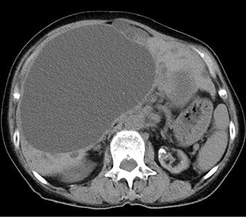
Interventional Radiology 2021; 6: 61-64
Figure 3. Non-contrast CT after fluid drainage of the hepat-
Figure 1. Non-contrast CT shows a large hepatic cyst, 17 × ic cyst shows the pig-tail drainage catheter (arrow) positioned
12 × 17 cm in dimensions, in segment four of the liver. in the liver hilum.
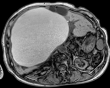
Figure 4. T1-weighted magnetic resonance image during
sclerotherapy shows homogeneous high-intensity fluid of the
Figure 2. X-ray fluoroscopy shows a 7F drainage catheter hepatic cyst.
inserted into the hepatic cyst through segment five of the liver
using right hypochondriac approach.
rate was 57 beats per minute. The laboratory test results on
mL of 3% polidocanol foam with 24 mL of room air. No admission were as follows: hemoglobin level, 8.6 g/dL; leu-
immediate issues were noted, and the catheter was kept in kocyte count, 4900 cells/μL; platelet count, 17.6 × 10⁴
place for 24 h and removed thereafter. No hemorrhagic fluid cells/μL; estimated glomerular filtration rate (eGFR), 10.3
was observed during the removal of the drainage catheter, mL/min; and CRP level, 9.69 mg/dL. The hemoglobin level
and the patient was discharged five days later. further dropped to 7.1 g/dL after two days. Non-contrast CT
One month after sclerotherapy, non-contrast CT showed a revealed enlargement of the treated hepatic cyst compared
34% reduction in the volume of the hepatic cyst compared with its size measured on images obtained seven days ear-
with its volume measured on pretreatment non-contrast CT lier. Contrast-enhanced CT could not be performed because
images, and the patient reported symptomatic relief. Labora- of poor renal function, and T1-and T2-weighted non-contrast
tory test results on the day were not abnormal, with a hemo- magnetic resonance imaging revealed high signal intensity
globin level of 9.9 g/dL, leukocyte count of 4800 cells/μL, of the treated hepatic cyst (Fig. 4). Intracystic hemorrhage
and C-reactive protein (CRP) level of 1.15 mg/dL. However, and infection were suspected. The patient had persistent
the patient presented to our emergency room with a seven- right hypochondrial pain and worsening anemia. The cause
day history of fever (body temperature: 38℃) and sudden of the intracystic hemorrhage was unknown; therefore, ab-
onset of abdominal pain in the right hypochondrium. On ar- dominal angiography was performed for transcatheter arte-
rival, her blood pressure was 112/57 mmHg, and her heart rial embolization (TAE) of the hepatic arteries feeding the
62Interventional Radiology 2021; 6: 61-64
a b c
d e
Figure 5. a. Left hepatic arteriography shows no extravasation form the middle hepatic artery. b.
Left hepatic arteriography shows embolization of the middle hepatic artery using pushable-coils. c.
Superior mesenteric arteriography shows a pseudoaneurysm (arrow) in the replaced right hepatic
artery originated from the superior mesenteric artery. d. Superior mesenteric arteriography after
coiling of the pseudoaneurysm shows embolization of the pseudoaneurysm using a detachable coil
(arrow) (Target XL; diameter, 6 mm; length, 20 cm; Stryker Corporation, Tokyo, Japan). e. Right
infraphrenic arteriography shows the presence of sufficient collateral arteries to feed the right he-
patic lobe via the right infraphrenic artery.
cystic wall under sufficient hydration. symptoms and complications such as pain, nausea, meteo-
Left hepatic arteriography showed no extravasation from rism, vomiting, early satiety, intracystic infection, and ob-
the middle hepatic arteries (Fig. 5a); however, the middle structive jaundice [1]. The long-term efficacy and safety of
hepatic arteries were embolized to reduce the cyst size (Fig. percutaneous drainage and sclerotherapy for symptomatic
5b). Superior mesenteric artery arteriography performed hepatic cysts have been demonstrated [4]. However, intra-
thereafter revealed a pseudoaneurysm in the right hepatic ar- cystic hemorrhage may occasionally occur as a life-
tery (Fig. 5c), and coil embolization using a detachable coil threatening complication in 2%-5% patients [5]. In a review
(Target XL; diameter, 6 mm; length, 20 cm; Stryker Corpo- of case reports on hemorrhagic hepatic cysts published in
ration, Tokyo, Japan) was immediately performed (Fig. 5d). 2015, 15 of 28 (54%) patients underwent surgical treatment,
Right infraphrenic arteriography revealed the presence of including partial hepatectomy (n = 8, 29%), cystectomy (n =
sufficient collateral arteries to feed the right hepatic lobe via 3, 11%), and fenestration (n = 4, 14%), whereas seven
the right infraphrenic artery (Fig. 5e). The right hypochon- (25%), two (7%), and four (14%) patients underwent tran-
drial pain and abdominal distention disappeared after TAE, shepatic percutaneous drainage, TAE, and conservative ther-
and no progression of anemia was observed. The patient apy, respectively [5-9]. The etiology and incidence of intra-
was discharged 14 days after TAE, and six months later, ab- cystic hemorrhage are unclear. The hepatic cystic wall in-
dominal ultrasound revealed a marked reduction in the cyst cludes three layers―an inner single layer comprising cuboi-
size, which was 9 × 6 cm in diameter. dal or columnar epithelium such as biliary epithelial cells, a
middle layer comprising compact connective tissue contain-
Discussion ing small blood vessels, and an outer layer comprising loose
connective tissue with large blood vessels, bile ducts, and
Most congenital hepatic cysts, including simple cysts and occasional von Meyenburg complexes. Increased intracystic
polycystic liver disease, which have an approximate preva- pressure due to an increase in cyst volume because of bili-
lence of 18% in adults, are asymptomatic [1]. Enlargement ary epithelial secretion causes necrosis and sloughing of the
of hepatic cysts during follow-up may be associated with biliary epithelium, leading to the exposure of blood vessels
63Interventional Radiology 2021; 6: 61-64
within the middle or outer cystic wall layer. Subsequent in- of the drainage catheter during overnight placement dam-
jury to the blood vessels in the fragile cystic wall may be aged the exposed hepatic artery surrounding the hepatic cys-
responsible for intracystic hemorrhage. Furthermore, rupture tic wall and caused pseudoaneurysm in the right hepatic ar-
of the fragile cystic wall due to increased intracystic pres- tery. In our case, the pseudoaneurysm was safely embolized
sure because of rapid intracystic hemorrhage can lead to se- using an isolation technique.
rious outcomes. In conclusion, interventional radiologists should be aware
TAE can produce therapeutic effects in patients with poor of the potential damage to the hepatic arteries in the middle
clinical conditions who cannot tolerate surgical treatment; or outer layer of the cystic wall during needle puncture or
however, bleeding may recur afterwards. Ishikawa et al. re- insertion of a drainage catheter. Urgent embolization may be
ported a case of a patient who underwent TAE for hepatic needed in patients with artery injuries. TAE of the damaged
intracystic hemorrhage; however, the volume of the cyst in- hepatic arteries feeding the hepatic cystic wall is an effective
creased three weeks after TAE, requiring simple cystectomy method for the treatment of intracystic hemorrhage.
[8]. Identifying the source of bleeding within the hepatic
cystic wall is often difficult using arteriography, and treat- Conflict of interest: The authors declare that they have no con-
ment with TAE may be unsuccessful. However, Takei et al . flicts of interest to report.
reported that TAE may be considered in patients with
autosomal dominant polycystic kidney disease and sympto- References
matic polycystic liver who are not candidates for surgical 1. Macedo FI. Current management of noninfectious hepatic cystic
lesions: a review of the literature. World J Hepatol 2013; 5: 462-
treatment because blood flow in the hepatic cystic wall is
469.
derived primarily from the hepatic artery [10]. Therefore, we 2. Spârchez Z, Radu P, Zaharie F, Al Hajjar N, Sparchez M. Percuta-
suggest that TAE might be an effective treatment for symp- neous treatment of symptomatic non-parasitic hepatic cysts: initial
tomatic hepatic cysts if all intrahepatic arteries feeding the experience with single-session sclerotherapy with polidocanol.
hepatic cystic wall are embolized; however, the efficacy of Med Ultrason 2014; 16: 222-228.
TAE for hemorrhagic hepatic cysts should be investigated in 3. Sekiguchi T, Hirose S, Hara T, Koizumi J. Complications during
future studies. sclerotherapy for symptomatic hepatic cysts: successful bailout for
To date, only two cases of iatrogenic intracystic hemor- penetrating the hepatic artery and portal vein. J Vasc Interv Radiol
2019; 30: 1048-1049.
rhage during percutaneous drainage and sclerotherapy have
4. Wijnands TM, Görtjes AP, Gevers TJ, et al. Efficacy and safety of
been reported [2, 3]. Spârchez et al . reported a case of pa- aspiration sclerotherapy of simple hepatic cysts: a systematic re-
tient who experienced intracystic bleeding with clot forma- view. AJR 2017; 208: 201-207.
tion during aspiration [2]. As sclerotherapy is contraindi- 5. Simon T, Bakker IS, Penninga L, Nellensteijn DR. Haemorrhagic
cated in patients with active bleeding, sclerotherapy was rupture of hepatic simple cysts. BMJ Case Rep 2015; 2015. doi:
postponed for one month, and the patient was successfully 10.1136/bcr-2014-208676.
treated. Percutaneous aspiration using an 18-G aspiration 6. Fong ZV, Wolf AM, Doria C, et al. Hemorrhagic hepatic cyst: re-
needle may lead to bleeding if the tip of the needle used port of a case and review of the literature with emphasis on clini-
cal approach and management. J Gastrointest Surg 2012; 16:
during the procedure sticks to the cystic wall. Therefore,
1782-1789.
small catheters measuring 6-7 Fr are preferred over aspira- 7. Marion Y, Brevartt C, Plard L, Chiche L. Hemorrhagic liver cyst
tion needles. Additionally, Sekiguchi et al . reported that rupture: an unusual life-threatening complication of hepatic cyst
complications such as penetration of the hepatic artery and and literature review. Ann Hepatol 2013; 12: 336-339.
portal vein occurred during sclerotherapy in a patient with 8. Ishikawa H, Uchida S, Yokokura Y, et al. Nonparasitic solitary
symptomatic hepatic cysts [3]. They embolized the left he- huge liver cysts causing intracystic hemorrhage or obstructive
patic artery from the arterial side with a micro-coil, injected jaundice. J Hepatobiliary Pancreat Surg 2002; 9:764-768.
1.1 mL of 50% n-butyl-cyanoacrylate into the hepatic cyst 9. Suzuki T, Kawata Y, Tanaka Y, et al. Ruptured nonparasitic liver
cyst caused by blunt abdominal trauma. Rinsho Hoshasen 1989;
from a 4-Fr straight catheter through the drainage catheter,
34: 905-908.
and sealed the portal vein hole from the outside with a glue
10. Takei R, Ubara Y, Hoshino J, et al. Percutaneous transcatheter he-
ball while removing the catheter system. In our case, the patic artery embolization for liver cysts in autosomal dominant
cause of the pseudoaneurysm in the replaced right hepatic polycystic kidney disease. Am J Kidney Dis 2007; 49: 744-752.
artery was unknown because intracystic hemorrhage was not
observed during the procedure, and the bleeding point was Interventional Radiology is an Open Access journal distributed under the Crea-
tive Commons Attribution-NonCommercial 4.0 International License. To view
different from the puncture line. To date, no study has re- the details of this license, please visit (https://creativecommons.org/licenses/by-
ported the formation of hepatic artery pseudoaneurysms nc/4.0/).
around the cystic wall. We assumed that contact stimulation
64You can also read