ISOLATED OSTEOCHONDROMA OF PROXIMAL FEMORAL METAPHYSIS PRESENTING AS A MECHANICAL BLOCK TO HIP RANGE OF MOTION AND PAIN
←
→
Page content transcription
If your browser does not render page correctly, please read the page content below
Article ID: WMC005566 ISSN 2046-1690 ISOLATED OSTEOCHONDROMA OF PROXIMAL FEMORAL METAPHYSIS PRESENTING AS A MECHANICAL BLOCK TO HIP RANGE OF MOTION AND PAIN Peer review status: No Corresponding Author: Dr. Pulak Vatsya, Senior Resident , PGIMER, Chandigarh , house no 259, sector 15A, 160015 - India Submitting Author: Dr. Pulak Vatsya, Senior Resident , PGIMER, Chandigarh , house no 259, sector 15A, 160015 - India Other Authors: Dr. Prashant Sharma , Senior Resident , Department of orthopedics, PGIMER, Chandigarh , sector 12 , 160012 - India Dr. Karthick Rangasamy , Senior Resident , Department of orthopedics, PGIMER, Chandigarh , Sector 12 , 160012 - India Dr. PEBUM SUDESH , associate proffesor , Department of orthopedics, PGIMER, Chandigarh , sector 12 , 160012 - India Dr. Gopinathan NirmalRaj, Additional Prefessor , Department of orthopedics, PGIMER, Chandigarh , sector 12 , 160012 - India Article ID: WMC005566 Article Type: Case Report Submitted on:28-Apr-2019, 11:53:54 AM GMT Published on: 30-Apr-2019, 05:05:38 AM GMT Article URL: http://www.webmedcentral.com/article_view/5566 Subject Categories:ORTHOPAEDICS Keywords:Osteochondroma, proximal femoral osteochondroma, symptomatic osteochondroma How to cite the article:Sharma P, Vatsya P, Rangasamy K, SUDESH P, NirmalRaj G. ISOLATED OSTEOCHONDROMA OF PROXIMAL FEMORAL METAPHYSIS PRESENTING AS A MECHANICAL BLOCK TO HIP RANGE OF MOTION AND PAIN. WebmedCentral ORTHOPAEDICS 2019;10(4):WMC005566 Copyright: This is an open-access article distributed under the terms of the Creative Commons Attribution License(CC-BY), which permits unrestricted use, distribution, and reproduction in any medium, provided the original author and source are credited. Source(s) of Funding: No source of funding WebmedCentral > Case Report Page 1 of 7
WMC005566 Downloaded from http://www.webmedcentral.com on 27-Jun-2019, 12:32:42 PM Competing Interests: No conflicting interests WebmedCentral > Case Report Page 2 of 7
WMC005566 Downloaded from http://www.webmedcentral.com on 27-Jun-2019, 12:32:42 PM
ISOLATED OSTEOCHONDROMA OF PROXIMAL
FEMORAL METAPHYSIS PRESENTING AS A
MECHANICAL BLOCK TO HIP RANGE OF MOTION
AND PAIN
Author(s): Sharma P, Vatsya P, Rangasamy K, SUDESH P, NirmalRaj G
Abstract of a large, pedunculated lesion arising from proximal
femoral metaphysis, which created a diagnostic
dilemma since it could be heterotopic ossification,
chondrosarcoma, synovial chondromatosis or an
Osteochondromas are the commonest bone tumors.
osteochondroma. We managed this with surgical
They cease to grow post skeletal maturity and are
excision and  fixation with  LCP as prophylaxis for
rarely malignant(1%), thus making the indications for
fracture  with good results and histopathology of the
their excision very minimal. Diagnosis is usually
lesion to get to a sure diagnosis.
confirmed on a radiograph, since most are
asymptomatic, a conservative protocol is followed. Case Report(s)
Rapid increase in size, especially after skeletal
maturity, pain, a cartilage cap of more than 2cm are all
indications of a malignant lesion and need definitive A 10-year-old male child presented to our outpatient
diagnosis and surgery. Our case, presented with a department with complaints of difficulty in squatting for
lesion in an unusual location for osteochondroma, 2 year and right hip pain for 6 monthsÂ
which was large enough to be symptomatic, thus
 The patient had visited multiple doctors previously
leading to our management being aggressive,
and was given pain relief medications, which would
including excision, biopsy with a definitive diagnosis
only provide temporary pain relief. The patient was
and plate fixation. Thus emphasizing the need to
referred to our center by the doctor, due to the
suspect osteochondromas in unusual locations and
appearance of a large bony lesion on x-ray which he
surge for definitive diagnosis in aggressive
suspected to be malignant. There was no history of
lesions. Â
any trauma or snapping sound from the hip while
 playing. The patient noticed difficulty in squatting from
Introduction 2 years due to a mehanical block of flexion which was
progressive. From 6 months Patient also complain
of pain in Right Hip which occurred on walking and
squatting. The pain was insidious in onset and
Osteochondroma is a benign osteo-cartilaginous
gradually progressive, no history of night pain or rest
tumor of originating from metaphysis of long bones,
pain. On examination, the patient had an antalgic gait,
usual sites being the knee, ankle, shoulder and
a bony, non-mobile swelling of size 10x8 x 4cm(AP,
forearm These are usually asymptomatic and
medial-lateral & proximal-distal respectively) on the
incidental findings. They are also mostly extra-articular
anteromedial aspect of the thigh, and restricted flexion,
and grow away from epiphysis1.They can rarely be
adduction and Internal rotation which are painful in
symptomatic, when they compress surrounding
extreme of motion.
structures( tendon, joint capsule, bursa), malignant
transformation, pathological fracture of stalk or Orthogonal radiographs of the pelvis with bilateral hips
compression of neurovascular structures 2 , 3 , 4 and right hip with femur were ordered. Radiographs
Intra-articular osteochondromas can be symptomatic showed a cauliflower-like growth from anterior femoral
earlier that extra-articular in the form of painful range cortex involving the greater trochanter, no cortical
of motion or joint stiffness or leg- length discrepancy. breach, no other lesions, no loose bodies in the joint,
Osteochondroma of or around the hip joint, causing and no pathological fracture.
block in range of motion and pain is a not so rare a Considering this a large and symptomatic lesion we
condition now in children. Management options in ordered for an MRI. MRI was suggestive of a lesion
such children are many ranging from conservative, originating from proximal femoral metaphysis, with
surgical excision and plate fixation. We present a case
WebmedCentral > Case Report Page 3 of 7
WMC005566 Downloaded from http://www.webmedcentral.com on 27-Jun-2019, 12:32:42 PM
medullary canal continuous with the femur, with the
involvement of greater trochanter and bone expansion
but did not breach the cortex. This was suggestive of
an osteochondroma in an unusual location.
We discussed the need for surgery since this was a
large and symptomatic osteochondroma of an unusual
location and even a minimal risk of transformation
needed to be negated. We also realized that excision
of lesion of this size, will weaken the bone and need a
prophylactic fracture stabilization procedure. We kept
options of a Dynamic Hip Screw(Pediatric), Angle
Blade Plate, LCDCP and a Distal femoral locking plate,
which we would use in a reverse fashion for
stabilization.
The surgical decision, procedure, and outcomes were
discussed at length with the patient and parents, who
consented for surgery and biopsy to reach a
confirmatory diagnosis.
An anterolateral Watson Jones approach was used to
expose the lateral and anterior aspect of the lesion. An Â
extended approach was used to ensure
extra-periosteal excision to avoid re-occurrences.
Once excision was complete, a void was created
which needed to be dealt with. As decided
pre-operatively, we used a distal femoral locking plate,
in a reverse order to adjust for the contour of the
greater trochanter and fixed the bone. For bridging the
void created by excision we filled the void with iliac
crest bone graft.
POST-OPERATIVE
Â
 The patient was kept no weight bearing for 6 weeks,
Â
after which partial and then full weight bearing was
gradually started over the next 4-6 weeks. The patient 2) Intraoperative images and specimen
at 6 months, was pain-free, with a 120 degree flexion
as compared to 130 degree of the normal side, 40
degree abduction as compared to 45 degree of normal
side, 25 degree adduction as compared to 30 degree
of normal side, 40 degree of internal and external
rotation which was comparable to other side. The
patient was able to sit cross-legged and squat.
Table 1 - Range of motion(in degrees) before and after
surgery
 Pre-operative ROM Post-operative ROM
Flexion 90 120
Extension 5 10
Abduction 30 40
Adduction 5 25
External Rotation 30 40
Internal Rotation 20 40
Â
Â
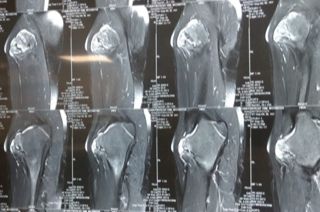
1) preoperative radiographs and MRI
WebmedCentral > Case Report Page 4 of 7
WMC005566 Downloaded from http://www.webmedcentral.com on 27-Jun-2019, 12:32:42 PM
transformation is one of the most severe complications
of an osteochondroma.
Being a large, symptomatic, and presentation at an
unusual location, all indicated towards an aggressive
lesion, needing an excision biopsy, a confirmatory
diagnosis, exclusion of malignant potential,
improvement in symptoms and stabilization.
Biopsy, though rarely needed, can be a core needle,
but is more often excisional, since it also relieves the
patient of cosmetic complaints of swelling or any
 compressive symptoms if present. A complete
 extra-periosteal excision, with complete removal of
 cartilage cap is a must to avoid any re-occurrences
which is approximated to be present in about 2%
DISCUSSION cases.10,11 Â Larger osteochondromas, especially in
proximal femoral metaphysis, present with the problem
of need of stabilization, being an area of high shear
Osteochondroma is the most common benign tumor of forces, and a void or weakening of bone in this area
bone, has both osteo-cartilaginous components, has a can lead to a very high risk of fracture. Multiple fixation
medullary canal continuous with that of parent bone, is devices in the form of threaded screws, blade plates,
usually extra-articular and grows away from the joint. LCDCP and locking plates along with bone graft for
Most common sites are the rapidly growing epiphysis, voids, if any created are available. Blade plates and
namely distal femur, proximal tibia, and proximal screws are good options if intra-articular or
humerus.(5) Growth usually stops at skeletal maturity, peritrochantric involvement are present, where as
and growth after this is considered to be a sign of LCDCP’s and locking plates can be used for
malignant transformation.(5) The proximal femur is an extra-articular fixation. We have had a good
uncommon site of osteochondroma. There are case experience with using a distal femoral locking plate, in
reports which emphasize that proximal femoral, a reverse fashion to act like a proximal femoral locking
acetabular and intra-articular osteochondromas can plate. This provides a well contoured fit for the
cause pain, stiffness, snapping and even sciatic nerve proximal femur and adequate fixation without
compression, making them an important entity to be disturbing the intra-articular anatomy.
considered for surgical excision before they become
symptomatic. Saglik et al had only 4.8% cases of
CONCLUSION
proximal femur out of all the 313 cases described.6
Multiple Hereditary Exostosis (MHE) is a hereditary
Osteochondromas are very common lesions in
form of multiple exostosis or osteochondromas,
skeletally immature, and can present in locations
although single lesions are found in about 85% of
where the orthopaedician might not keep them as a
those diagnosed with osteochondroma. 5 Hip
first differential. Also these unusual sites of
involvement in MHE is common and presents with a
 presentation need newer methods of management,
different set of complications like coxa Valga,
biopsy and fixation. The surgeon should always
increased femoral ante-version or overgrowth of
approach such lesions with suspicion and manage
femoral neck.4 Rather in solitary osteochondroma of
them aggressively to provide symptomatic relief as
hip present with compressive complaints of bursitis,
well as negate any evidence of malignancy.
snapping2, sciatic nerve palsy7,3 or restriction of motion
due to mechanical blockade or femoro-acetabular Â
impingement7.
REFRENCES
The risk of malignant transformation is indicated by
raid increase in size, especially after skeletal maturity,
new onset pain, pathological fracture or a cartilage cap 1.Azar FM, Beaty JH, Canale ST. Campbell’s
thickness of >2cm. solitary lesions have a risk of 1-2%8 Operative Orthopaedics. 13th editi. Elsevier; 2017.
whereas MHE have a risk of 1-25%. 9 Any doubts 938-942 p.
about malignant transformation need to be negated on
2.Inoue S, Noguchi Y, Mae T, Rikimaru S, Hotokezaka
histopathology after biopsy, as malignant
WebmedCentral > Case Report Page 5 of 7WMC005566 Downloaded from http://www.webmedcentral.com on 27-Jun-2019, 12:32:42 PM
S. An external snapping hip caused by Assistant in surgery and planning, Concept and design
osteochondroma of the proximal femur. Mod Â
Rheumatol. 2005 Dec 20;15(6):432–4.
Â
3.Turan Ilica A, Yasar E, Tuba Sanal H, Duran C,
Guvenc I. Sciatic nerve compression due to femoral illustrations
neck osteochondroma: MDCT and MR findings. Clin
Rheumatol. 2008 Mar 17;27(3):403–4.
4.Am El-Fiky T, Chow W, Li YH, To M. Hereditary Illustration 1: Clinical picture of child showing
multiple exostoses of the hip. Vol. 17, Journal of
rocker- bottom foot.
Orthopaedic Surgery. 2009.
5.Czerniak B. Bone Tumors. 2nd editio. Elsevier; 2016.
6.Saglik Y, Altay M, Unal VS, Basarir K, Yildiz Y.
Manifestations and management of osteochondromas:
a retrospective analysis of 382 patients. Acta Orthop
Belg. 2006 Dec;72(6):748–55.
7.Mondal S, Chowdhury A, Mandal PK, Roy D, Pal S,
Gazi E, et al. Osteochondroma of femoral neck-a rare
cause of femoro-acetabular impingement and sciatic
nerve compression. Vol. 13, IOSR Journal of Dental
and Medical Sciences (IOSR-JDMS) e-ISSN. 2014.
8.Garrison RC, Unni KK, McLeod RA, Pritchard DJ,
Dahlin DC. Chondrosarcoma arising in
osteochondroma. Cancer. 1982 May 1;49(9):1890–7.
9.Peterson HA. Multiple hereditary osteochondromata. Â
Clin Orthop Relat Res. 1989 Feb;(239):222–30.
Illustration 2: Lateral view with forced plantar flexion
10.Humbert ET, Mehlman C, Crawford AH. Two cases showing vertical talus (talar- first metatarsal axis angle
of osteochondroma recurrence after surgical resection. > 35?) on left foot.
Am J Orthop (Belle Mead NJ). 2001 Jan;30(1):62–4.
Â
11.Bottner F, Rodl R, Kordish I, Winklemann W,
Gosheger G, Lindner N. Surgical treatment of
symptomatic osteochondroma. A three- to eight-year
follow-up study. J Bone Joint Surg Br. 2003
Nov;85(8):1161–5.
Â
author contributions
Dr.Prashant Kumar Sharma
Acquisition of data, assistant in surgery and planning Â
 Dr.Pulak Vatsya
Illusration 3: AP radiograph of foot showing Angle X
Drafting of article, Critical revision of article, Assistant – Talar first metatarsal axis angle and Angle Y –
in surgery and planning Talo calcaneal angle.
 Dr.Pebum Sudesh
Primary surgeon, Concept and design
Dr.G.Nirmal Raj
Acquisition of data, Revision of article
 Dr.Karthick Rangaswamy
WebmedCentral > Case Report Page 6 of 7WMC005566 Downloaded from http://www.webmedcentral.com on 27-Jun-2019, 12:32:42 PM Illustration 4: Post op Xray showing Talo navicular joint reduced and fixed with a K wire. Â Â Â WebmedCentral > Case Report Page 7 of 7
You can also read