Infected Pancreatic Necrosis Mimicking Pancreatic Cancer
←
→
Page content transcription
If your browser does not render page correctly, please read the page content below
Case Rep Gastroenterol 2020;14:436–442
                                DOI: 10.1159/000510161                            © 2020 The Author(s)
                                Published online: August 26, 2020                 Published by S. Karger AG, Basel
                                                                                  www.karger.com/crg

                                This article is licensed under the Creative Commons Attribution-NonCommercial 4.0
                                International License (CC BY-NC) (http://www.karger.com/Services/OpenAccessLicense).
                                Usage and distribution for commercial purposes requires written permission.

Single Case

Infected Pancreatic Necrosis
Mimicking Pancreatic Cancer
Jun Heoa, b
a Department
           of Internal Medicine, Kyungpook National University Hospital,
Daegu, Republic of Korea; b School of Medicine, Kyungpook National University,
Daegu, Republic of Korea

Keywords
Pancreatitis · Pancreatic cancer · Endoscopic ultrasonography

Abstract
Although infected pancreatic necrosis can develop as a result of rare conditions involving
trauma, surgery, and systemic infection with an uncommon pathogen, it usually occurs as a
complication of pancreatitis. Early phase of acute pancreatitis can be either edematous inter-
stitial pancreatitis or necrotizing pancreatitis. The late complications of pancreatitis can be di-
vided into pancreatic pseudocyst due to edematous interstitial pancreatitis or walled-off ne-
crosis due to necrotizing pancreatitis. During any time course of pancreatitis, bacteremia can
provoke infection inside or outside the pancreas. The patients with infected pancreatic necrosis
may have fever, chills, and abdominal pain as inflammatory symptoms. These specific clinical
presentations can differentiate infected pancreatic necrosis from other pancreatic diseases.
Herein, I report an atypical case of infected pancreatic necrosis in which abdominal pain, ele-
vation of white blood cell, and fever were not found at the time of admission. Rather, a 10-kg
weight loss (from 81 to 71 kg) over 2 months nearly led to a misdiagnosis of pancreatic cancer.
The patient was finally diagnosed based on endoscopic ultrasound-guided fine-needle aspi-
ration. This case highlights that awareness of the natural course of pancreatitis and infected

                                  Jun Heo
                                  Department of Internal Medicine
                                  Kyungpook National University Hospital
                                  130 Dongdeok-ro, Jung-gu, Daegu 41944 (Korea)
                                  hero797@hanmail.net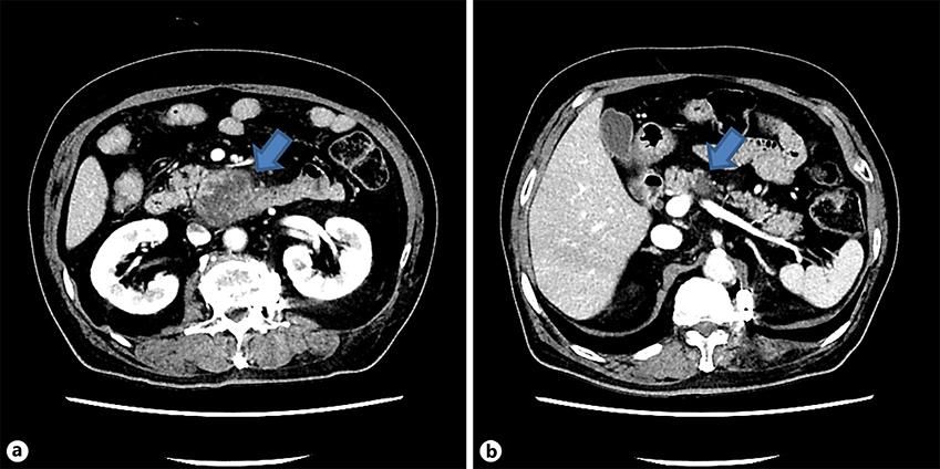
Case Rep Gastroenterol 2020;14:436–442 437
DOI: 10.1159/000510161 © 2020 The Author(s). Published by S. Karger AG, Basel
www.karger.com/crg
Heo: Infected Pancreatic Necrosis Mimicking Pancreatic Cancer
pancreatic necrosis is important. In addition, endoscopic ultrasound-guided fine-needle aspi-
ration should be recommended for the diagnosis and treatment of indeterminate pancreatic
lesions in selected patients. © 2020 The Author(s)
Published by S. Karger AG, Basel
Introduction
Pancreatic mass-forming, inflammatory, benign lesions can masquerade as pancreatic
cancer. These cases include pancreatic tuberculosis, autoimmune pancreatitis, eosinophilic
pancreatitis, and infected pancreatic necrosis [1]. Among them, infected pancreatic necrosis
is a kind of complication of pancreatitis. It usually requires treatment with antibiotics and/or
drainage. Most patients with infected pancreatic necrosis complain of abdominal pain, fever,
chills, nausea, and leukocytosis. These specific clinical presentations can differentiate infected
pancreatic necrosis from other pancreatic diseases. However, when we meet this infected
pancreatic necrosis at a specific time of the pancreatitis course at which no infectious symp-
toms/sign are present, an infected wall-off pancreatic necrosis might be misdiagnosed as a
pancreatic malignancy. Herein, I present a case of infected pancreatic necrosis mimicking ma-
lignancy.
Case Presentation
A 67-year-old male was referred from a local medical center and admitted to our hospital
for body weight loss of approximately 10 kg over 2 months (from 81 to 71 kg). He denied fever
or chills and had no abdominal pain. Abdominal computed tomography (CT) performed at a
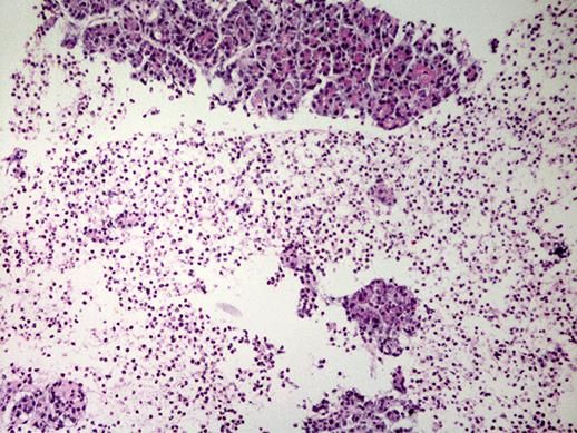
local medical center showed a 53 × 39 mm, exophytic low-density mass in the uncinate pro-
cess of the pancreas (Fig. 1a) and a 19 × 14 mm, low-density mass in the body of the pancreas
(Fig. 1b). The patient was taking medication for hypertension and type 2 diabetes mellitus. He
reported consuming one bottle of an alcoholic beverage per day and had never smoked. Vital
signs were as follows: blood pressure 110/65 mm Hg, pulse rate 77/min, respiratory rate
18/min, and body temperature 36.4°C. On physical examination, the patient appeared to have
anicteric sclera, and his abdomen was soft and flat without tenderness. The laboratory find-
ings were as follows: white blood cell count 7,060/µL (neutrophil 69.3%) , hemoglobin 12.3
g/dL, platelet count 300,000/µL, aspartic acid aminotransferase 14 IU/L, alanine aminotrans-
ferase 17 IU/L, blood urea nitrogen 12.4 mg/dL, creatinine 0.79 mg/dL, C-reactive protein
2.62 mg/dL, carcinoembryonic antigen 5.9 ng/mL, carbohydrate antigen 19-9 48.35 U/mL,
fasting serum glucose 367 mg/dL, and hemoglobin A1c 11.6%.
Since pancreatic cancer was suspected, the patient was evaluated for surgical treatment
based on the CT findings. The mass in the pancreatic body seemed to abut the superior mes-
enteric artery. On the day after admission, magnetic resonance imaging (MRI) was performed
to further characterize the pancreatic lesion. The interval between the MRI and the previous
CT was 21 days. On MRI, the mass in the uncinate process decreased from 53 to 35 mm, while
the mass in the body increased from 19 mm to 40 mm (Fig. 2). Based on the MRI findings, we
predicted that the pancreatic lesions are atypical inflammations, such as tuberculosis.
Case Rep Gastroenterol 2020;14:436–442                                                  438
                                  DOI: 10.1159/000510161         © 2020 The Author(s). Published by S. Karger AG, Basel
                                                                 www.karger.com/crg

                                  Heo: Infected Pancreatic Necrosis Mimicking Pancreatic Cancer

     Endoscopic ultrasound-guided fine-needle aspiration (EUS-FNA) was planned to differ-
entiate this atypical inflammatory lesion. On the 6th day of hospitalization, one day prior to
EUS-FNA, the patient developed a fever of 37.9°C and was treated with intravenous third-gen-
eration cephalosporin. The next day, EUS-FNA (GF-UCT 260; Olympus Co., Tokyo, Japan, and
22-gauge needle, Mediglobe; Mediglobe Co., Achenmuehle, Germany) was performed accord-
ing to our previous plan. The mass of the body of the pancreas was irregular in shape, and the
margin was unclear; moreover, a low echoic lesion was identified in the center. Pus-like fluid
was aspirated from the tissue by EUS-FNA. However, the abscess of the pancreatic body lesion
was ruptured to the peripancreatic area just after EUS-FNA. The EUS-FNA procedure was
aborted, and the patient was closely monitored in the ward. The aspirated fluid was not sent
for culture sensitivity. Later, a cytological examination only revealed the presence of inflam-
matory cells within the pancreas parenchyma (Fig. 3). Fever had subsided after 4 days of in-
travenous antibiotic treatment. After treatment with intravenous antibiotics for 11 days, the
patient was discharged with per-oral antibiotics for 7 days. After 1 month, the follow-up ab-
dominal CT demonstrated that the mass in the uncinate process decreased to 8 mm, and the
mass in the body was no longer present. The patient has been compliant and has continued
with follow-up as of now.

    Discussion

      According to the revised 2012 Atlanta classification of acute pancreatitis, the early phase
of acute pancreatitis can be either edematous interstitial pancreatitis or necrotizing pancrea-
titis. The late complications of pancreatitis can be divided into pancreatic pseudocyst due to
edematous interstitial pancreatitis or walled-off necrosis due to necrotizing pancreatitis.
These late complications usually develop approximately 4 weeks after the onset of acute pan-
creatitis [2]. During any time course of pancreatitis, bacteremia can provoke infection inside
or outside the pancreas. Patients with infected pancreatic necrosis usually complain of fe-
ver/chills and present with an increased white blood cell count. However, in this case, leuko-
cytosis and fever were not found at the time of admission. Meanwhile, a 10-kg weight loss over
2 months, which was the result from poorly controlled diabetes, led to a misdiagnosis as pan-
creatic cancer. Immediately after FNA, the detailed medical history of the patient revealed that
severe epigastric pain had persisted for several days at 6 weeks prior to admission. Therefore,
in the case of pancreatic masses, it is important to identify the history and specific symptoms
of pancreatitis in order to rule out pancreatitis-related inflammatory lesions.
      External wall formation, a septum, or liquid-liquid interface can be observed inside the
infected pancreatic necrosis. The characteristics and morphology of an abscess can change
according to the degree of maturity. In the very early stages of infected pancreatic necrosis,
the focal pancreatitis can appear as an acutely inflamed area without wall formation. Depend-
ing on the imaging modality, both pancreatic cancer and the early stage of infected pancreatic
necrosis have the same features, which are as follows: hypoechoic appearance on ultrasonog-
raphy, hypodense appearance on CT, low-signal intensity on a T1-weighted image by MRI, and
high-signal intensity on a T2-weighted image by MRI. Moreover, some focal pancreatitis also
may present findings of malignancy, such as the double duct sign, ductal strictures, and infil-
tration of adjacent fat. Although there are some differential features in image findings such as
Case Rep Gastroenterol 2020;14:436–442 439
DOI: 10.1159/000510161 © 2020 The Author(s). Published by S. Karger AG, Basel
www.karger.com/crg
Heo: Infected Pancreatic Necrosis Mimicking Pancreatic Cancer
calcification inside the mass, “duct-penetrating sign,” and an abrupt interruption of a smoothly
dilated pancreatic duct with upstream pancreatic atrophy [1], these are not always present,
as in the case reported herein.
In cases of clinically suspected pancreatic cancer that is at an operable stage, surgical re-
section might be preferred in the absence of any other invasive pathologic diagnosis. However,
in clinical practice, there is the possibility of misdiagnosis and subsequent unnecessary surgi-
cal resection for benign pancreatic disease. It was known that benign pancreatic disease ac-
counts for 5–21% of all pancreatectomy-presumed cancers [3, 4]. Several case reports on pan-
creatic abscesses (infected pancreatic necrosis) have been published that would come close
to misdiagnosis as malignancy. These lesions were finally diagnosed based on diagnostic lap-
aroscopy, CT-guided percutaneous aspiration, and EUS-FNA (Table 1) [5–8]. Among these mo-
dalities, EUS with FNA has an advantage because it is less invasive in diagnosing pancreatic
disease. It has also shown a higher diagnostic yield for indeterminate pancreatic masses com-
pared with multidynamic CT [9]. In addition, EUS-FNA is used not only for diagnosis but also
as a useful treatment modality for infected pancreatic necrosis. In general, drainage catheter
placement is usually needed in addition to antibiotic treatment. However, if the amount of
abscess aspirate is sufficient to minimize the abscess cavity, drainage with aspiration only
might be sufficient for treatment [10].
Infected pancreatic necrosis may be present and can be confused with pancreatic malig-
nancy in an early stage of development. Therefore, awareness of the natural course of pancre-
atitis and infection/abscess formation is important. In addition, EUS-FNA should be recom-
mended for the diagnosis and treatment of indeterminate pancreatic lesions in selected pa-
tients.
Statement of Ethics
I have reported this case in compliance with the Declaration of Helsinki. Consent was ob-
tained from the patient for publication of the clinical data.
Disclosure Statement
I have no conflicts of interest to disclose.
Funding Sources
No funding was obtained for this study.Case Rep Gastroenterol 2020;14:436–442 440
DOI: 10.1159/000510161 © 2020 The Author(s). Published by S. Karger AG, Basel
www.karger.com/crg
Heo: Infected Pancreatic Necrosis Mimicking Pancreatic Cancer
References
1 Torres US, Matsumoto C, de Macedo Neto AC, Caldana RP, Motoyama Caiado AH, Tiferes DA, et al. Common
and Uncommon Benign Pancreatic Lesions Mimicking Malignancy: Imaging Update and Review. Semin
Ultrasound CT MR. 2018 Apr;39(2):206–19.
2 Trikudanathan G, Wolbrink DRJ, van Santvoort HC, Mallery S, Freeman M, Besselink MG. Current Concepts in
Severe Acute and Necrotizing Pancreatitis: An Evidence-Based Approach. Gastroenterology. 2019
May;156(7):1994-2007.e3.
3 Gerritsen A, Molenaar IQ, Bollen TL, Nio CY, Dijkgraaf MG, van Santvoort HC, et al.; Dutch Pancreatic Cancer
Group. Preoperative characteristics of patients with presumed pancreatic cancer but ultimately benign
disease: a multicenter series of 344 pancreatoduodenectomies. Ann Surg Oncol. 2014 Nov;21(12):3999–
4006.
4 Birnbaum DJ, Gaujoux S, Berbis J, Dokmak S, Hammel P, Vullierme MP, et al. Surgery for pancreatic
neoplasms: how accurate are our surgical indications? Surgery. 2017 Jul;162(1):112–9.
5 Chase MP, Yarze JC, Gumustop B, Leach RP. Endoscopic ultrasound-guided aspiration and oral antibiotic
therapy as definitive treatment of an asymptomatic pancreatic abscess. Pancreas. 2009 May;38(4):475–6.
6 Chong VH. Isolated pyogenic pancreatic abscess mimicking a neoplasm. JOP. 2008 May;9(3):309–12.
7 Shulik O, Cavanagh Y, Grossman M. Pancreatic Lesion: malignancy or Abscess? Am J Case Rep. 2016
May;17:337–9.
8 Kim MJ, Seo EK, Kang ES, Kim KM, Oh YM, Cho BH, et al. Pyogenic pancreatic abscess mimicking pancreatic
neoplasm: a four-case series. Korean J Gastroenterol. 2015 Apr;65(4):252–7.
9 Krishna SG, Rao BB, Ugbarugba E, Shah ZK, Blaszczak A, Hinton A, et al. Diagnostic performance of
endoscopic ultrasound for detection of pancreatic malignancy following an indeterminate multidetector CT
scan: a systemic review and meta-analysis. Surg Endosc. 2017 Nov;31(11):4558–67.
10 Jo HG, Amarbat B, Jeong JW, Song HY, Song SR, Kim TH. Could Transgastric Endoscopic Ultrasound-Guided
Aspiration Alone Be Effective for the Treatment of Pancreatic Abscesses? Clin Endosc. 2015 Jul;48(4):345–7.Case Rep Gastroenterol 2020;14:436–442 441
DOI: 10.1159/000510161 © 2020 The Author(s). Published by S. Karger AG, Basel
www.karger.com/crg
Heo: Infected Pancreatic Necrosis Mimicking Pancreatic Cancer
Fig. 1. Abdominal CT. a 53 × 39 mm, heterogeneously enhancing, low-attenuating mass in the pancreatic
uncinate process (arrow). b 19 × 14 cm, heterogeneously enhancing, low-attenuating mass in the pancre-
atic body (arrow).
Fig. 2. Liver acquisition with volume acceleration dynamic image (MRI). Peripheral enhancement was also
observed in the pancreatic masses. a The mass in the uncinate process decreased from 53 to 35 mm (ar-
row). b The mass in the body increased from 19 to 40 mm (arrow).Case Rep Gastroenterol 2020;14:436–442 442
DOI: 10.1159/000510161 © 2020 The Author(s). Published by S. Karger AG, Basel
www.karger.com/crg
Heo: Infected Pancreatic Necrosis Mimicking Pancreatic Cancer
Fig. 3. Microscopic examination. Diffuse neutrophil and lymphocyte infiltration near the pancreatic paren-
chyma (H&E stain. ×200).
Table 1. Review of published cases of pancreatic abscesses (infected pancreatic necrosis) mimicking pan-
creatic cancer
First author [ref.], Age, Symptoms Location Fever WBC/µL Size, cm Comorbidities Diagnostic
year years/ modality
Sex
Chase [5], 2009 48/M None Body and No n.a. 8.0 × 10.0 DM, alcoholics EUS-FNA
tail
Shulik [7], 2016 67/M Jaundice Head No Within 3.1 × 2.4 DM EUS-FNA
normal limits
Chong [6], 2008 72/M Abdominal Head No n.a. 3 DM CT-guided
discomfort percutaneous
aspiration
Kim [8], 2015 51/F Abdominal Body No 9,100 2.7 DM Laparoscopy
pain
This case, 2020 67/M Body weight Body and No 7,060 5 and 2 DM, alcoholics EUS-FNA
loss uncinate
process
DM, diabetes mellitus; EUS-FNA, endoscopic ultrasound-guided fine-needle aspiration.You can also read