TNF-α inhibitors used as steroid-sparing maintenance monotherapy in parenchymal CNS sarcoidosis
←
→
Page content transcription
If your browser does not render page correctly, please read the page content below
Neuro-i nflammation
J Neurol Neurosurg Psychiatry: first published as 10.1136/jnnp-2020-325665 on 8 June 2021. Downloaded from http://jnnp.bmj.com/ on October 26, 2021 by guest. Protected by copyright.
Original research
TNF-α inhibitors used as steroid-sparing maintenance
monotherapy in parenchymal CNS sarcoidosis
Frédéric Hilezian ,1 Adil Maarouf,1,2 Clemence Boutiere,1,2 Audrey Rico,1,2
Sarah Demortiere ,1,2 Philippe Kerschen,3 Thomas Sene,4 Caroline Bensa-Koscher,5
Claire Giannesini,6,7 Jean Capron,6,7 Arsene Mekinian,7,8
Jean-Philippe Camdessanché ,9 Géraldine Androdias,10 Romain Marignier,11,12
Nicolas Collongues ,13,14 Olivier Casez,15 Catalina Coclitu,15 Mathieu Vaillant,15
Guillaume Mathey ,16,17 Jonathan Ciron,18 Jean Pelletier,1,2 Bertrand Audoin,1,2
Under the aegis of the French Multiple Sclerosis Society
For numbered affiliations see Abstract 5% of patients3 4 and may be inaugural in 70%.5
end of article. Objective To assess the efficacy of tumour necrosis Cranial nerve involvement and particularly facial
factor-α (TNF-α) inhibitors used as steroid-sparing palsy are the most common manifestations and
Correspondence to
monotherapy in central nervous system (CNS) occur in 70% of cases.3 Aseptic meningitis is the
Professor Bertrand Audoin,
Hôpital de la Timone, Pôle parenchymal sarcoidosis. second most common manifestation. The central
de Neurosciences Cliniques, Methods The French Multiple Sclerosis and nervous system (CNS) parenchymal form is less
Service de Neurologie, 13005, Neuroinflammation Centers retrospectively identified common but is associated with worse prognosis.5
Assistance Publique Hopitaux de patients with definite or probable CNS sarcoidosis
Treatment strategies for neurosarcoidosis are
Marseille, Marseille, Provence-
Alpes-Côte d’Azur, France; treated with TNF-α inhibitors as steroid-sparing mainly based on expert opinion and small retro-
bertrand.audoin@ap-hm.fr monotherapy. Only patients with CNS parenchymal spective studies because the disorder is extremely
involvement demonstrated by MRI and imaging rare.6 For CNS sarcoidosis, the first-line therapy
Received 28 November 2020 follow-up were included. The primary outcome was is steroids. However, the therapy response is
Revised 12 April 2021
the minimum dose of steroids reached that was not frequently incomplete and associated with frequent
Accepted 14 April 2021
associated with clinical or imaging worsening during a side effects, which motivates the use of steroid-
minimum of 3 months after dosing change. sparing agents. Several agents, such as metho-
Results Of the identified 38 patients with CNS trexate, mycophenolate mofetil, cyclophosphamide
sarcoidosis treated with TNF-α inhibitors, 23 fulfilled and azathioprine, have been used in CNS sarcoid-
all criteria (13 females). Treatments were infliximab osis and could be partially efficient. More recently,
(n=22) or adalimumab (n=1) for a median (IQR) of 24 monoclonal antibodies against tumour necrosis
(17–40) months. At treatment initiation, the mean (SD) factor-α (TNF-α) have been proposed: several
age was 41.5 (10.5) years and median (IQR) disease studies have highlighted the involvement of TNF-α
duration 22 (14–49.5) months. Overall, 60% of patients in the pathogenesis of sarcoidosis.7–10 Also, several
received other immunosuppressive agents before a case series have suggested the potential high efficacy
TNF-α inhibitor. The mean (SD) minimum dose of steroids of monoclonal antibodies against TNF-α in neuro-
was 31.5 (33) mg before TNF-α inhibitor initiation and sarcoidosis.11–19 Because most patients included in
6.5 (5.5) mg after (p=0.001). In all, 65% of patients these studies received a TNF-α inhibitor combined
achieved steroids dosing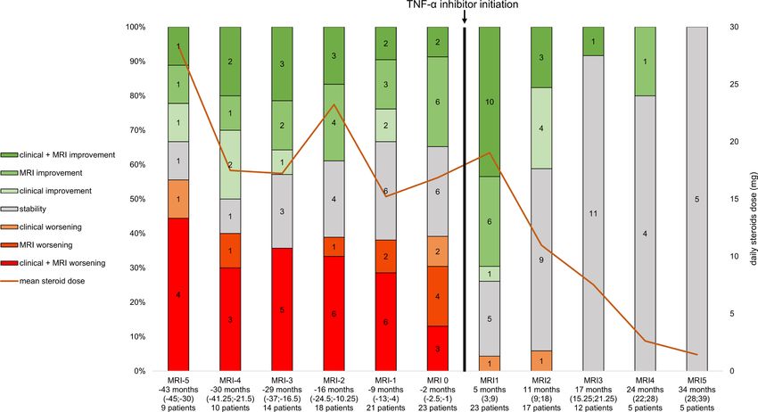
Neuro-i nflammation
J Neurol Neurosurg Psychiatry: first published as 10.1136/jnnp-2020-325665 on 8 June 2021. Downloaded from http://jnnp.bmj.com/ on October 26, 2021 by guest. Protected by copyright.
Methods
Protocol and participants
The project aimed to assess the clinical and MRI evolution of
patients with a parenchymal form of CNS sarcoidosis treated with
a TNF-α inhibitor used as monotherapy in the French Multiple
Sclerosis and Neuroinflammation centres. The project was
presented, discussed and initiated at the annual meeting of the
French Multiple Sclerosis Society in September 2019, at which
all French Multiple Sclerosis and Neuroinflammation centres
were represented. Inclusion criteria were (1) diagnosis of prob-
able or confirmed CNS sarcoidosis according to the consensus
diagnostic criteria for neurosarcoidosis20; (2) parenchymal CNS
lesion attributed to sarcoidosis demonstrated by MRI; (3) treat-
ment with a TNF-α inhibitor and (4) at least 6 months’ follow-up
after TNF-α inhibitor initiation. Exclusion criteria were (1) no
CNS MRI in the 12 months preceding the TNF-α inhibitor initi-
ation; (2) no available MRI data after TNF-α inhibitor initiation
and (3) the association of a TNF-α inhibitor and other steroid-
sparing maintenance immunotherapy.
Outcomes
The primary outcome was the minimum dose of steroids (MDS)
(converted to equivalent doses of prednisolone) reached that
was not associated with any clinical or MRI-evidenced wors-
ening of CNS sarcoidosis during a minimum of 3 months
after dosing change. These minimum doses of steroids were
compared between before TNF-α inhibitor initiation (if appli-
cable) and after. The secondary outcome was the proportion of
patients with clinical and/or MRI- evidenced CNS sarcoidosis
activity after TNF-α inhibitor initiation. Activity was defined
Figure 1 Flow chart of the study. CNS, central nervous system; TNF-α,
as clinical worsening or progression of MRI-evidenced lesions
tumour necrosis factor-α
compared with previous imaging. All MRI data available before
and after TNF-α inhibitor initiation were collected. Worsening,
stabilisation or improvement of CNS lesion(s) were assessed on
each MRI image and compared with the previous image. For signed-rank test with continuity correction. Potential changes in
the sake of readability, each MRI was numbered with reference the proportion of patients with CNS sarcoidosis activity after
to the MRI performed just before TNF-α inhibitor initiation, TNF-α inhibitor initiation were assessed with the Cochran q test.
arbitrarily named MRI0. MRI+1 was the first MRI performed
after the TNF-α inhibitor initiation, MRI+2 the second, etc.
MRI−1 was the MRI performed before MRI0, MRI−2 the MRI
Results
performed before MRI−1, etc. According to inclusion criteria, Study population
MRI0 and MRI+1 were available for all patients. In total, 56 patients with parenchymal CNS sarcoidosis
All adverse events (AEs) ≥grade 2 according to the Common were identified in most of the French Multiple Sclerosis and
Terminology Criteria for Adverse Events (CTCAE) V.5.0 after Neuroinflammation centres: 38 received a TNF-α inhibitor.
TNF-α inhibitor initiation mentioned in the medical record were In these centres, the mean ratio of patients with CNS paren-
registered. chymal sarcoidosis to those with multiple sclerosis ranged
MRI changes were evaluated by the neurologist of each from 1/100 to 1/150.
neuroinflammatory centre in charge of the patient. MRI improve- After excluding patients not fulfilling all inclusion and
ment or worsening was defined as a reduction or increase in exclusion criteria, 23 patients were included in the analysis
the extent of T2 abnormalities and/or gadolinium enhancement (figure 1). Demographic, clinical and MRI characteristics
compared with the last MRI. of patients are in table 1. The median (IQR) time between
diagnosis and TNF-α inhibitor initiation was 22 months
(14–49.5). The median (IQR) follow-u p after TNF-α inhib-
Data availability itor initiation was 24 months (17–40). A total of 22 patients
The corresponding author has full access to all the data in the received infliximab (5–10 mg/kg every 4–10 weeks, most
study. He takes full responsibility for the integrity of the data, the commonly 5 mg/kg every 8 weeks) and 1 adalimumab (40 mg
accuracy of the data analysis and interpretation, and the conduct every 2 weeks).
of the research. The authors have the right to publish any and Before TNF-α inhibitor treatment, 9 of 23 patients fulfilled
all data, separate and apart from the guidance of any sponsor. criteria of refractory parenchymal CNS sarcoidosis, defined as
persistence of clinical and/or MRI activity after at least 6 months
Statistical analyses of immunosuppressive maintenance therapy or the need to use
Statistical analyses were performed with JMP V.9.0.0 (SAS Insti- at least 20 mg/day of steroids associated with immunosuppres-
tute). Potential changes of the MDS between pre-TNF-α and sive maintenance therapy to be stable. Before TNF-α inhibitor
post-TNF-α inhibitor periods were assessed with a Wilcoxon treatment, 15 of the 23 patients presented steroid dependency,
2 Hilezian F, et al. J Neurol Neurosurg Psychiatry 2021;0:1–7. doi:10.1136/jnnp-2020-325665Neuro-i nflammation
J Neurol Neurosurg Psychiatry: first published as 10.1136/jnnp-2020-325665 on 8 June 2021. Downloaded from http://jnnp.bmj.com/ on October 26, 2021 by guest. Protected by copyright.
(p=0.002). In the subgroup of patients with refractory CNS
Table 1 Characteristics of the included patients (n=23)
parenchymal sarcoidosis (n=9), the magnitude of TNF-α
Neurosarcoidosis diagnosis, n (%) inhibitor efficacy was similar to that for other patients.
Probable 21 (91.3) The mean MDS in these patients was 23.7 (18.9) mg/day
Definite 2 (8.7) before TNF-α inhibitor treatment and 5.8 (5.9) mg/day
Age at TNF-α inhibitor initiation, years, mean (SD) 41.5 (10.4) after (pNeuro-i nflammation
J Neurol Neurosurg Psychiatry: first published as 10.1136/jnnp-2020-325665 on 8 June 2021. Downloaded from http://jnnp.bmj.com/ on October 26, 2021 by guest. Protected by copyright.
Figure 2 Dynamics of disease evolution before and after TNF-α inhibitor. TNF-α, tumour necrosis factor-α.
patients: the proportion with clinical or MRI activity at vs 33% at MRI0 (p=0.046). MRI+4 data were available for
MRI+2 (median 10.5 months after TNF-α inhibitor initia- five patients: the proportion with clinical or MRI activity at
tion, IQR 8.75–18.75) was 6% vs 35% at MRI0 (p=0.042). MRI+4 (median 24 months after TNF-α inhibitor initiation,
MRI +3 data were available for 12 patients: the proportion IQR 22–28) was 0% vs 40% at MRI 0 (p=0.22). MRI+5 data
with clinical or MRI activity at MRI+3 (median 17 months were available for five patients: the proportion with clinical
after TNF-α inhibitor initiation, IQR 15.25–21.25) was 0% or MRI activity at MRI+5 (median 34 months after TNF-α
Figure 3 Characteristics of MRI evolution before and after TNF-α inhibitor. TNF-α, tumour necrosis factor-α.
4 Hilezian F, et al. J Neurol Neurosurg Psychiatry 2021;0:1–7. doi:10.1136/jnnp-2020-325665Neuro-i nflammation
J Neurol Neurosurg Psychiatry: first published as 10.1136/jnnp-2020-325665 on 8 June 2021. Downloaded from http://jnnp.bmj.com/ on October 26, 2021 by guest. Protected by copyright.
Figure 4 Complete stability and steroids withdrawn after TNF-α inhibitor initiation in a patient with an isolated cerebral sarcoidosis with previous disease
worsening after steroids were tapered. TNF-α, tumour necrosis factor-α.
inhibitor initiation, IQR 28–39) was 0% vs 40% at MRI 0 Likewise, we found that neurosarcoidosis was refractory
(p=0.22) (figure 2). to steroids and second-line therapies combined in many of
our 23 patients before TNF-α inhibitor initiation. About
Safety of TNF-α inhibitors 40% of our patients showed active disease despite a steroid
In total, 10 AEs ≥grade 2 of the CTCAE occurred in 10 dose >15 mg/day and second-line therapy in 60%, including
patients. No patient died. Three AEs grade 3 and one AE methotrexate, cyclophosphamide and azathioprine. This
grade 4 were reported. All AEs were related to infection: relatively large proportion of patients with refractory
one diverticulitis (grade 2), two urinary infections (grade neurosarcoidosis is probably related to the characteristics
2), two lower respiratory tract infections (one grade 3 and of the patients recruited. All patients included had CNS
one grade 4), one dermatophytosis (grade 2), one isolated parenchymal sarcoidosis; thus, patients with potential more
fever (grade 2), one hepatitis E (grade 3), one post-traumatic benign forms of neurosarcoidosis such as isolated cranial
infection of a finger (grade 2) and one dermohypodermitis nerve or isolated meningeal involvement were excluded.
(grade 3). Only patients with the more severe form of neurosarcoidosis
were included in the present study, but we observed substan-
Discussion tial clinical and MRI-e videnced improvement after TNF-α
The parenchymal form of CNS sarcoidosis is an extremely inhibitor initiation. Crucially, improvement occurred while
rare disorder with a potentially devastating prognosis. In the steroids were being tapered in 21 of 23 patients, and the
present French nationwide study, we report the evolution of mean dose of steroids decreased to 6.5 mg/day. This favour-
CNS parenchymal sarcoidosis—mostly refractory—treated able evolution despite marked reduction of steroids dosing
with a TNF-α inhibitor. We found fast and striking clinical strongly argues for the high efficacy of this treatment in
and radiological improvement in most patients after treat- CNS parenchymal sarcoidosis.
ment initiation. Importantly, improvement was evidenced Of note, we included only patients receiving TNF-α inhib-
when steroids had been tapered rapidly after treatment itors used as steroid-s paring monotherapy in the present
onset, reaching a doseNeuro-i nflammation
J Neurol Neurosurg Psychiatry: first published as 10.1136/jnnp-2020-325665 on 8 June 2021. Downloaded from http://jnnp.bmj.com/ on October 26, 2021 by guest. Protected by copyright.
by clinical progression without any imaging changes. Thus, Contributors BA initiated the study, planned and conducted the study,
TNF-α inhibitor used as monotherapy may be efficient in conducted statistical analysis and wrote the manuscript. AM played a major role
in the acquisition of the data and in the formatting of the figures and revised the
neurosarcoidosis. Importantly, that good outcomes occur manuscript. FH played a major role in the acquisition of the data and the gathering
in patients with parenchymal involvement allows for more the data. CB, AR, SD, PK, TS, CB-K, CG, JC, AM, J-PC, GA, RM, NC, OC, CC, MV,
easily generalising the potential efficacy to all forms of CNS GM, JC and JP played a major role in the acquisition of the data and revised the
sarcoidosis. manuscript. As reported in the method section of the manuscript, all the authors
As reported in several studies of neurosarcoidosis, 14 19 listed have contributed to the conception of the present retrospective study. Indeed,
the conception of the study has been deeply discussed between all the co-authors
infections were encountered here in a significant number listed in a specific meeting during the annual meeting of the French Multiple
of patients receiving a TNF-α inhibitor. Ten infections Sclerosis Society in September 2019. Then, all the authors have participated to the
including two serious infections occurred in 10 of 23 draft and/or the revision of the paper and checked the accuracy or integrity of all
patients. No specific patterns of infection were evidenced. aspects of the work. Finally, all the co-authors have approved the final version of
the manuscript. In that way, we consider that all the names listed meet the ICMJE
Finally, follow-up data were available for 7 of 9 patients authorship requirements.
who stopped TNF-α inhibitors: 5 experienced clinical and
Funding The authors have not declared a specific grant for this research from any
MRI worsening in the few months (mean 7.6) after stopping funding agency in the public, commercial or not-for-profit sectors.
arguing for active surveillance of patients in case of treat-
Competing interests None declared.
ment discontinuation.
The present study has some limitations. First, the sample Patient consent for publication Not required.
size is small, inherent to the rarity of the disease and the Ethics approval The authors obtained ethical approval of the institutional review
stringent inclusion criteria. As mentioned above, the char- board of the University Hospital of Marseille, France (number of the approval, RGPD/
AP-HM 2020-76) to conduct the present study based on retrospective chart review.
acteristics of the population were highly homogeneous,
which allowed for describing the effect of TNF-α inhibitors Provenance and peer review Not commissioned; externally peer reviewed.
in the most aggressive form of neurosarcoidosis. Second, Data availability statement Data are available upon reasonable request. The
we cannot fully exclude that regression to the mean effect corresponding author has full access to all the data in the study. He takes full
responsibility for the integrity of the data, the accuracy of the data analysis and
partly contributes to the dramatic decrease of disease interpretation, and the conduct of the research. The authors have the right to publish
activity measured after TNF-α inhibitor. However, in the any and all data, separate and apart from the guidance of any sponsor. Individuals
present study, in order to rule out a simple regression to participant data underlying the results reported in this article will be available for the
the mean effect, we have collected the disease activity since reviewers. Proposals should be directed to bertrand.audoin@ap-hm.fr for access to
those data.
the first MRI available in all the patients and evidenced
in the large majority of the patients an important disease Open access This is an open access article distributed in accordance with the
Creative Commons Attribution Non Commercial (CC BY-NC 4.0) license, which
activity for several years and a clear breaking point in this
permits others to distribute, remix, adapt, build upon this work non-commercially,
disease activity since the onset of TNF-α inhibitor. Third, and license their derivative works on different terms, provided the original work is
the follow-up was short, which may underestimate a poten- properly cited, appropriate credit is given, any changes made indicated, and the use
tial medium-term return of disease activity or side effects. is non-commercial. See: http://creativecommons.org/licenses/by-nc/4.0/.
Fourth, the retrospective design may have contributed to
ORCID iDs
underreporting side effects. Frédéric Hilezian http://orcid.org/0000-0002-5854-9325
The present study suggests that TNF-α inhibitor treat- Sarah Demortiere http://orcid.org/0000-0002-8513-5443
ment is associated with clinical and imaging improvement in Jean-Philippe Camdessanché http://orcid.org/0000-0002-5282-6707
patients with CNS parenchymal sarcoidosis and allows for Nicolas Collongues http://orcid.org/0000-0002-3683-5582
Guillaume Mathey http://orcid.org/0000-0002-5747-9169
tapering steroids toNeuro-i nflammation
J Neurol Neurosurg Psychiatry: first published as 10.1136/jnnp-2020-325665 on 8 June 2021. Downloaded from http://jnnp.bmj.com/ on October 26, 2021 by guest. Protected by copyright.
14 Gelfand JM, Bradshaw MJ, Stern BJ, et al. Infliximab for the treatment of CNS 21 Fritz D, van de Beek D, Brouwer MC. Clinical features, treatment and outcome in
sarcoidosis: a multi-institutional series. Neurology 2017;89:2092–100. neurosarcoidosis: systematic review and meta-analysis. BMC Neurol 2016;16:220.
15 Cohen Aubart F, Bouvry D, Galanaud D, et al. Long-Term outcomes of refractory 22 Lower EE, Broderick JP, Brott TG, et al. Diagnosis and management of neurological
neurosarcoidosis treated with infliximab. J Neurol 2017;264:891–7. sarcoidosis. Arch Intern Med 1997;157:1864–8.
16 Sodhi M, Pearson K, White ES, et al. Infliximab therapy rescues cyclophosphamide 23 Agbogu BN, Stern BJ, Sewell C, et al. Therapeutic considerations in patients with
failure in severe central nervous system sarcoidosis. Respir Med 2009;103:268–73. refractory neurosarcoidosis. Arch Neurol 1995;52:875–9.
17 Moravan M, Segal BM. Treatment of CNS sarcoidosis with infliximab and
24 Marangoni S, Argentiero V, Tavolato B. Neurosarcoidosis. Clinical description of 7
mycophenolate mofetil. Neurology 2009;72:337–40.
cases with a proposal for a new diagnostic strategy. J Neurol 2006;253:488–95.
18 Santos E, Shaunak S, Renowden S, et al. Treatment of refractory neurosarcoidosis with
25 Scott TF, Yandora K, Valeri A, et al. Aggressive therapy for neurosarcoidosis: long-term
infliximab. J Neurol Neurosurg Psychiatry 2010;81:241–6.
19 Lord J, Paz Soldan MM, Galli J, et al. Neurosarcoidosis: longitudinal experience in follow-up of 48 treated patients. Arch Neurol 2007;64:691.
a single-center, academic healthcare system. Neurol Neuroimmunol Neuroinflamm 26 Bitoun S, Bouvry D, Borie R, et al. Treatment of neurosarcoidosis: a comparative study
2020;7:e743. of methotrexate and mycophenolate mofetil. Neurology 2016;87:2517–21.
20 Stern BJ, Royal W, Gelfand JM, et al. Definition and consensus diagnostic criteria for 27 Doty JD, Mazur JE, Judson MA. Treatment of corticosteroid-resistant
neurosarcoidosis: from the neurosarcoidosis Consortium consensus group. JAMA neurosarcoidosis with a short-course cyclophosphamide regimen. Chest
Neurol 2018;75:1546. 2003;124:2023–6.
Hilezian F, et al. J Neurol Neurosurg Psychiatry 2021;0:1–7. doi:10.1136/jnnp-2020-325665 7You can also read