3D Visualization of the Dental Anatomy of the Narwhal
←
→
Page content transcription
If your browser does not render page correctly, please read the page content below
3D Visualization of the Dental Anatomy of the Narwhal,
                               Monodon monoceros

                Ethan M. Tyler, Martin T. Nweeia, Brent R. Whitaker, Charles W. Potter,
                         Timothy H. Phelps, Elliot K. Fishman, Gary P. Lees

                                                                     Dunn 1990). The specimens that were used in this study exhibit
Computed tomography and digital three-                               dental anatomy that is representative of typical narwhals from a
dimensional imaging software were used to create                     selected population. In addition to known dental structures, the
a comprehensive visual record of the dental                          imagery produced in this investigation identified paired vestigial
anatomy of the narwhal, Monodon monoceros.                           teeth in all specimens examined in this study. This investigation
This multi-institutional study was conducted with                    was part of an ongoing project by Narwhal Tusk Research (NTR)
cooperation between the Johns Hopkins Medical                        to describe the dental and cranial anatomy of the narwhal. This
Institution (JHMI), Harvard University School of                     study at the JHMI created a comprehensive record of the dental
                                                                     anatomy of M. monoceros in cooperation with NTR and Dr. Brent
Dental Medicine, the Smithsonian Institution (SI),
                                                                     Whitaker, DVM, Deputy Director for Biological Programs at the
the National Aquarium in Baltimore, and scientists                   National Aquarium in Baltimore.
with Narwhal Tusk Research (NTR). Digital
reconstructions provide an undisturbed view of the
teeth in situ, and reveal anatomical information                     Materials and Methods
that has been inaccessible and undescribed in                           Narwhal Tusk Research (NTR) legally purchased, and
previous studies. This investigation produced novel                  transported, the two adult narwhal heads—one male and one
information on the presence of paired vestigial                      female—from the Inuit in July 2004 during a field expedition to
                                                                     Pond Inlet, Baffin Island, Canada. The specimens were frozen
teeth in the narwhal, and discovered developmental
                                                                     and stored at Woods Hole, Massachusetts, until NTR transferred
changes in the relative positions of the vestigial                   them to the large specimen freezer at the Smithsonian Institution
teeth to the tusks in the maxillae. Frozen specimens                 National Museum of Natural History (SINMNH) in October 2005.
examined in this study included one male and one
female complete head sectioned posteriorly at the
occipital condyle, and a complete fresh female fetus.
Results include images and descriptions of the teeth
and associated bony structures.

Introduction
  The narwhal, Monodon monoceros, is a coastal cetacean in
the family Monodontidae (Figure 1). Narwhals have northern
circumpolar distribution, and are rarely observed below 60°N.
longitude. Males may grow up to 4.7m (15.42 f) in length and
weigh 1600kg (3,527 lbs) (Heide-Jorgensen 2002). The most
conspicuous characteristic of the male narwhal is its left tusk,
which emerges horizontally through the maxilla and upper lip
and can extend up to 3m (9.84 ft) beyond the head of the animal.
The left tusk is straight and spirals counterclockwise in its long
axis. The right tusk of the male typically remains embedded
in the skull (Brear et al 1993). The paired tusks of the female
narwhal usually remain embedded in the skull (Roberge and
                                                                     Figure 1. Taxonomy of the narwhal, M. monoceros.

JBC Vol. 34, No. 3 2008                                         E44                                      www.jbiocommunication.org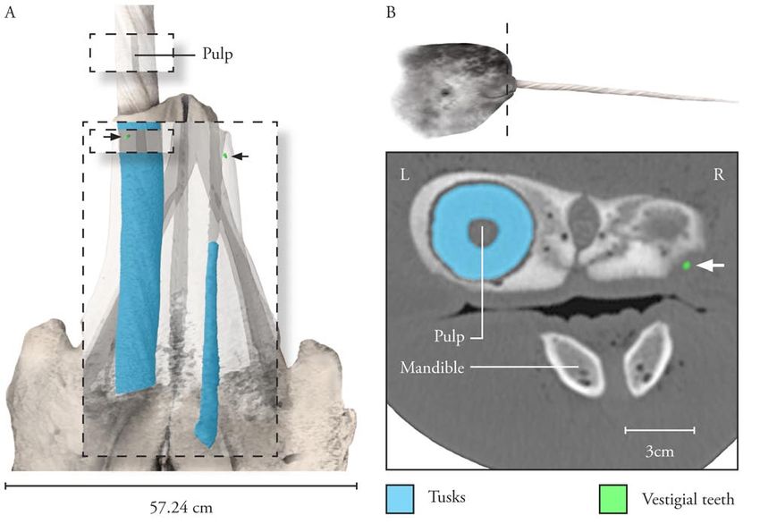
3D Visualization of the Dental Anatomy of the Narwhal, Monodon monoceros
On November 10, 2005, the frozen specimens were transported JHH. Following CT imaging, all of the specimens were returned
to Baltimore, Maryland for computed tomography (CT) at The to the SINMNH laboratory in Washington, D.C. With a secure
Johns Hopkins Hospital (JHH) Russell H. Morgan Department understanding of the anatomy obtained from the CT images, a
of Radiology and Radiological Science. Harvard University team dissected both adult heads at the SINMNH. Prior to the
also obtained one fetal female narwhal specimen, between four dissection, the data from radiography was used to create digital
and six months in its development, from the Inuit during a field three-dimensional (3D) models of narwhal dental anatomy. The
expedition in August 2005. The NTR immediately shipped the CT data collected in the Department of Radiology at JHH was
ice-packed fetal narwhal from the Arctic to Baltimore for CT at conducted using a Siemens Medical Solutions Sensation Cardiac
64. This 64-slice scanner—optimized for cardiac, thoracic, and
vascular imaging—generated 0.5mm-thick slices for each of
the three specimens as follows: 2916 for the male, 1460 for the
female, and 2500 for the fetus. Dr. Elliot Fishman supervised
the procedure and saved the images as Digital Imaging and
Communications in Medicine (DICOM) files. The original data
has been archived at the SINMNH in Washington, D.C.
The DICOM files from CT were imported into Mimics® 8.0 on
a Dell Dimension 8400 desktop computer. Mimics® enabled the
creation of segmentation masks to select specific areas of density.
These were saved as discrete parts of anatomy and named and as
exportable layers. Value thresholds were manipulated to select
distinct target areas of data, such as bone, and then segmentation
masks were created from each of these selections. For some
structures, such as embedded teeth, contours were hand-
selected with the Edit Tool in order to guarantee accuracy for
surface models. Finally, the segmentation masks were saved
and exported as high-quality stereolithography (STL) files.
Each specimen was composed of three registered STL files:
soft tissue, bone, and teeth. Autodesk 3D Studio Max® 8.0 was
used to create digital 3D models. Registered layers of STL data
were imported into 3D Studio Max® at full resolution for each
specimen. In 3D Studio Max®, standard views of the structures
were selected and a virtual photometric light apparatus was used
to light the models and to highlight the important structures. A
single color selection (R=247,G=229,B=206) was made for bone
and teeth. Each of the 3D models was also rendered at 50%
opacity. Because the STL data retains information on internal
structures, reducing the opacity of a specimen’s model reveals
embedded anatomy and cavities. At 50% opacity, hollow regions
and channels appear darker in value than the surrounding bone.
This process was used for each of the three specimens. The final
renders were saved as Tagged Image File Format (TIFF) files at
300 dots per inch and Adobe Photoshop CS2® was used to adjust
colors and compose final images. Photoshop® was also used to
colorize discrepant parts of bony anatomy (Figure 2).
QuickTime® movies of CT data for each of the three narwhal
specimens were generated with files exported from Mimics®.
JPEG files were exported from Mimics® and imported into
Adobe After Effects® 6.5. Three consecutive film compositions
were created for each specimen—axial, coronal, and sagittal—
and the JPEG files were sequenced at 30 frames per second with
QuickTime® Animation compression type. (These movie files
can be viewed on the electronic version of this article on the JBC
site).
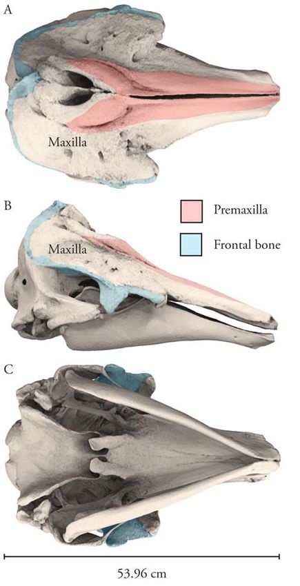
Figure 2. Anatomy of skull of adult female narwhal. (A) Dorsal, (B)
lateral, and (C) ventral views.
JBC Vol. 34, No. 3 2008 E45 www.jbiocommunication.org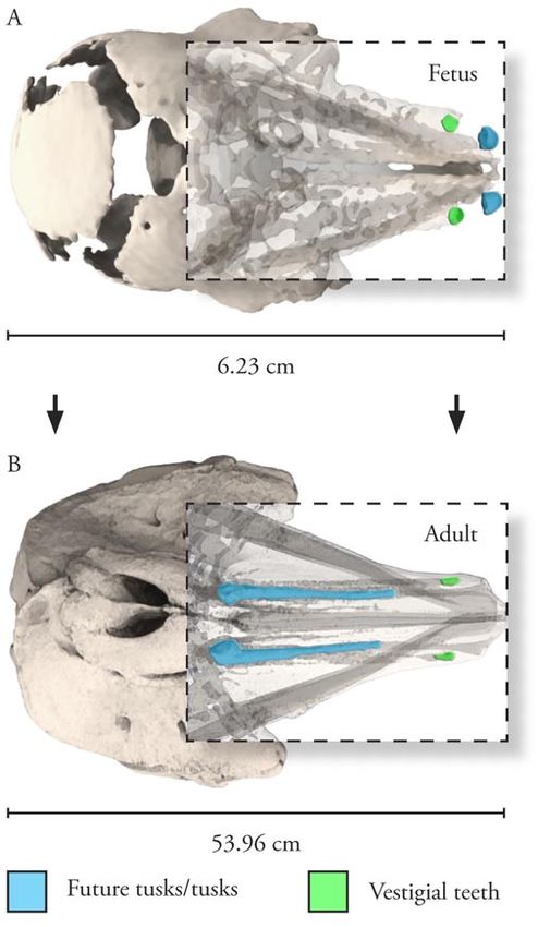
3D Visualization of the Dental Anatomy of the Narwhal, Monodon monoceros

Results and Discussion                                                     pair of teeth. This investigation revealed that the future tusks
                                                                           move in a posterior direction during development and will form
   Digital 3D reconstructions of the three narwhal specimens
                                                                           the two large tusks of the adult narwhal (Figure 4). Growth
provided an undisturbed view of the teeth and associated bony
                                                                           and development of the vestigial pair of teeth terminate later in
structures in situ.
                                                                           development. The future tusks and the vestigial pair of teeth
Fetus                                                                      both exhibit asymmetry.
   The skull of the fetus is 6.23cm in length and 4.96cm in width          Female
at the most distal points on its frontal bones. Calcification of
                                                                             The sectioned head of the female narwhal was 55.82cm in
major bones of the skull is incomplete, but most of the main
                                                                           length and 47.90cm in width at its base. The skull of the specimen
membrane bones are present. This is not the youngest narwhal
                                                                           was 53.96cm in length and 35.08cm in width at the most distal
fetus recorded in the literature (Eales 1950). The maxillae are
                                                                           points on its frontal bones. Like that of most cetaceans, the skull
excavated antero-ventrally to form two conspicuous pairs of
tooth sockets, or alveoli. Pre- and post-orbital processes are
well developed in this specimen. Two pairs of teeth—future
tusks and vestigial—are evident in the upper jaw. Both pairs are
located in their respective sockets in the maxillary bone (Figure
3). The future tusks are located antero-medially to the vestigial

Figure 3. Dentition of fetal female narwhal. (A) Lateral view of           Figure 4. Migration of dental structures during development. (A)
complete fetus and (B) lateral view of skull and teeth; fetus four-six     Dorsal fetal female and (B) adult female views; skulls rendered to
months in its development. Lateral view of fetus 3D surface model          same length of base. Rostrums are transparent for complete
from CT.                                                                   visualization of the teeth.

JBC Vol. 34, No. 3 2008                                                  E46                                       www.jbiocommunication.org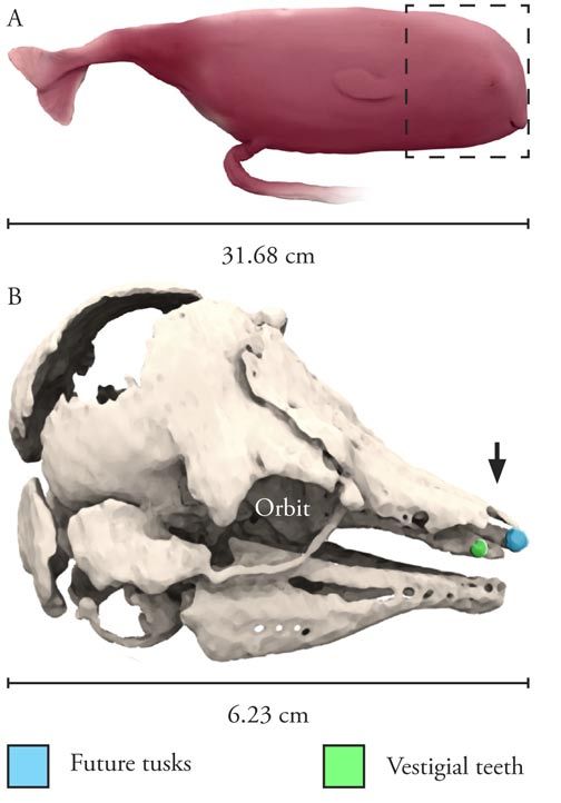
3D Visualization of the Dental Anatomy of the Narwhal, Monodon monoceros

Figure 5. Dentition of adult male narwhal. (A) Dorsal view of skull and teeth; left vestigial tooth is inferior to tusk, as shown in inset. Detail shows
tuskal pulp cavity in erupted tusk. (B) Lateral view of male narwhal head with plane of section and coronal CT slice. Lateral view colorized 3D
surface model from CT data.

of this specimen was asymmetrical, and bony structures were                   literature, perhaps because the vestigial teeth may erupt from the
skewed toward the left side of the head (Berta 2003). In digital              bone and are easily lost during dissection and preparation.
3D reconstructions, two pairs of teeth—tusks and vestigial—are
                                                                              Male
visible in the upper jaw of the female narwhal. Both pairs are
found in their respective sockets in the maxillae. The tusks                     The sectioned head of the male narwhal was 62.33cm in length
are located postero-medially to the vestigial pair of teeth. In               and 49.70cm in width at its base. The skull of the specimen
the female, the paired tusks typically remain embedded in                     was 57.24cm in length and 35.01cm in width at the most distal
the maxillae, as is the case with this specimen. The tusks are                points on its frontal bones. Like the female narwhal specimen,
asymmetrical. Transparent 3D models reveal that sockets for the               the skull of the male narwhal was asymmetrical. In digital 3D
tusks in the skull begin at the bases of the teeth and terminate in           reconstructions, two pairs of teeth–tusks and vestigial–are visible
the most distal part of each respective maxillary bone.                       in the upper jaw of the male narwhal specimen. The paired tusks
   Transparent 3D models also reveal previously undocumented                  are located postero-medially to the vestigial pair of teeth. As
sockets for the vestigial teeth. Sockets for the vestigial teeth              is typical with the species, the right tusk remains embedded in
begin near the bases of the tusks and terminate in the most distal            the skull and the left tusk erupts from the left maxilla. Sockets
part of the maxillae. Well-developed sockets for the vestigial                for the tusks begin at the bases of the teeth and terminate in the
teeth in the adult narwhal are significant, as they provide clues             most distal part of each respective maxillary bone. The vestigial
about the evolution of the modern dentition of the narwhal. The               pair of teeth also exhibits asymmetry. The left tusk is 89.62cm in
vestigial pair of teeth also exhibits asymmetry. The left tusk is             length and the right tusk is 20.81cm in length. Digital 3D images
18.33cm in length and the right tusk is 17.47cm in length.                    reveal that, unlike the vestigial teeth of the female specimen,
   In the adult female specimen, the right vestigial tooth protruded          the vestigial teeth of the male narwhal are not embedded in
slightly from the bone. The presence of vestigial teeth in the                bone; instead, they are suspended in the soft tissue lateral to the
adult narwhal is largely without extensive documentation in the               maxillae (Figure 5).

JBC Vol. 34, No. 3 2008                                                  E47                                            www.jbiocommunication.org
3D Visualization of the Dental Anatomy of the Narwhal, Monodon monoceros
Conclusion Authors
A comprehensive visual record of the dental anatomy of the Ethan Tyler is currently a medical illustrator and animator at
narwhal, M. monoceros, was created with CT data and digital the National Institutes of Health, Department of Medical Arts,
3D imaging via Mimics®. Digital reconstructions of the two in Bethesda, Maryland. He has a Master of Arts in Medical
adult specimen heads–male and female–and a complete fetal and Biological Illustration from The Johns Hopkins University
specimen revealed novel information on the dental anatomy of School of Medicine, and a Bachelor of Arts in Biology from
this animal. Concordia University at Austin. tylereth@od.nih.gov
The first major finding was the presence of paired vestigial
teeth in all three specimens. Although a previous study Martin T. Nweeia, DMD, is a Clinical Instructor at the Harvard
documents the presence of a few single vestigial teeth found School of Dental Medicine, and a Research Associate in the
in a small collection of adult narwhal skulls, no other known Marine Mammal Program at the Smithsonian Institution. Dr.
investigation has found paired vestigial teeth (Fraser 1938). The Nweeia is also Expedition Leader and Principal Investigator for
lack of prior documentation on vestigial teeth may be because Narwhal Tusk Research. He has a Doctorate of Dental Surgery
the vestigial teeth are easily lost or damaged during dissection from Case Western Reserve University, in Cleveland, OH, and
and preparation. In the case of the male narwhal, the vestigial a Doctorate of Dental Medicine from Case. He has a full time
teeth are not embedded in bone. Radiography and digital 3D general practice in Sharon, CT.
reconstructions provided an undisturbed view of the vestigial
teeth in situ. Brent R. Whitaker, DVM, is the Deputy Executive Director of
The second major finding was that changes occur in the positions Biological Programs at the National Aquarium in Baltimore,
of the two pairs of teeth–tusks and vestigial–in the maxillae MD. Dr. Whitaker has a Master of Veterinary Science and a
during development. The two pairs of teeth reverse positions. Doctor of Veterinary Medicine degree from the University of
In the fetus, the future tusks are located antero-medially to the Flordia, College of Veterinary Medicine.
vestigial pair of teeth at four-six months in development. The
fully developed tusks are located postero-medially to the vestigial Charles Potter is the Collection Manager, Marine Mammals, at
pair of teeth in the adult narwhal. the Smithsonian Institution National Museum of Natural History.
He has a Bachelor of Science (Zoology) from SUNY College of
Forestry and Environmental Science.
References Timothy H. Phelps, MS, Associate Professor in, and Assistant
Berta, Annalisa and James L. Sumich. Marine Mammals: Director of the Department of Art as Applied to Medicine, Johns
Evolutionary Biology. London: Elsevier Science, 2003. Hopkins University, School of Medicine, Baltimore, MD.
Brear, K., J.D. Currey, M.C.S. Kingsley, and M. Ramsay. “The Elliot K. Fishman, M.D., Professor of Radiology and
Mechanical Design of the Tusk of the Narwhal (Monodon Radiological Science and Professor of Oncology at Johns
Monoceros: Cetacea).” Journal of the Zoological Society of
London 230 (1993): 411-423. Hopkins Hospital, Baltimore, MD.
Eales, Nellie B. “The Skull of the Foetal Narwhal, Monodon Gary P. Lees, MS, CMI, Associate Professor, Chair and Director
Monoceros.” Philosophical Transactions of the Royal Society of of the Department of Art as Applied to Medicine, Johns Hopkins
London 235 (1950): 1-33. University, School of Medicine, Baltimore, MD.
Fraser, F.C. “Vestigial Teeth in the Narwhal.” Proceedings of
the Linnean Society of London 24 (1938): 155-161
Heide-Jorgensen, M.P. “Narwhal”. Encyclopedia of Marine
Mammals. (2002): 784.
Roberge, M.M. and J.B. Dunn. “Assessment of the subsistence
harvest and biology of narwhal (Monodon monoceros L.)
from Admirality Inlet, Baffin Island, Northwest Territories
(Canada).” Proceedings of Canadian Fish and Aquatic Science
Organization (1990): 1-32.
JBC Vol. 34, No. 3 2008 E48 www.jbiocommunication.orgYou can also read