Inflammatory Implant Periapical Lesion: Etiology, Diagnosis, and Treatment-Presentation of 7 Cases

Page created by Arthur Owen
 
CONTINUE READING
Inflammatory Implant Periapical Lesion: Etiology, Diagnosis, and Treatment-Presentation of 7 Cases
DENTAL IMPLANTS
J Oral Maxillofac Surg
67:168-173, 2009

                 Inflammatory Implant Periapical
                  Lesion: Etiology, Diagnosis, and
                Treatment—Presentation of 7 Cases
                                              Miguel Peñarrocha-Diago, DDS, PhD,*
                                                  Araceli Boronat-Lopez, DDS,†
                                               and Berta García-Mira, DDS, PhD‡
     Purpose: To investigate implant periapical lesions, and to describe their treatment. The hypothesis of this
     evaluation is that implant periapical lesions are disorders of the area surrounding the apex of a dental implant,
     and that their etiology can be multifactorial (ie, vascular impairment, vascular ischemia, overheating of bone
     during drilling, and implant surface contamination). The diagnosis is based on the clinical manifestations and
     x-ray findings. The x-ray findings usually involve a periapical radiotransparency.
     Materials and Methods: Seven patients with implant periapical lesions (3 in the upper jaw, and 4 in
     the mandible) after implant placement are described. All patients reported pain, and 3 suffered from
     inflammation. Upon percussion, the 3 nonsubmerged implants produced a dull sound, with no mobility.
     A panoramic x-ray study showed periapical transparencies around 5 implants, whereas in 1 case,
     computed tomography showed a maxillary sinus reaction. The diagnosis was acute apical peri-implantitis
     (nonsuppurative in 2 cases, and suppurative in 5 cases).
     Results: The clinical manifestations did not subside with antibiotics. In all cases, treatment consisted of
     implant periapical surgery, after which the symptoms disappeared. The radiotransparencies showed
     progressive resolution.
     Conclusion: The possibility of implant periapical lesions must be taken into account. A rapid diagnosis
     should be established to treat the lesions at an early stage, hence preventing the need for implant extraction.
     © 2009 American Association of Oral and Maxillofacial Surgeons
     J Oral Maxillofac Surg 67:168-173, 2009

In 1993, Sussman and Moss1 introduced the concept                          close to an infected maxillary sinus.5 The incidence of
of implant periapical pathology as an infectious-                          implant periapical lesions is very low, and almost all
inflammatory disorder of the tissues surrounding the                       studies in the literature are limited to 1 or 2 cases.2,3,5
apex of a dental implant. The underlying causes may                           We describe 7 patients with implant periapical pa-
include vascular impairment, overheating of bone dur-                      thology, whose treatment was provided in the form of
ing drilling, and vascular ischemia, all of which can                      periapical surgery. A Medline review is also presented
result in bone loss around the apex of the implant.                        of the literature on implant periapical lesions span-
Other etiological factors may comprise overheating of                      ning the past 10 years.
bone during drilling,2 overdrilling of the site, implant
surface contamination,3,4 pre-existing bone pathology,
the presence of root remains, and implant placement                        Materials and Methods
                                                                              Between 1996 and 2002, the same surgeon in the
Received from the Faculty of Medicine and Dentistry, University of         same operating room conventionally placed approxi-
Valencia, Valencia, Spain.                                                 mately 2,500 implants under abundant irrigation with
  *Professor of Oral Surgery.                                              saline solution. We documented all cases in which
  †Associate Professor of Oral Surgery.                                    implant periapical surgery was performed. The crite-
  ‡Master of Oral Surgery and Implantology.                                ria for performing such surgery included: 1) acute
   Address correspondence and reprint requests to Dr Penarrocha-           pain after implant placement that failed to respond
Diago: Clinicas Odontologicas, Gasco Oliag 1, 46021 Valencia,              to antibiotic treatment (amoxicillin 500 mg, 3 times
Spain; e-mail: Miguel.Penarrocha@uv.es                                     a day, for 7 days); 2) absence of implant mobility;
© 2009 American Association of Oral and Maxillofacial Surgeons             3) dull percussion of nonsubmerged implants; and
0278-2391/09/6701-0025$34.00/0                                             4) presence or absence of implant periapical radi-
doi:10.1016/j.joms.2007.12.022                                             olucency. In the presence of implant mobility, non-

                                                                     168
Inflammatory Implant Periapical Lesion: Etiology, Diagnosis, and Treatment-Presentation of 7 Cases
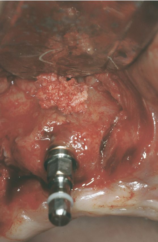
PENARROCHA-DIAGO, BORONAT-LOPEZ, AND GARCÍA-MIRA                                                                    169

dull percussion, or radiolucency along the longitu-        surgery was performed after 4 weeks, with implant
dinal axis of the implant, peri-implant surgery was        loading 3 months later.
not considered.
                                                             CASE 7
  CASE 1                                                      A 60-year-old woman developed pain and gingival
  A 32-year-old woman with no relevant medical his-        swelling in the region of an implant placed 10 days
tory presented with a partially edentulous upper jaw.      previously in the left mandibular lateral incisor. An
Ten days after placement of a submerged 14 ⫻ 4.1           x-ray study revealed a radiolucent image. The symp-
mm ITI implant (Straumann, Waladent, Switzerland)          toms failed to subside with antibiotic treatment, and
in the zone of the left maxillary central incisor, she     periapical surgery was performed, followed by a fa-
developed pain in the apical region that failed to         vorable course and implant loading after 3 months.
subside with antibiotics. Periapical surgery was per-         Peri-implant surgery was performed in 7 cases (3 in
formed, and the course of the fixation proved favor-       the upper jaw and 4 in the mandible). In 6 cases we
able, allowing for implant loading after 3 months.         used sandblasted and acid-etched (SLA) surface ITI im-
                                                           plants (Straumann), and in one case, an Avantblast sur-
  CASE 2                                                   face Defcon implant (Implandent, Senmenat, Barcelona,
   A 32-year-old woman presented with a totally eden-      Spain). Four submerged and 3 nonsubmerged implants
tulous lower jaw. Seven days after surgery, she devel-     were involved.
oped pain in the implant periapical zone, and percus-         The patients included 5 women and 2 men (age
sion proved dull, with identification of an apical         range, 32 to 78 years). In all cases, oral and radiologic
radiolucency. Antibiotic treatment was provided, and 2     examinations were performed, based on periapical
weeks after implant placement, periapical surgery was      and extraoral panoramic x-rays. In addition, com-
performed, allowing for implant loading after 4 months.    puted axial tomographic imaging was performed in
                                                           one patient with suspected maxillary sinusitis.
  CASE 3
   A 78-year-old woman developed pain 12 days after          SURGICAL PROCEDURE
implant placement in the region of the right mandib-          Surgery was performed under local anesthesia (4%
ular first premolar, with dull percussion. Periapical      articaine with 1:100,000 adrenalin), raising a full-
x-rays showed radiolucency in the zone of the implant      thickness flap in the affected zone. A tungsten drill
apex. Antibiotics were prescribed, and periapical sur-     was used to perform the ostectomy to gain access to
gery was performed 3 weeks after the intervention.         the implant apex, under abundant irrigation with ster-
Her subsequent course proved favorable, and implant        ile saline solution. Curettage of the inflamed tissue
loading could be performed after 2 months.                 was performed, followed by a histologic study of the
                                                           samples collected. An ultrasound tip was used to irrigate
  CASE 4                                                   the surgical bed with sterile saline solution. When nec-
   A 70-year-old woman developed implant apical pain       essary, bone shavings from the surgical field or synthetic
in the region of the right mandibular lateral incisor, 1   bone (Bio-Oss; Geistlich, Wolhusen, Switzerland) were
week after placement, with dull percussion and an          placed in the bone defect. After 8 to 12 weeks, second-
apical radiolucent image. Antibiotic treatment proved      step surgery was performed, and prosthetic rehabilita-
ineffective, and periapical surgery was performed,         tion of the implants was carried out (Figs 1-6).
with implant loading after 3 months.

  CASE 5
   A 49-year-old man with no relevant medical history
developed pain and gingival swelling in the region of
an implant placed 7 days previously in the zone of the
left maxillary first premolar. A sinus reaction was
suspected and confirmed by computed tomography.
After 1 week of antibiotic treatment, periapical sur-
gery successfully resolved the lesion, and placement
of the prosthesis proved possible after 3 months.

  CASE 6                                                   FIGURE 1. Case 6. Extraoral panoramic x-ray view after dental
                                                           implant placement: 4 in the anterior sector, 2 trans-zygomatic
  A 56-year-old man presented with a totally edentu-       implants, and 2 pterygoid implants.
lous upper jaw. Fifteen days after implant placement,      Penarrocha-Diago, Boronat-Lopez, and García-Mira. Inflammatory
he developed pain and gingival swelling. Periapical        Implant Periapical Lesions. J Oral Maxillofac Surg 2009.
Inflammatory Implant Periapical Lesion: Etiology, Diagnosis, and Treatment-Presentation of 7 Cases
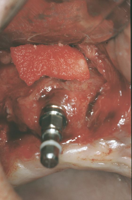
170                                                                                 INFLAMMATORY IMPLANT PERIAPICAL LESIONS

                                                                          manifestations failed to improve in our patients, and
                                                                          all underwent peri-implant surgery.
                                                                             The diagnosis of inflammatory implant periapical
                                                                          lesions is based on clinical findings (pain, possible
                                                                          swelling, and dull percussion) and radiologic evi-
                                                                          dence, since at some stage, a radiolucent image may
                                                                          appear at implant periapical level, reflecting the exis-
                                                                          tence of suppuration. Table 2 lists 8 implant periapi-
                                                                          cal lesions described by different authors.2,3,5,7-9 Bretz
                                                                          et al9 reported that maxillary sinusitis was associated
                                                                          with the implant apical zone, whereas one of our
                                                                          patients developed a sinusal reaction. Unlike other
                                                                          investigators,2,3,7,8 we recorded no fistulas. The ap-
FIGURE 2. Case 6. After 15 days, the patient reported constant            pearance of a fistula probably corresponds to a more
pain in the zone of tooth 3. Computed tomography showed loss of
the vestibular layer, with the identification of an peri-implant radio-   advanced phase of periapical disease (periapical ab-
transparency. The maxillary sinuses presented artifacts attributable      scess), and in this sense, our lesions were diagnosed
to the trans-zygomatic implants.                                          and treated at an early stage of acute apical periim-
Penarrocha-Diago, Boronat-Lopez, and García-Mira. Inflammatory            plantitis. Five lesions were suppurative, and 2 were
Implant Periapical Lesions. J Oral Maxillofac Surg 2009.                  nonsuppurative but not fistular.
                                                                             Such periapical lesions may be secondary to con-
                                                                          tamination during instrumentation, as suggested by
Results
   The patients’ characteristics are summarized in Ta-
ble 1. Approximately 7 to 15 days after implant place-
ment, all patients reported pain, with gingival inflam-
mation of the affected zone in 3 cases. No fistulas
were observed; no implants presented mobility; and 3
implants showed dull percussion (in 4 cases, this
could not be evaluated because the implants were sub-
merged). A radiologic study revealed radiolucency
around the apex of 5 implants, and in one case, com-
puted tomography showed a maxillary-sinus reaction.
   In all cases, the diagnosis was acute apical peri-
implantitis, as based on the symptoms. In 3 cases,
sufficient material could be collected for a histologic
study (using hematoxylin-eosin staining). An acute
inflammatory infiltrate was identified in all cases.
   One week after periapical surgery, the pain and
inflammation had subsided. The radiolucencies grad-
ually resolved entirely. Between 2 and 4 months later,
implant loading started, and after a minimum fol-
low-up of 1 year, all 7 implants were seen to remain
functional in the mouth, with no clinical or radiologic
alterations.

Discussion
   Implant periapical lesions are infrequent, with an
incidence of 10 cases per 3,800 implants, according
to Reiser and Nevins.6 In our series of implant peria-
pical lesions, the symptoms appeared 7 to 15 days
after surgery, in concordance with the report of Ro-                      FIGURE 3. Case 6. After raising a full-thickness flap, destruction of
dríguez and Rodríguez.7 However, other authors2,8                         the vestibular layer is observed in the apical zone of the implant.
described implant radiolucencies even months later.                       Penarrocha-Diago, Boronat-Lopez, and García-Mira. Inflammatory
Despite systemic antibiotic medication, the clinical                      Implant Periapical Lesions. J Oral Maxillofac Surg 2009.
PENARROCHA-DIAGO, BORONAT-LOPEZ, AND GARCÍA-MIRA                                                                               171

Scarano et al.5 According to other authors,2,3 these
lesions result from excessive heating of bone during
surgery. Ayangco and Sheridan8 described 3 cases of
implant periapical lesions in patients with a history of
endodontic treatment and failed apicoectomy before
implant placement. According to these authors, de-
spite curettage, socket cleaning, and a long waiting
time, bacteria would have persisted in the bone, caus-
ing implant periapical alterations as a result. Suss-
man10 classified such lesions as implant-to-tooth (type
I) when produced during instrumentation of the im-
plant bed, and as tooth-to-implant (type II) when
originating from apical lesions of the teeth adjacent to
the implant. In our cases, there was no previous
periapical pathology, and the lesions may have been a
consequence of contamination of the apical region of
the bed during surgery.
   None of the cases responded to antibiotic therapy
alone, indicating that bacteria were not the only cause of
the condition, therefore necessitating surgery. In our
cases, the histopathologic study identified an acute in-

                                                                      FIGURE 5. Case 6. An absorbable collagen membrane is placed
                                                                      on the surgical field.
                                                                      Penarrocha-Diago, Boronat-Lopez, and García-Mira. Inflammatory
                                                                      Implant Periapical Lesions. J Oral Maxillofac Surg 2009.

                                                                      flammatory infiltrate, in concordance with other inves-
                                                                      tigators, eg, Piattelli et al3 and Balshi et al.11
                                                                         Bretz et al9 successfully performed periapical sur-
                                                                      gery with curettage and chlorhexidine irrigation to

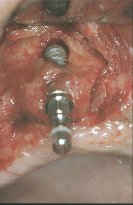
FIGURE 4. Case 6. Careful curettage of the zone was performed,
with elimination of all inflammatory tissue. Bone shavings from the   FIGURE 6. Case 6. Panoramic x-ray view after placement of the
surgical field were placed in the bone defect.                        definitive screwed prostheses.
Penarrocha-Diago, Boronat-Lopez, and García-Mira. Inflammatory        Penarrocha-Diago, Boronat-Lopez, and García-Mira. Inflammatory
Implant Periapical Lesions. J Oral Maxillofac Surg 2009.              Implant Periapical Lesions. J Oral Maxillofac Surg 2009.
172
 Table 1. CHARACTERISTICS OF PATIENTS WITH IMPLANT PERIAPICAL LESIONS

                                                  Case 1                   Case 2                Case 3             Case 4            Case 5              Case 6           Case 7

Age (yr)                                            32                      32                     78                 70                49               56                60
Gender                                     F                       F                       F                  F                M                    M                F
Edentulism                                 Partial upper           Total                   Total              Total            Partial upper        Total upper      Partial lower
Location                                             2.1                     3.2                   4.4                4.2                2.4              1.3               3.2
Implant
   Type                                    ITI                     ITI                     ITI                ITI              ITI                  ITI              Defcon
   Length                                        14 mm                    14 mm               16 mm              14 mm               12 mm             12 mm            16 mm
   Diameter                                      4.1 mm                   4.1 mm             4.1 mm             4.1 mm               4.1 mm           4.1 mm           4.2 m
Submerged                                  Yes                     No                      No                 No               Yes                  Yes              Yes
Time to symptoms (days)                            10                        7                 12                   7                   7               15               10
Time to treatment (weeks)                           4                        2                   3                  5                   3                 4                3
Symptoms
   Pain                                    Yes                     Yes                     Yes                Yes              Yes                  Yes              Yes
Signs
   Percussion                                        ⫺             Dull                    Dull               Dull                      ⫺                   ⫺                ⫺
   Mobility                                          ⫺             No                      No                 No                        ⫺                   ⫺                ⫺
   Inflammation                            No                      No                      No                 No               Yes                  Yes              Yes

                                                                                                                                                                                      INFLAMMATORY IMPLANT PERIAPICAL LESIONS
                                                                                                                               Sinusal reaction:
  Apical radiotransparency                 No                      Yes                     Yes                No                 CT                 Yes              Yes
Treatment                                  Apical surgery          Apical surgery          Apical surgery     Apical surgery   Apical surgery       Apical surgery   Apical surgery
                                           Acute inflammatory      Acute inflammatory                                          Acute inflammatory
Histology                                    infiltrate              infiltrate                                                  infiltrate
Course                                                ⫹                       ⫹                     ⫹                  ⫹                  ⫹                 ⫹                ⫹
Loading after periapical surgery (mo)                 3                       4                     2                  3                  3                 3                3
Control                                             6 years              15 months                1 year             1 year          18 months            1 year           1 year
  Abbreviations: ITI, Straumann, Waladent, Switzerland; Defcon, Impladent, Senmenat, Barcelona, Spain; ⫺, not evaluable, because implant is submerged; ⫹, positive course;
CT, computed tomography.
Penarrocha-Diago, Boronat-Lopez, and García-Mira. Inflammatory Implant Periapical Lesions. J Oral Maxillofac Surg 2009.
PENARROCHA-DIAGO, BORONAT-LOPEZ, AND GARCÍA-MIRA                                                                                                                                                                                                                                                                                                     173

                                                                                                                                                                                                                                                                                      treat an implant periapical lesion, with the placement

                                                                          Success

                                                                                             Yes
                                                                                             Yes

                                                                                                                                 Yes
                                                                                             No
                                                                                             No
                                                                                             No
                                                                                                                                                                                                                                                                                      of demineralized bone and a collagen membrane cov-
                                                                                                                                                                                                                                                                                      ering. Ayangco and Sheridan8 also performed periapi-
                                                                                                                                                                                                                                                                                      cal surgery on an implant, with the additional place-

                                                                                             Curettage and tetracycline
                                                                                                                                                                                                                                                                                      ment of tetracycline in the zone for 1 minute, for local

                                                                                             Curettage and irrigation

                                                                                                                                 Curettage and irrigation
                                                                                                                                                                                                                                                                                      disinfection. As an alternative to treatment, these au-
                                                                          Treatment                                                                                                                                                                                                   thors mentioned the possibility of sectioning the im-
                                                                                             Implant removal
                                                                                             Implant removal
                                                                                             Implant removal
                                                                                                                                                                                                                                                                                      plant apex in those cases where complete removal of
                                                                                                                                                                                                                                                                                      the granular tissue is not assured, and when the loca-
                                                                                                                                                                                                                                                                                      tion involves the maxillary sinus or nasal cavity. We
                                                                                                                                                                                                                                                                                      performed periapical surgery, gaining access to the
                                                                                                                                                                                                                                                                                      apex through an ostectomy window. If the vestibular
                                                                                                                                                                                                                                                                                      bone wall was found to be intact, the inflammatory
                                                                                                                                                                                                                                                                                      tissue was removed, and chlorhexidine irrigation was
                                                                 Radiotransparency

                                                                                                                                                                                                                                                                                      performed. Posteriorly, we introduced the implant a
                                                                                                                                                                                                                                                                                      further 1 to 2 mm in the bone, to improve primary
                                                                       Apical

                                                                                             Yes
                                                                                             Yes
                                                                                             Yes
                                                                                             Yes
                                                                                             Yes
                                                                                             Yes
                                                                                             Yes
                                                                                             Yes

                                                                                                                                                                                                                                                                                      fixation and ensure better soft-tissue closure.
                                                                                                                                                                                                                                                                                         The treatment of implant periapical pathology de-
                                                                                                                                                                                                                                                                                      pends on the stage of the lesion. In the case of an
                                                                                                                                                                                                                                                                                      acute lesion (suppurative or otherwise), we perform
                                                                                                                                                                                                                                                                                      periapical surgery with curettage and irrigation,8-10
                                                                                                                                                            Penarrocha-Diago, Boronat-Lopez, and García-Mira. Inflammatory Implant Periapical Lesions. J Oral Maxillofac Surg 2009.
                                                                          Fistula

                                                                                             Yes
                                                                                             Yes

                                                                                             Yes
                                                                                             Yes

                                                                                             Yes

                                                                                                                                                                                                                                                                                      whereas in the case of a periapical abscess affecting
                                                                                             No

                                                                                             No
                                                                                             No

                                                                                                                                                                                                                                                                                      the bone surface or involving the loss of primary fixa-
                                                                                                                                                                                                                                                                                      tion, implant removal is the only management op-
                                                                                                       Yes
                                                                                                       Yes

                                                                                                                          Yes
                                                                          Pain

                                                                                                                                                                                                                                                                                      tion.2,3,12 For this reason, Reiser and Nevins6 contend
                                                                                                                                                                                                                                                                                      that implant periapical pathology treatment should be
                                                                                                                                                                                                                                                                                      immediate, to avoid the need for extraction.
                                                           Diagnosis
                                                            Time to

                                                             (mo)

                                                                                              7
                                                                                              5
                                                                                              6
                                                                                              8
                                                                                             18
                                                                                              9
                                                                                              1
                                                                                              6

                                                                                                                                                                                                                                                                                      References
                                                                                                                                                                                                                                                                                       1. Sussman HI, Moss SS: Localized osteomyelitis secondary to
                                                                                             Maxillary second premolar

                                                                                                                                                                                                                                                                                          endodontic-implant pathosis. A case report. J Periodontol 64:
                                                                                             Maxillary first premolar

                                                                                             Maxillary lateral incisor

                                                                                                                                                                                                                                                                                          306, 1993
                                                                          Implant Location

                                                                                                                                                                                                                                                                                       2. Piattelli A, Scarano A, Balleri P, et al: Clinical and histologic
                                                                                             Premolar mandible

                                                                                             Canine mandibular
                                                                                             Maxillary premolar

                                                                                                                                                                                                                                                                                          evaluation of an active “implant periapical lesion:” A case
                                                                                                                                                                                                                                                                                          report. Int J Oral Maxillofac Implants 13:713, 1998
                                                                                                                                                                                                                                                                                       3. Piattelli A, Scarano A, Piattelli M, et al: Implant periapical
                                                                                                                                                                                                                                                                                          lesions: Clinical, histologic and histochemical aspects. A case
                                                                                             Mandible

                                                                                             Mandible

                                                                                                                                                                                                                                                                                          report. Int J Periodont Restor Dent 18:181, 1998
        Table 2. STUDIES WITH IMPLANT PERIAPICAL LESIONS

                                                                                                                                                                                                                                                                                       4. Chaffee NR, Lowden K, Tiffee JC, et al: Periapical abscess
                                                                                                                                                                                                                                                                                          formation and resolution adjacent to dental implants: A clinical
                                                                                                                                                                                                                                                                                          report. J Prosthet Dent 85:109, 2001
                                                                                                                                                                                                                                                                                       5. Scarano A, Di Domizio P, Petrone G, et al: Implant periapical
                                                                                                                                                                                                                                                                                          lesion: A clinical and histologic case report. J Oral Implantol
                                                           Periapical Lesions

                                                                                                                                                                                                                                                                                          26:109, 2000
                                                              Number of

                                                                                                                                                                                                                                                                                       6. Reiser GM, Nevins M: The implant periapical lesion: Etiology,
                                                                Implant

                                                                                                                                                                                                                                                                                          prevention and treatment. Compend Contin Educ Dent 16:768,
                                                                                             1
                                                                                             1
                                                                                             1
                                                                                             1
                                                                                             3

                                                                                                                                 1

                                                                                                                                                                                                                                                                                          1995
                                                                                                                                                                                                                                                                                       7. Rodríguez A, Rodríguez F: Proceso periapical implantológico.
                                                                                                                                                                                                                                                                                          Rev Esp Odontostomatol Implantes 3:159, 1995
                                                                                                                                                                                                                                                                                       8. Ayangco L, Sheridan PJ: Development and treatment of retro-
                                                                                                                                                                                                                                                                                          grade peri-implantitis involving a site with a history of failed
                                                                                                                                                                                                                                                                                          endodontic procedures: A series of reports. Int J Oral Maxillo-
                                                                                             Rodríguez and Rodríguez7

                                                                                                                                                                                                                                                                                          fac Implants 16:412, 2001
                                                                                             Ayangco and Sheridan8

                                                                                                                                                                                                                                                                                       9. Bretz WA, Matuck AN, de Oliveira G, et al: Treatment of
                                                                                                                                                                                                                                                                                          retrograde peri-implantitis: A clinical report. Implant Dent
                                                                                                                                                                                                                                                                                          6:287, 1997
                                                                          Authors

                                                                                                                                                                                                                                                                                      10. Sussman HI: Periapical implant pathology. J Oral Implantol
                                                                                             Scarano et al5
                                                                                             2

                                                                                             Piattelli et al3

                                                                                                                                                                                                                                                                                          24:133, 1998
                                                                                             Piattelli et al

                                                                                                                                Bretz et al9

                                                                                                                                                                                                                                                                                      11. Balshi J, Pappas E, Wolfinger J, et al: Management of an abscess
                                                                                                                                                                                                                                                                                          around the apex of a mandibular root-form implant: Clinical
                                                                                                                                                                                                                                                                                          report. Implant Dent 3:815, 1994
                                                                                                                                                                                                                                                                                      12. Oh TJ, Yoon J, Wang HL: Management of the implant periapical
                                                                                                                                                                                                                                                                                          lesion: A case report. Implant Dent 12:41, 2003
You can also read