Osteoid osteoma of the ribs-Is image intensifier or bone scintigraphy a mandatory diagnostic tool-A case report with Review of literature - OAText
←
→
Page content transcription
If your browser does not render page correctly, please read the page content below
Otorhinolaryngology-Head and Neck Surgery
Case Report ISSN: 2398-4937
Osteoid osteoma of the ribs-Is image intensifier or bone
scintigraphy a mandatory diagnostic tool-A case report
with Review of literature
Suraj Wasudeo Nagre* and Saptarshi Paul
Department of CVTS, Grant Medical College, India
Abstract
Osteoid osteoma (OO) is a benign osteogenic tumour first reported by Jaffe in 1935. OOs may affect any bone, but more than half of the tumors occur in the long
bones of the lower extremities. The frequency of OOs affecting the ribs is extremely low (1-1.4%), with only 14 reported cases with surgical intervention to date.
Complete surgical excision is the standard treatment method for osteoid osteoma and is usually offered to patients experiencing chronic and substantial pain that is
not relieved by conservative treatment. In this report we present a case of osteoid osteoma of the posterior part of the shaft of the sixth rib affecting a 30-year-old
male, who had presented with symptoms of severe pain over the affected area and underwent surgical resection. Excised rib segment showed no osteosclerotic lesion
on X ray so immediately extended resection of sixth rib was done. Here we have tried to evaluate the importance of the presence of the skeletal scintigraphy or C Arm
image intensifier intra operatively by comparing our experience with the available literature.
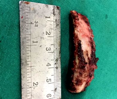
Introduction Incision closed in layers after keeping negative suction drain (Figure
2b). Patient extubated and shifted to ward.The resected bone segments
Osteoid osteoma is a type of benign bone tumor, which was first of both sixth and seventh rib externally looked same (Figure 4a and 4b)
described by Jaffe in 1935 [1]. It is a rare condition, which accounts so we sent them for histopathological examination.
for only 3% of primary bone tumors. Individuals aged between 5 and
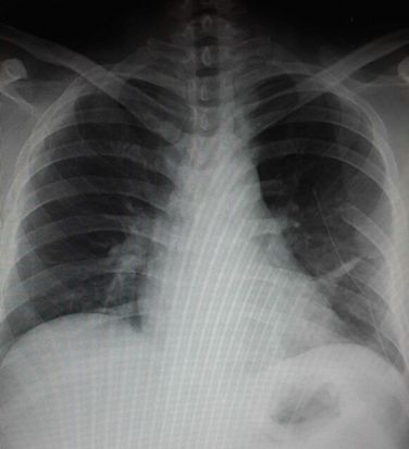
24 years old are the most commonly affected [2]. The main symptom Post operatively patient was comfortable. Chest X ray done and
of the disease is pain that worsens at night, which may be alleviated by compared with preopt Chest X ray (Figure 5a and 5b). Drain removed
NSAIDs (non-steroidal anti-inflammatory drugs). Current treatment after 48 hours and patient was discharged on fifth postopt day.Stich
modalities include surgical excision, as well as less invasive techniques. removal was done on fiftinth postopt day .
After treatment, symptoms can be controlled. The most commonly
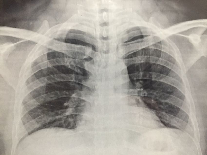
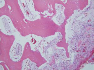
affected sites are the long bones of the lower extremities and the patient Histopathological examination of sixth rib was normal but seventh
outcome is good [3]. rib revealed the presence of osteoid tissue in a background stroma of
fibrovascular tissue and thin trabeculae inter-anastomosing with a
Case report single layer of osteoblasts (Figure 6).
A 30-year-old male complained of nocturnal pain in his left Discussion
posterior chest which had persisted for 2 years. Conservative treatment
was performed for a period of 12 months with administration of NSAIDs OO is a benign osteogenic tumour first reported by Jaffe [1] in
at a nearby hospital. His symptoms were refractory to medication. 1935. OOs may affect any bone, but more than half of the tumours
Therefore, he was referred to our hospital for further treatment. At the occur in the long bones of the lower extremities. The frequency of
initial visit to our hospital, there was no local tenderness, swelling, or OOs affecting the ribs is extremely low (1-1.4%), with only 14 reported
redness, but the patient complained of severe pain over the left side of cases with surgical intervention to date [3-16]. Our case had presented
the back, inferior to the left scapula. A plain radiograph showed mild itself at an age which is beyond the normal age group of incidence [3].
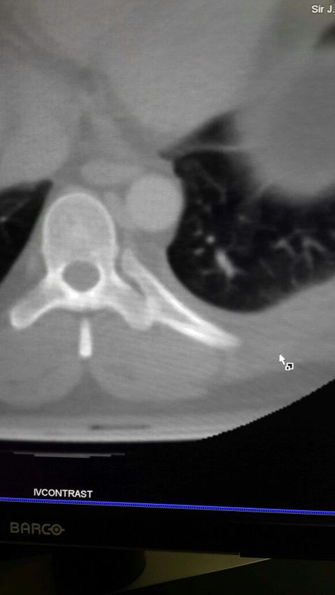
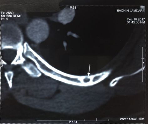
bone sclerosis of the posterior part of the left 6th rib. CT examinations Osteoid osteomas are characterized by a well-demarcated core with a
revealed a small (6 × 6 mm) well demarcated osteosclerotic lesion in typical size of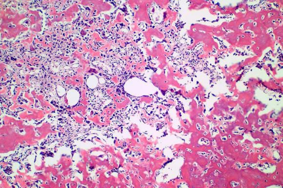
Nagre SW (2018) Osteoid osteoma of the ribs-Is image intensifier or bone scintigraphy a mandatory diagnostic tool-A case report with Review of literature

                 a                                                                           b
                  Figure 1. a) Small (6 × 6 mm) well demarcated osteosclerotic lesion in the posterior end of the left 6th rib in propt CT scan b) Post opt CT scan.

                   a                                                                           b
                                                         Figure 2. a) Intra Operative sixth rib, b) After closure of incision.

                                                   Figure 3. a) No osteosclerotic lesion 6 rib b) Osteosclerotic lesion seen 7 rib.

                                                           Figure 4. Resected rib segments a) Sixth rib b) Seventh rib.

Otorhinolaryngol Head Neck Surg, 2018         doi: 10.15761/OHNS.1000163                                                                                               Volume 3(1): 2-4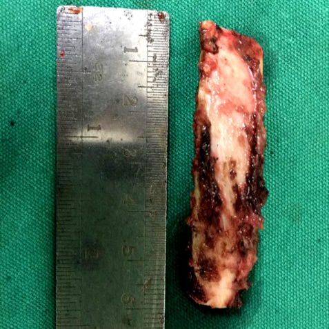
Nagre SW (2018) Osteoid osteoma of the ribs-Is image intensifier or bone scintigraphy a mandatory diagnostic tool-A case report with Review of literature

                                                            Figure 5. Chest X ray a) Preopt   b) Postopt.

                                                      Figure 6. Histopath seventh rib under low and high power.

drugs and salicylates. The lesions most commonly involve the posterior                   As per the available literature of the 15 cases of rib osteomas
or posterolateral shaft of the rib and, upon imaging, present with                   reported so far, we have come to the following consensus [3-16] as in
similar features to tumours located elsewhere in the body. Imaging                   Table 1.
typically reveals a small radiolucent lesion with a thick sclerotic
                                                                                         As per the above Table 1, a total of 8 authors used bone
margin of reactive bone. Due its accuracy in detecting the nidus, CT
                                                                                     scintigraphy; of which Pirayesh et al. [16] used bone scintigraphy pre,
is the preferred imaging technique used for the assessment of osteoid                intra as well as post operatively [16]. A total of 3 authors used both
osteomas [3,7,15,18]. When the osteoid osteoma occurs in the posterior               intra operative C arm image intensifier as well as pre-operative skeletal
portion of the rib near the spine, it may lead to scoliosis [19]                     scintigraphy [3,4,8]. C arm was used by 3 authors, but that was always
    Both conservative and operative techniques have been reported                    in combination with the skeletal scintigraphy [3,4,12]. No localizing
for the treatment of OO. Conservative treatment consists of NSAID                    diagnostic modality was used by 3 authors [8,15]. Pre-operative CT
administration for a prolonged period of time, which reportedly                      imaging was used by all authors [3-16]. No data was available in case
leads to alleviation of pain in select cases. The common principle for               of 4 reports [5,9,11,13]. In the case reported by Veluvolu et al. [7], CT
operative treatment of OO is thorough resection of the nidus, but                    scan had missed the lesion pre-operatively [7], that later had to be
incomplete resection could lead to local recurrence[20]. Although the                confirmed by scintigraphy.
ribs are easily accessed, the reported lengths of en bloc resection of the               The limited data available online shows that preoperative skeletal
nidus ranged from 5 to 9.5 cm, which is significantly wider than the size            scintigraphy, first described in 1980 by Rinsky et al. [17], is the
of the nidus, that have led to varying reports of functional impairments             most relied modality of investigation that authors worldwide have
[3,5-7]. Klonecke et al. [20] reported that intraoperative scanning has              resorted to in order to localize the nidus, and accordingly reduce the
evolved because of the excessively wide excision necessary in the past               amount of material resected. Although some authors have reported
that had to be planned to help guarantee complete removal of the lesion.             the importance of CT scan as being the preferred imaging modality
Here we shall discuss about the importance of the image intensifier or               [3,7,15,18], yet there has been a single instance in which the lesion
skeletal scintigraphy in accurately localising the lesion and leading to             has been missed entirely by CT scan [7]. Intra operative C arm image
minimal excision, instead of a wide local excision.                                  intensifier was always used in combination with pre-operative skeletal

Otorhinolaryngol Head Neck Surg, 2018     doi: 10.15761/OHNS.1000163                                                                         Volume 3(1): 3-4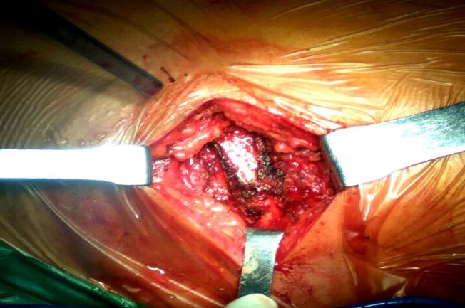
Nagre SW (2018) Osteoid osteoma of the ribs-Is image intensifier or bone scintigraphy a mandatory diagnostic tool-A case report with Review of literature

Table 1. Comparision of various modalities used by surgeon.
 Year of study          Authors                Scintigraphy        C arm image intensifier     CT Scan No used (C arm/scintigraphy)                           No data
     2018              Nagre [19]                    -                         -                   -                     Yes                         Used intra opt Xray of rib
     2015            Deng et al. [3]                Yes                      Yes                  Yes                     -                                      -
     1983            Kehl et al. [4]                Yes                      Yes                  Yes                     -                                      -
     1984          McGuire et al. [5]                -                         -                  Yes                     -                                     Yes
     1988          Mehdian et al. [6]               Yes                      No                   Yes                     -                                      -
     1992          Veluvolu et al. [7]              Yes                      No                   Yes                     -                                      -
     1958           Mauer et al. [8]                 -                         -                  Yes                    Yes                                     -
     1986           Lynch et al. [9]                 -                         -                  Yes                     -                                     Yes
     1987          Nelson et al. [10]               Yes                        -                  Yes                     -                                      -
     1989         Kozlowski et al. [11]              -                         -                  Yes                     -                                     Yes
     1991          Cossetto et al. [12]             Yes                      Yes                  Yes                     -                                      -
     1993          Hoeffel et al. [13]               -                         -                  Yes                     -                                     Yes
     1996        McDermott et al. [14]              Yes                        -                  Yes                     -                                      -
     1996          Kargar et al. [15]                -                         -                  Yes                    Yes                                     -
     2012          Pirayesh et al. [16]             Yes                        -                  Yes                     -                                      -

scintigraphy, but never in an isolated fashion [3,4,12]. This shows the                      4. Kehl DK, Alonso JE, Lovell WW (1983) Scoliosis secondary to an osteoid-osteoma of
                                                                                                the rib. A case report. J Bone Joint Surg Am 65: 701-703. [Crossref]
growing reliance of authors on the efficacy of pre-operative skeletal
scintigraphy, while intra operative C arm imaging remains as a guiding                       5. Nagre SW, Bhosle KN, Gawali R, Ravikumar V (2017) Innovative use of contegra
factor for surgeons inside the operating room.                                                  valved conduit in left iliocaval stent thrombosis. Indian J Thorac Cardiovasc Surg: 1-4.

                                                                                             6. Mehdian H, Summers B, Eisenstein S (1988) Painful scoliosis secondary to an osteoid
    As far as our case is concerned, we are in the league of 2 previous                         osteoma of the rib. Clin Orthop Relat Res: 273-276. [Crossref]
authors who have attempted to resect the lesion with the guidance
                                                                                             7. Nagre SW (2016) Hurdles for Starting Ministernotomy Aortic Valve Replacement
of only computed tomography [8,15]; though we have been able to                                 Program. J Cardiovasc Med Cardiol 3: 035-037.
resect the tumour with negative margins, yet we had to do an extended
                                                                                             8. MAUER I (1958) Osteoid osteoma of the 12th rib; resection under local anesthesia; a
resection, in the absence of intra operative guidance.
                                                                                                case report. Mil Med 122: 194. [Crossref]

Conclusion                                                                                   9. Nagre SW Bendre S (2016) New four patch repair [Modified Brom’s] technique for
                                                                                                supravalvular aortic stenosis. J Cardiovasc Dis Res 7: 39.
     We feel that more comprehensive data regarding the efficacy and
                                                                                             10. Nelson MC, Brower AC, Ragsdale BD (1987) Case report 448. Osteoid osteoma of
reliability of skeletal scintigraphy and intra operative C arm imaging                           left 6th rib with inflammatory reaction in the adjacent pleura and hyperostosis of the
needs to be published, with a comparison of both with computed                                   adjacent ribs. Skeletal Radiol 16: 601-603. [Crossref]
tomography. This data might in future establish skeletal scintigraphy,                       11. Nagre SW, Bhosle KN, Bendre S Vignesh R (2017) Surgical Management of Medium
with or without the associated intra operative C arm image intensifier,                          to Large Size Pulmonary AV Malformations- A Case Series. J Clin Respir Dis Care 3: 129.
as the default and most preferred diagnostic modality for this rare
                                                                                             12. Cossetto D, Cummine J (1991) Rapidly growing twelfth rib osteoid osteoma. Aust N Z
tumour. It was always better to use these modalities intraoperatively to                         J Surg 61: 862-864. [Crossref]
prevent morbidity to patient as well as to the surgeon. So our conclusion
                                                                                             13. Nagre SW (2017) Mobile Left Atrial Mass-Clot or Left Atrial Myxoma. J Cardiovasc
was - use any of available modality intraoperatively was compulsory.                             Dis Res 8: 31-34.

Consent                                                                                      14. McDermott MB, Kyriakos M, McEnery K (1996) Painless osteoid osteoma of the rib in
                                                                                                 an adult. A case report and a review of the literature. Cancer 77: 1442-1449. [Crossref]
    Informed consent has been obtained
                                                                                             15. Nagre SW, Nagre MS (2015) Observational Study of Surgical Closure of Ostium Primum
                                                                                                 Atrial Septal Defect in Thirty Paediatric Patients. Ann Woman Child Health 1: 1-4
Ethical approval
                                                                                             16. Pirayesh E, Amoui M (2012) A rare presentation of osteoid osteoma in a rib and
    All procedures performed in studies involving human participants                             unexpected “double density sign”: A case report and review of literature. Iran J Radiat
were in accordance with the ethical standards of the institutional and/                          Res 10: 197-199.
or national research committee and with the 1964 Helsinki declaration                        17. Nagre SW (2015) Surgical removal of embolised atrial septal defect device from
and its later amendments or comparable ethical standards.                                        pulmonary artery. J Thorac Cardiovasc Surg 150: e55-e57.

                                                                                             18. Touraine S, Emerich L, Bisseret D, Genah I, Parlier-Cuau C, et al. (2014) Is pain
References
                                                                                                 duration associated with morphologic changes of osteoid osteomas at CT? Radiology
1. Jaffe HL (1935) Osteoid osteoma. A benign osteoblastic tumor composed of osteoid              271: 795-804. [Crossref]
   and atypical bone. Arch Surg 31: 709-728.
                                                                                             19. Nagre SW, Bhosle KN, Shaikh A, Ravikumar V (2018) Pleuropulmonary blastoma-a
2. Unni KK (1996) Osteoid osteoma. In: Dahlin’s bone tumors: general aspects and data            case report and review of literature. Indian J Thorac Cardiovasc Surg 3: 72-75.
   on 1,087 cases. 5th edition. Lippincott-Raven, Philadelphia, PA, USA, pp: 121-130.
                                                                                             20. Mizuno S, Anazawa U, Hotta H, Asano N, Susa M, et al. (2015) A Rare Case of an
3. Deng Z, Ding YI, Hao L, Yang F, Gong L, et al. (2015) Osteoid osteoma of the rib: A           Osteoid Osteoma of the Rib Treated under Computed Tomography Guidance: A Case
   report of two cases. Oncol Lett 9: 1857-1860. [Crossref]                                      Report and Review of the Literature. Case Rep Oncol 8: 509-514. [Crossref]

Copyright: ©2018 Nagre SW. This is an open-access article distributed under the terms of the Creative Commons Attribution License, which permits unrestricted
use, distribution, and reproduction in any medium, provided the original author and source are credited.

Otorhinolaryngol Head Neck Surg, 2018           doi: 10.15761/OHNS.1000163                                                                                           Volume 3(1): 4-4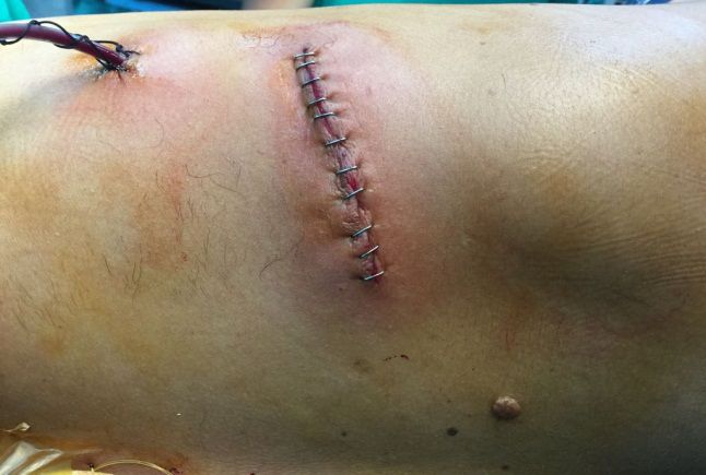
You can also read