Rapidly Expanding Pediatric Post Radiation Brainstem Cavernoma Presenting with Singultus - Cureus
←
→
Page content transcription
If your browser does not render page correctly, please read the page content below
Open Access Case
Report DOI: 10.7759/cureus.4157
Rapidly Expanding Pediatric Post Radiation
Brainstem Cavernoma Presenting with
Singultus
Troy Dawley 1 , Gary Rajah 2 , William Kupsky 3 , Abilash Haridas 2
1. Neurosurgery, St. John Providence Hospital, Southfield, USA 2. Neurosurgery, Wayne State University
School of Medicine, Detroit, USA 3. Pathology, Wayne State University School of Medicine, Detroit, USA
Corresponding author: Troy Dawley, tcdawley@gmail.com
Disclosures can be found in Additional Information at the end of the article
Abstract
Here we present a pediatric patient status post resection of a primitive neuroectodermal tumor
(PNET) with cranial/spinal radiation and development of a medullary cavernoma seven years
after radiation therapy. The patient’s cavernoma demonstrated rapid symptomatic growth in
six weeks resulting in the presentation of intractable hiccups (singultus). The patient
underwent resection of the cavernoma with cessation of the hiccups. We also review the
pathology and possible mechanisms of such rapid growth of this post-radiation cavernoma as
well as advise surveillance for patients with such lesions, as their course may be different from
that of sporadic cavernomas.
Categories: Pediatrics, Radiation Oncology, Neurosurgery
Keywords: post radiation, cavernoma, singultus, pnet
Introduction
There is a well-described link between early radiation therapy in children (prior to 10 years)
and future cavernoma development with a median time to development of post radiation
cavernoma of ten and a half years (3000 cGy) [1].
Medullary cavernomas have been previously reported to be associated with singultus [2]. Here
we describe a pediatric patient with an intramedullary cavernoma presenting with singultus,
and discuss why post-radiation brainstem lesions may follow a natural history different from
that of incidental brainstem cavernous malformations. Informed consent was obtained from all
individual participants included in the study.
Received 12/05/2017
Review began 03/04/2018 Case Presentation
Review ended 02/25/2019
Published 02/28/2019
A 14-year-old male presented with medically refractory hiccups and vomitus with a history of a
© Copyright 2019 post-radiation medullary cavernoma that acutely enlarged with significant surrounding edema.
Dawley et al. This is an open access Originally, he presented at five years of age after a fall, and was incidentally found to have a
article distributed under the terms of
right temporo-parietal and posterior fossa melanotic primitive neuroectodermal tumor (PNET,
the Creative Commons Attribution
License CC-BY 3.0., which permits Figure 1). He underwent gross total resection and was treated with adjuvant chemotherapy and
unrestricted use, distribution, and radiation.
reproduction in any medium, provided
the original author and source are
credited.
How to cite this article
Dawley T, Rajah G, Kupsky W, et al. (February 28, 2019) Rapidly Expanding Pediatric Post Radiation
Brainstem Cavernoma Presenting with Singultus. Cureus 11(2): e4157. DOI 10.7759/cureus.4157
FIGURE 1: Presenting magnetic resonance imaging (MRI) prior
                                     to cavernoma formation
                                     (a) T2 axial MRI demonstrating a large R parietal lesion consistent with melanotic primitive
                                     neuroectodermal tumor (PNET), which did undergo gross total resection. (b) T2 axial MRI from
                                     2007 demonstrating no lesions in the right medullary region.

                                   The amount of radiation received was 3600 cGy to the entire neuroaxis with a 5580 cGy boost to
                                   the tumor field. Approximately seven years after radiation, he presented with intermittent
                                   hiccups for two weeks. A brain magnetic resonance imaging (MRI) revealed a 4 mm medullary
                                   cavernoma that had minimal mass effect or edema present (Figure 2). The hiccups were
                                   managed medically for over three years. This included an extensive gastrointestinal workup,
                                   thoracic bracing, and behavioral modifications, and several medications.

                                     FIGURE 2: Right medullary cavernoma initially diagnosed on

2019 Dawley et al. Cureus 11(2): e4157. DOI 10.7759/cureus.4157                                                                     2 of 6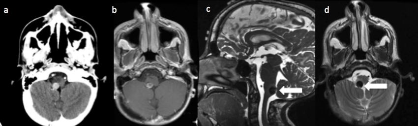
routine follow-up imaging seven years after the initial
                                     resection of the primitive neuroectodermal tumor (PNET)
                                     Right medullary cavernoma size over time. (a/b): Axial T1 and T2 images depicting first
                                     radiographic evidence of the right medullary cavernoma (white arrow), with some T1 with contrast
                                     enhancement seven years after initial surgery/radiation for melanotic PNET. (c/d): Axial T1 and T2
                                     images depicting one-year follow-up study after initial cavernoma findings demonstrating less T1
                                     with contrast enhancement, and small increase in size on T2 image (white arrow). (e/f): T1 and T2
                                     images depicting cavernoma six weeks prior to presentation with intractable hiccups. The pattern of
                                     enhancement on T1 image is now ring like, but the overall size on T2 image is slightly smaller.
                                     (g): Post-resection T1 with contrast revealing complete resection of previous cavernoma (white
                                     arrow).

                                   At the age of 14, he then presented to our emergency department with singultus and vomiting
                                   for three days. His singultus was refractory to medical management and hindered his ability for
                                   oral intake. This disrupted his normal breathing synchrony as well as sleep pattern. Repeat MRI
                                   imaging (Figure 3) showed that the cavernoma had acutely enlarged from 6 mm to 10 mm over a
                                   six-week period with significant surrounding edema. The persistent hiccups and radiological
                                   growth prompted surgical intervention. He underwent a midline suboccipital craniotomy and
                                   partial C1 laminectomy.

                                     FIGURE 3: Presenting magnetic resonance imaging (MRI) after
                                     worsening hiccups
                                     (a) Computed tomography (CT) with contrast showing 1 cm enhancing lesion in R posterior medulla
                                     (40% increase in size from six weeks prior). (b) T1 with contrast axial MRI showing enhancing
                                     portion of cavernoma. (c) T2 high-resolution sagittal MRI depicting cavernoma (white arrow)
                                     location in lower posterior, right medulla. (d) T2 axial MRI showing the cavernoma (white arrow) with
                                     surrounding brainstem edema.

                                   The lesion was approached using a right lazy hockey stick durotomy and a subtonsillar
                                   approach. Arachnoid dissection of the right tonsil allowed elevation off the medulla. Pial
                                   representation was seen on the lateral wall of the obex and the right lower mid-medulla. This
                                   corridor was opened sharply and circumferential dissection of the cavernoma was performed
                                   with motor and somatosensory evoked potential monitoring. The surrounding hemosiderin-
                                   stained tissue was left in place. Please refer to Video 1.

2019 Dawley et al. Cureus 11(2): e4157. DOI 10.7759/cureus.4157                                                                         3 of 6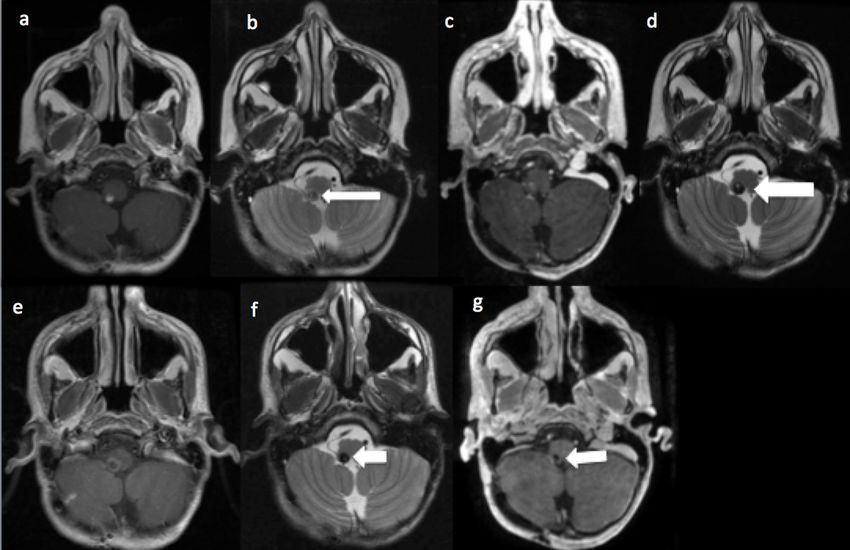
VIDEO 1: Surgical video
View video here: https://youtu.be/mSzrxC79W0k
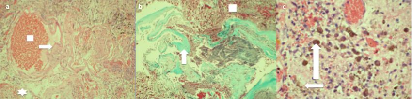
Histopathological examination revealed single layer endothelium-lined channels with
intervening glial tissue and immature granulation tissue displaying the growing nature of this
lesion (Figure 4). Post-operative imaging revealed total resection of the cavernoma and his
hiccups ceased immediately. The post-operative course was routine and he was discharged
home in two days.
FIGURE 4: Histopathological examination
(a) Hematoxylin and eosin stain from resected medullary cavernoma displaying large single layered
endothelium-lined channels (arrow) with red blood cells within (box). Small amounts of intervening
glial tissue are present suggesting the growing nature of the lesion (star). (b) Gomori trichrome
stain with collagen stained green showing large sclerotic vascular channels (arrow). Surrounding
immature granulation tissue suggests a growing nature of the lesion (square). (c) High power
hematoxylin and eosin field revealing granulation tissue (small arrow), with abundant hemosiderin-
laden macrophages (long arrow).
Discussion
Prior brain radiation has several long-term effects including secondary tumors and cavernomas.
One large series found a 3.4% incidence of cavernomas following radiotherapy for brain tumors
[3]. Brainstem cavernomas can present with a variety of symptoms, rarely including
singultus. Spontaneous and familial medullary cavernomas resulting in singultus, while not
common, have also been described with one review by Lee et al. reporting six patients with a
mean age of 34 years (26-40) undergoing surgery [2]. This review found an incidence of hiccups
ranging from 3 to 27% with medullary lesions, lesion size ranged from under 1 cm to 2.2 cm and
hiccup duration ranged from 15 days to three years, with all patients having a sudden onset. All
hiccups resolved post operatively in this review.
2019 Dawley et al. Cureus 11(2): e4157. DOI 10.7759/cureus.4157 4 of 6Surgery is the treatment of choice for symptomatic brainstem cavernomas coming to the pial
surface. A large series of 100 patients by Porter et al. [4] reported 87% of patients undergoing
resection were the same or better. Permanent morbidity occurred in 12%. Surgical approaches
to the brainstem are numerous, and in a study of 300 cases, the most common approach is the
suboccipital craniotomy with or without the telovelar approach. Brown et al. have described the
selection of a surgical approach as a “two-point method” where the first point is in the middle
of the lesion and the next is the area where it comes to/nearest to the pial surface. An extension
of this line is the approach to the lesion [5]. Radiation therapy has been reported for deep
seated lesions, however, its efficacy is debated given a two-year latency period where the
hemorrhage rate in one study was 8.8% per year [6].
The mechanism of medullary cavernoma and hiccups is debated, but many theories exist
including (1) disruption of the GABA inhibitory control via the raphe magnus nucleus of the
hiccup arc [7], (2) irritation of the medullary reticular activating system, near nucleus ambiguus
and the obex [8], and lastly (3) involvement of Mollaret’s myoclonic triangle and the olive [2].
Our surgical approach was planned with regards to this anatomy. The cavernoma described
herein came to the surface at the obex and more posteriorly in the region of the inferior
cerebellar peduncle, dorsal vagal motor nuclei, and nucleus tractus solitarius. We decided to
avoid entering though the obex to avoid further disruption at the area postrema and leave the
ventricular surface intact. The location allowed for access via the cerebellomedullary fissure,
and pial entry just below the inferior loop of the tonsillomedullary portion of the right posterior
inferior cerebellar artery (PICA).
A large literature review recommends only offering surgery to patients who have at least one
episode of hemorrhage and have pial representation [9]. Many factors that have been
determined to help predict outcomes include large lesion size, crossing midline, older age,
presence of developmental venous anomaly, and a larger time interval from hemorrhage to
surgical intervention [10].
Conclusions
This report has detailed a rare rapidly expanding post radiation medullary cavernoma in a
pediatric patient resulting in singultus. This can be extremely distressing and debilitating and
prompt treatment is necessary. Although surgery in the brainstem is technically challenging, it
remains a safe and viable option for symptomatic cavernomas that come to the pial surface.
Additional Information
Disclosures
Human subjects: Consent was obtained by all participants in this study. Wayne State
University issued approval N/A. The IRB approval is waived for this since it is a retrospective
case presentation. There is no identifying information within the article. Patient approval was
obtained before submission. Conflicts of interest: In compliance with the ICMJE uniform
disclosure form, all authors declare the following: Payment/services info: All authors have
declared that no financial support was received from any organization for the submitted work.
Financial relationships: Abilash Haridas, MD declare(s) royalties from UptoDate. Educational
Royalty. Other relationships: All authors have declared that there are no other relationships
or activities that could appear to have influenced the submitted work.
References
1. Heckl S, Aschoff A, Kunze S: Radiation-induced cavernous hemangiomas of the brain: a late
effect predominantly in children. Cancer. 2002, 94:3285-3291. 10.1002/cncr.10596
2. Lee K-H, Moon K-S, Jung M-Y, Jung S: Intractable hiccup as the presenting symptom of
2019 Dawley et al. Cureus 11(2): e4157. DOI 10.7759/cureus.4157 5 of 6cavernous hemangioma in the medulla oblongata: a case report and literature review. J
Korean Neurosurg Soc. 2014, 55:379-382. 10.3340/jkns.2014.55.6.379
3. Burn S, Gunny R, Phipps K, Gaze M, Hayward R: Incidence of cavernoma development in
children after radiotherapy for brain tumors. J Neurosurg. 2007, 106:379-383.
10.3171/ped.2007.106.5.379
4. Porter RW, Detwiler PW, Spetzler RF, Lawton MT, Baskin JJ, Derksen PT, Zabramski JM:
Cavernous malformations of the brainstem: experience with 100 patients . J Neurosurg. 1999,
90:50-58. 10.3171/jns.1999.90.1.0050
5. Brown A, Thompson B, Spetzler RF: The two-point method: evaluating brain stem lesions .
BNI. 1996, 12:
6. Kondziolka D, Lunsford LD, Flickinger JC, Kestle JR: Reduction of hemorrhage risk after
stereotactic radiosurgery for cavernous malformations. J Neurosurg. 1995, 83:825-831.
10.3171/jns.1995.83.5.0825
7. Oshima T, Sakamoto M, Tatsuta H, Arita H: GABAergic inhibition of hiccup-like reflex
induced by electrical stimulation in medulla of cats. Neurosci Res. 1998, 30:287-293.
10.1016/S0168-0102(98)00011-X
8. Arita H, Oshima T, Kita I, Sakamoto M: Generation of hiccup by electrical stimulation in
medulla of cats. Neurosci Lett. 1994, 175:67-70. 10.1016/0304-3940(94)91079-0
9. Gross BA, Batjer HH, Awad IA, Bendok BR, Du R: Brainstem cavernous malformations: 1390
surgical cases from the literature. World Neurosurg. 2013, 80:89-93.
10.1016/j.wneu.2012.04.002
10. Walcott BP, Choudhri O, Lawton MT: Brainstem cavernous malformations: natural history
versus surgical management. J Clin Neurosci. 2016, 32:164-165. 10.1016/j.jocn.2016.03.021
2019 Dawley et al. Cureus 11(2): e4157. DOI 10.7759/cureus.4157 6 of 6You can also read