Clinical presentation and prognostic indicators in 100 adults and children with neurofibromatosis 1 associated non-optic pathway brain gliomas
←
→
Page content transcription
If your browser does not render page correctly, please read the page content below
J Neurooncol (2017) 133:609–614
DOI 10.1007/s11060-017-2475-z

    CLINICAL STUDY

Clinical presentation and prognostic indicators in 100 adults
and children with neurofibromatosis 1 associated non-optic
pathway brain gliomas
Susan Byrne1,2,3,4 · Steve Connor5,6 · Karine Lascelles1,3 · Ata Siddiqui5,6 ·
Darren Hargrave7 · Rosalie E. Ferner1,2,4

Received: 7 August 2016 / Accepted: 14 May 2017 / Published online: 7 June 2017
© The Author(s) 2017. This article is an open access publication

Abstract Type 1 Neurofibromatosis (NF1) is a common                    5 years of diagnosis of glioma. Serial neuroimaging was
autosomal dominant condition, with a major impact on the               undertaken in 88 patients. In 66 (75%), the lesion on the
nervous system, eye, bone, and skin, and a predisposition              scan was stable or had improved at follow-up. High-grade
to malignancy. At present it is not possible to predict clini-         lesions were present in five patients and were strongly
cally or on imaging, whether a brain tumour will remain                associated with tumours in the thalamus (p = 0.001). Five
indolent or undergo high-grade change. There are no con-               patients died during follow-up. The diagnosis of high-grade
sensus guidelines on the follow-up of non-optic pathway                glioma had a HR of 99.7 (95% CI 11.1–898.9, p < 000.1)
glioma (non-OPG) tumours in NF1. One hundred patients                  on multivariate Cox regression to evaluate predictive fac-
from the National NF1 Service with generalised NF1 and                 tors related to death. In our cohort of 100 patients with
a diagnosis of non-OPG glioma were followed up for a                   NF1, we have shown that tumours in the thalamus are more
median time of 63 months after glioma detection. Forty-                likely to be associated with radiological progression, high-
two patients underwent surgical intervention. Ninety-                  grade tumours, and surgical intervention. As a result of this
one percent (38) of those requiring surgery did so within              finding, heightened surveillance with more frequent imag-
                                                                       ing should be considered in thalamic involvement. We have
                                                                       also demonstrated that over 40% of patients underwent sur-
Electronic supplementary material The online version of this           gery, and did so within 5 years of tumour diagnosis. Serial
article (doi:10.1007/s11060-017-2475-z) contains supplementary
material, which is available to authorized users.
                                                                       imaging should be undertaken for at the very least, 5 years
                                                                       from tumour detection.
* Susan Byrne
  Suabyrne@gmail.com                                                   Keywords Neurofibromatosis type 1 (NF1) · Brain
1                                                                      tumour · Pilocytic astrocytoma · Epidemiology ·
     National Neurofibromatosis Service, Guy’s and St. Thomas’
     NHS Foundation Trust, London, UK                                  Neurooncology · Thalamic tumour
2
     Department of Neurology, Guy’s and St. Thomas’ NHS
     Foundation Trust, London, UK
3                                                                      Background and aims
     Department of Paediatric Neurology, Evelina Children’s
     Hospital, Guy’s and St. Thomas’ NHS Foundation Trust,
     London, UK                                                        Type 1 Neurofibromatosis (NF1) is a common autosomal
4
     Institute of Psychiatry, Psychology, and Neuroscience, Kings      dominant condition, with a major impact on the nervous
     College London, London, UK                                        system, eye, bone, and skin. NF1 is a tumour suppressor
5
     Department of Neuroradiology, Guy’s and St. Thomas’ NHS           disorder that is typified by benign nerve sheath tumours
     Foundation Trust, London, UK                                      and a predisposition to malignancy. Lack of the NF1 gene
6
     Department of Neuroradiology, King’s College Hospital             product neurofibromin arising from gene mutation causes
     NHS Foundation Trust, London, UK                                  activation of the ras pathway and unrestrained cell growth
7
     Department of Paediatric Oncology, Great Ormond Street            [1, 2]. The natural history of NF1-associated tumours
     Hospital, London, UK

                                                                                                                         13
                                                                                                                    Vol.:(0123456789)
610 J Neurooncol (2017) 133:609–614 differs from the general population [3], and NF1 individu- the anatomical location of the tumour (2) unrelated—if the als require specialist assessment and management. MRI was undertaken by the referring institution for screen- Low-grade brain gliomas are a well-recognized com- ing purposes (although this is not our practice) or for symp- plication of NF1 and they may remain indolent over many toms unrelated to anatomical location of the tumour. years; however, a small number of low-grade gliomas trans- Extended disability scores (EDSS) [6] were calculated form into more aggressive tumour types [4]. At present it is using reported examination at each visit. Information col- not possible to predict clinically or on imaging, whether a lected is outlined in supplementary table 1. tumour will remain indolent or undergo high-grade change. Data analysis was carried out using SPSS 22 [IBM Another small subset of patients may present acutely with SPSS Statistics for Macintosh, Version 22.0]. Parametric a high-grade brain tumour without ever having had a low- tests were carried out where the data were normally dis- grade glioma identified. A study by Guillamo et al. looked tributed, and non-parametric tests were used if the data at brain gliomas in NF1 and identified that extra-optic loca- were not normally distributed. Chi Square testing or Fish- tion, tumours diagnosed in adulthood, and symptoms at er’s exact testing was used to compare two the relation- diagnosis are associated with a higher mortality [4]. ship between two categorical variables. Survival analysis, Two decades ago it was common practice to treat these and time to event analysis was carried out using univari- low-grade tumours aggressively, however, more recent ate and Cox multivariate survival analysis, with time epidemiological studies have demonstrated an association variable being time from glioma diagnosis to the event between radiotherapy and subsequent malignant transfor- in question (e.g. surgery, death) or censor date. Statistical mation [3], endocrine sequelae, neurovascular, and cogni- significance was set at
J Neurooncol (2017) 133:609–614 611
Location of tumours can be seen in Fig. 1. Diagnosis
was made on neuroimaging only in 58/100, and on histo-
logical analysis and neuroimaging in 42/100.
Surgical intervention
Forty-two of the 100 patients underwent surgical interven-
tion during follow-up; initial surgical intervention was total
or subtotal resection of tumour in 29, biopsy only in 6, and
shunt or ventriculostomy only in 7 individuals.
Twenty-four patients had surgery for relief of symptoms,
ten patients had surgery because of concern regarding the
radiological features of the tumour, and in eight cases the
specific reason for surgery was not stated in the medical
notes.
Ten patients went on to have further surgical interven-
tion at a later stage. Of the ten who had further surgery, five
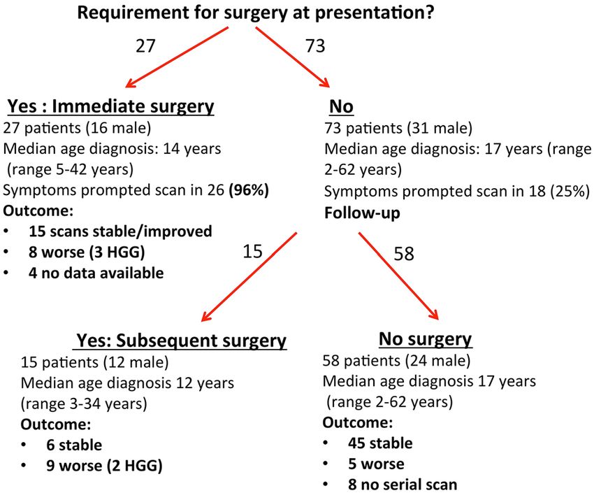
required debulking (initial surgery was debulking in three, Fig. 2 Flow diagram of the requirement for surgery in 100 patients
with NF1 related glioma (immediate surgery refers to surgery within
initial surgery was shunt/ventriculostomy in two), three 2 months of initial presentation, HGG high grade glioma)
required shunt revision (initial surgery was ventriculos-
tomy/shunt for all three), one required a shunt (initial sur-
gery was a biopsy), and one required a shunt and Omaya Histology
reservoir insertion (initial surgery was a biopsy).
The requirement for surgical intervention was much Formal histology results were available on 32 patients: 23
higher in the cohort of patients who had a scan because of pilocytic astrocytoma, 5 glioblastoma multiforme, 3 gangli-
glioma-related symptoms at presentation (31 of 44; 71%), oglioma, and 1 pilomyxoid glioma (grade 2). One of our
compared to those who had an MRI for screening or other patients underwent molecular analysis and had a BRAF
reasons (11 of 56), p < 0.0001. Symptomatic reasons for V600E mutation.
surgical intervention included seizures, raised intracranial High-grade lesions were present in five patients and
pressure, rapid increase in size of tumour, and hemiparesis. were strongly associated with tumours in the thalamus
Twenty-seven of 42 patients (64%) requiring surgical compared with other regions (4/12 compared to 1/88;
intervention did so within 2 months of presentation, and p = 0.001). Three patients with HGG presented acutely with
38/42 (91%) had surgery within 5 years of diagnosis of gli- symptoms; two with headache and signs of papilloedema,
oma. Only four patients (9%) had surgery beyond 5 years, and one with a new cranial nerve palsy and ataxia. Only
three because of radiological progression and one because two of the patients with HGG had previous imaging, both
of symptoms (see Fig. 2). of which showed lesions involving the thalamus—one of
Multivariate Cox regression was performed looking at the patients had previous debulking of a frontal/thalamic
predictive factors for surgery. Significant variables were lesion, and the histology showed a pilomyxoid astrocytoma
thalamic involvement [Hazard ratio (HR) 2.18, 95% CI before transforming to a HGG.
1.01–4.6, p = 0.049], frontal involvement (HR 3.36, 95% Details of the five patients with high-grade glioma are
CI 1.49–7.6, p = 0.004), and cerebellar involvement (HR outlined in Table 1.
4.44, 95% CI 2.0–9.8, p < 0.0001). Presence of a high-
grade glioma was also a significant risk factor for surgery
(HR 3.46, 95% CI 1.33–9, p = 0.011). Age at diagnosis was Survival analysis and outcome
not a significant variable (HR 0.975, 95% CI 0.94–1.004,
p = 0.087). Five of the 100 patients died during follow-up; two from
Three patients required surgical intervention, radio- progression of a low grade tumour to a high-grade glioma,
therapy and chemotherapy, four had surgery and chemo- two presented acutely with a high grade tumour in the first
therapy, and four had surgery and radiotherapy. None had instance, and one patient developed cerebellar necrosis fol-
chemotherapy alone. Of the 11 patients who received more lowing radiotherapy. The survival rate from diagnosis was
than one treatment modality, eight patients had multimodal 95% at 5 years (95% CI 92.6–97.4%). On multivariate Cox
treatment initially, and three patients had additional treat- regression to evaluate predictive factors related to death,
ments added after progression. the diagnosis of high grade glioma had a HR of 99.7 (95%
13612
13
Table 1 Clinical presentation, histology, treatment and outcome in five NF1 patients with high-grade gliomas
Age glioma Reason for scan Location Intervention Histology Alive/time to death from diagnosis
diagnosis
28 years Facial nerve palsy Left medial frontal cortex, right Craniotomy Grade 2 pilomyxoid astrocytoma Alive, 20 months after grade 4
mid thalamic, right superior Further surgery, temozolomide, Grade 4 astrocytoma GBM diagnosed
colliculus radiotherapy
8 years Baseline imaging Initially asymmetrical thalamic Debulking surgery, temozolomide Grade 4 GBM Died, 9 months after grade 4 GBM
myelination/possible pilocytic diagnosed
astrocytoma on imaging
Headache (2 years later) Bilateral thalamic grade 4 GBM
on next scan
17 years Headaches with papilloedema Right thalamic mass extending to Subtotal resection, temozolomide, Initial histology pilocytic astro- Died, 9 months after presentation
midbrain and into ventricle radiotherapy cytoma with high proliferation
index and atypical features,
revised to grade 4 GBM
6 years New onset ataxia with supe- Mass in left thalamus, caudate Empiric treatment with vincris- Grade 4 GBM Died, 6 months after presentation
rior oblique palsy nucleus, cerebellum, brainstem tine and carboplatin but biopsy
when did not respond
32 years Headaches, ataxia, seizure Right cerebellar mass Ventriculostomy, subsequent Astrocytoma grade 3 Died, 16 months after presentation
debulking, radiotherapy
J Neurooncol (2017) 133:609–614J Neurooncol (2017) 133:609–614 613
CI 11.1–898.9, p < 000.1). There was no significant asso- progressive tumours were not pilocytic astrocytomas but
ciation between age at tumour detection and death. rather other types of astrocytoma, that are associated with
At last follow-up 29/95 survivors had symptoms related a more aggressive histological subtype. Guillamo and
to glioma with an EDSS of 2.2 (SD 1.6). Fifty-seven per- colleagues showed that of 19 progressive tumours; only
cent (21/37) of patients who had surgery had symptoms at nine were pilocytic astrocytomas, with the remaining ten
follow up compared to 14% of patients who did not require tumours comprising two low grade astrocytomas, two ana-
surgery (p < 0.0001). Patients who were symptomatic at the plastic astrocytomas, two glioblastomas, and one dysplastic
time of diagnosis had a higher mean EDSS at most recent neuroepithelial tumour [7].
review (1.5) compared to individuals who had a glioma Leonard and colleagues reported that over half of
diagnosed incidentally (0.5), p = 0.01. patients with unusual clinical presentation or radiographic
Serial neuroimaging was undertaken in 88 patients. features were not classic pilocytic astrocytomas on histo-
The median time between first and most recent imaging logical analysis, and highlighted the value of biopsy in
was 59 months (range 2–453). In 66/88 (75%) the lesion those cases [8].
on the scan was stable or had improved, and 22/88 (25%) This study is limited by the fact that, histological diag-
showed radiological progression. Lesions in the thalamus nosis was only obtained in the minority of cases (32/100),
(p = 0.004) were more likely to undergo radiological pro- with the remaining diagnoses were suspected radiologi-
gression, however this was related to the transformation to cally. Radiological progression (improved/stable/worse)
high grade malignancy. was assessed by experienced neuroradiologists, using linear
Previous history of OPG was present in 25/100 but was measurement and qualitative assessment rather than volu-
not associated with poorer outcome or high-grade tumour. metric analysis.
In NF1 individuals brain lesions that have irregular
enhancement, have marked mass effect, or show restricted
Discussion diffusion are probable gliomas that require judicious clini-
cal and neuro-radiological follow up. There are currently
We have shown that tumours in the thalamus are more no guidelines on the surveillance of NF1 patients once a
likely to be associated with high-grade tumours in NF1. non-optic pathway glioma has been identified. Lack of evi-
As a result of this finding, heightened surveillance with dence makes determining a time interval for serial imag-
more frequent imaging should be considered in patients ing extremely difficult, especially when patient anxiety is
with NF1 who have thalamic involvement. Individuals heightened by an underlying disorder that predisposes to
with tumours in the thalamus, frontal lobes, and cerebel- tumours. We found that all high-grade gliomas and nearly
lum were more likely to require surgical intervention than all tumours requiring surgery did so within 5 years of
patients with tumours in other anatomical locations. At the tumour detection, and that the majority were symptomatic
current time there are no consensus guidelines, and height- at presentation. Serial imaging should be undertaken for at
ened surveillance in this subset of patients may lead to a the very least, 5 years from tumour detection.
decrease in morbidity and mortality. We also found that tumours located in the thalamus
Forty-two percent of patients (n = 42) underwent surgi- were more likely to be associated with high-grade tumours
cal intervention. Predictors of the requirement for surgical (2 cases) and high-grade transformation (2 cases), so that
intervention included symptoms that necessitated cranial location of tumour should direct one toward more frequent
imaging, and thalamic, cerebellar and frontal lesions. We imaging. Krishnatry and colleagues reviewed LGG in over
identified that 64% of patients requiring intervention had 1200 patients and found an association between mortality
surgery within 2 months, and 91% of patients who needed and thalamic lesion, however only 10% of this cohort had
intervention had surgery within 5 years of tumour detec- NF1 and the majority of patients with NF1 had an optic
tion. Overall the outcome was very favourable with 95% pathway glioma, and an excellent outcome [9]. As up to
survival at 5 years, and 92% at 10 years. 65% of patients 85% of children with NF1 have abnormal signal on their
were asymptomatic at follow-up. We did not find an asso- MRI scans (FASIs), and the thalamus is a common location
ciation between the age at tumour diagnosis and need for to see these changes [10], it is important to discuss cases
surgery and there was not association with death; however with an experienced neuroradiologist to ensure that FASI
there was a trend towards significance and this may reflect don’t have any atypical features that may suggest the pres-
the small size of out cohort. ence of a low grade glioma.
The majority of tumours biopsied were pilocytic astro- The patients in this series were referred to a national
cytomas, and only five patients (5%) in this cohort devel- neurofibromatosis centre and may represent a more com-
oped a high-grade glioma. Our findings are supported by plex case mix, which may not be reflective of the NF1
three studies [3, 4, 7] that reported that the majority of population as a whole. However, from analysis of the data
13614 J Neurooncol (2017) 133:609–614
we found that over half of the gliomas were diagnosed Ethical approval This study was approved as a clinical evaluation
when patients were scanned for another reason, and that with study number 5099 by the Clinical Audit Group Committee at
Guy’s and St. Thomas’ NHS Foundation Trust, London.
fewer than one-fifth of patients identified in that way ever
required surgery. These patients also had a better outcome
in terms of EDSS at most recent review. It is difficult to Open Access This article is distributed under the terms of the
Creative Commons Attribution 4.0 International License (http://
tell if symptoms at follow up in those who had surgical creativecommons.org/licenses/by/4.0/), which permits unrestricted
interventions are secondary to tumour or to side effects of use, distribution, and reproduction in any medium, provided you give
surgery. Our practice is to scan patients with clinical signs appropriate credit to the original author(s) and the source, provide a
or symptoms and not to perform baseline neuroimaging, link to the Creative Commons license, and indicate if changes were
made.
which may reveal incidental gliomas.
Under current WHO grading, brain tumours are given
a Grade of I–IV depending on histological features [11].
References
In future the use of molecular genetic findings will aid in
determining the course and prognosis of low-grade tumour 1. Ferner RE (2007) Neurofibromatosis 1 and neurofibromatosis 2:
types and also allow allocation of specific, targeted onco- a twenty first century perspective. Lancet Neurol 6:340–351
logical therapy in these patients. One of our patients was 2. Rauen KA, Schoyer L, McCormick F et al (2010) Proceedings
diagnosed with a cerebellar ganglioglioma and had molec- from the 2009 genetic syndromes of the Ras/MAPK pathway:
from bedside to bench and back. Am J Med Genet A 152A:4–24
ular genetic analysis that revealed a V600E BRAF muta- 3. Pollack IF, Shultz B, Mulvihill JJ (1996) The management of
tion. The BRAF pathway is a key regulator of the MAPK brainstem gliomas in patients with neurofibromatosis 1. Neurol-
pathway [12], and targeted treatment with a BRAF-MEK ogy 46:1652–1660
pathway inhibitor would be a potential therapy. Molecular 4. Guillamo JS, Creange A, Kalifa C et al (2003) Prognostic factors
of CNS tumours in Neurofibromatosis 1 (NF1): a retrospective
genetic analysis of tumours may aid in decisions regarding study of 104 patients. Brain 126:152–160
therapy. 5. Rodriguez FJ, Perry A, Gutmann DH et al (2008) Gliomas
In conclusion, MRI should be undertaken in patients in neurofibromatosis type 1: a clinicopathologic study of 100
with NF1 who have signs and symptoms suggestive of gli- patients. J Neuropathol Exp Neurol 67:240–249
6. Kurtzke JF (1983) Rating neurologic impairment in multiple
oma. Once a glioma is diagnosed any new symptoms indi- sclerosis: an expanded disability status scale (EDSS). Neurology
cating progression should be investigated promptly. Patient 33:1444–1452
education regarding ‘red flag’ symptoms may hasten refer- 7. Molloy PT, Bilaniuk LT, Vaughan SN et al (1995) Brainstem
ral to neurosurgical services. Heightened surveillance with tumors in patients with neurofibromatosis type 1: a distinct clini-
cal entity. Neurology 45:1897–1902
more frequent imaging should be considered in patients 8. Leonard JR, Perry A, Rubin JB, King AA, Chicoine MR, Gut-
with thalamic involvement, and also in those with atypical mann DH (2006) The role of surgical biopsy in the diagnosis
histological findings for at least 5 years. Overall the out- of glioma in individuals with neurofibromatosis-1. Neurology
come is very favourable with the majority of patients being 67:1509–1512
9. Krishnatry R, Zhukova N, Guerreiro Stucklin AS et al (2016)
symptom free at most recent follow-up. Clinical and treatment factors determining long-term outcomes
for adult survivors of childhood low-grade glioma: a population-
Acknowledgements We are most grateful to Dr. Catherine Walker based study. Cancer 122(8):1261–1269
for her help with this project and to Mr. Bhangoo and the neuro- 10. Rosenbaum T, Engelbrecht V, Krolls W, van Dorsten FA,
oncology team for help in clinical management of our patients. Hoehn-Berlage M, Lenard HG (1999) MRI abnormalities in
neurofibromatosis type 1 (NF1): a study of men and mice. Brain
Author contributions SB collected the clinical data, analysed the Development 21:268–273
data and wrote and edited the manuscript. SC analysed the neuroim- 11. Louis DN, Ohgaki H, Wiestler OD et al (2007) The 2007 WHO
aging data, wrote and edited the manuscript. KL collected the clinical classification of tumours of the central nervous system. Acta
data, wrote and edited the manuscript. AS analysed the neuroimaging Neuropathol 114:97–109
data, wrote and edited the manuscript. DH collected the clinical data, 12. Nageswara Rao AA, Packer RJ (2014) Advances in the manage-
and wrote and edited the manuscript. REF runs the National Neurofi- ment of low-grade gliomas. Curr Oncol Rep 16:398
bromatosis Service, designed the study, collected the data, anaylsed
the data and wrote and edited the manuscript.
Compliance with ethical standards
Conflict of interest None of the authors have any conflicts of interest
to declare in relation to this project.
13You can also read