Impacts of Artificial Intelligence and New Technology for Melanoma Detection
←
→
Page content transcription
If your browser does not render page correctly, please read the page content below

Impacts of Artificial Intelligence and New Technology
               for Melanoma Detection
                     S USAN M . S WETTER, M D
                     Professor of Dermatology
                     Director, Pigmented Lesion & Melanoma Program
                     Physician Leader, Cutaneous Oncology Program
                     Stanford University Medical Center and Cancer Institute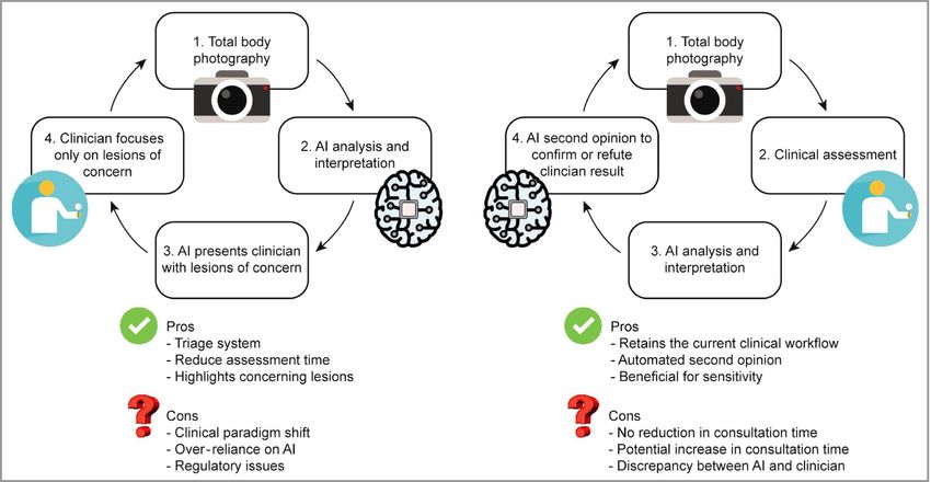
2
    How do we know what we know?

     • Unconscious pattern matching
     • Rules-based systems

                           Systems drive the way we think:
                           System 1 – fast, intuitive, emotional
                           System 2 – slower, more deliberate and logical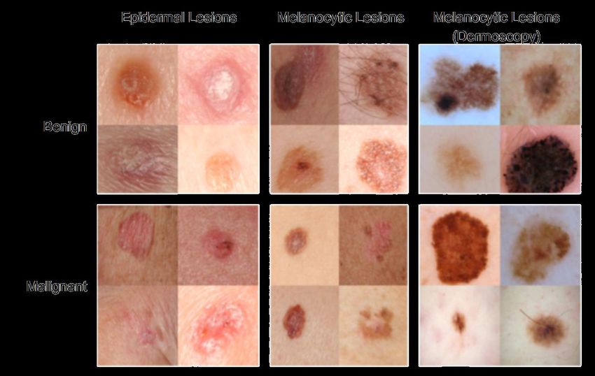
Dual-process theory applied to Dermatologists
System 1 (intuitive) “visual intelligence” – rapid, operates below level
  of perceptible consciousness (“gut feeling”)
  - focuses on pattern recognition: “blink, think” and “10-second rule”                        (Giuseppe
  Argenziano, Naples)
  - why “ugly duckling” rule discriminates melanoma more accurately than
  ABCD clinical warning signs (Gaudy-Marquest C et al. JAMA Dermatol 2017)

System 2 (analytical) – deliberate judgment, based on conscious
  applications of rules acquired through learning

Transition from intuitive to analytical reasoning can hinder clinical
  reasoning and increase diagnostic error

                Pelaccia T et al. Med Educ Online 2011. Stolper E et al. BMC Fam Pract 2009.
                Norman GR, Eva KW. Med Educ 2010.
Skin Cancer Facts
● Skin cancer - most common cancer in the US
● 1 in 5 Americans will develop skin cancer in their lifetime
● Latest estimate (2012): >5.4 million cases BCC/SCC treated in >3.3 million
  persons in the US (Rogers HW et al. Arch Dermatol 2015)
● In 2019 –
   ● estimated >96K new cases of invasive melanoma and >95K melanomas in situ
      in the US
   ● nearly 8000 melanoma-associated deaths
● Survival rate for melanoma is >95% if detected early

                    Siegal RL et al. Cancer Statistics, 2019. CA Cancer J Clin 2019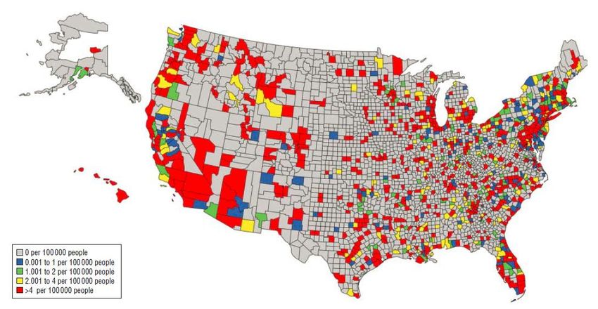
“MELANOMA WRITES ITS MESSAGE IN THE
       SKIN WITH ITS OWN INK
     AND FOR ALL OF US TO SEE”
         -Neville Davis, Queensland, Australia

   …so why is early detection so hard?
Global Health Care Accessibility

       Global access to quality health care
       has improved in the last two
       decades, but the gap between
       countries with the most and least
       effective treatments has grown

          Barber R et al. The Lancet 2017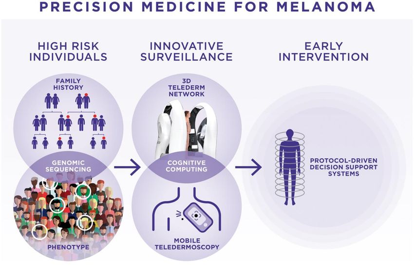
7
         How do we democratize health care access?

    ▪ 6.3 billion smartphones
      globally by 2021
    ▪ Data collection at scale
    ▪ Diagnostics at scale
    ▪ Diminish health disparities

                       Can we use AI to expand access to dermatologists?

                             Aneja S et al. Arch Dermatol 2012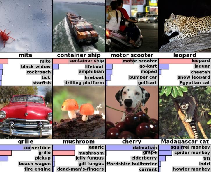
8

    AI has changed our world

                          •    Driverless cars
                          •    Translation capabilities
                          •    Mortgage lending
                          •    Financial markets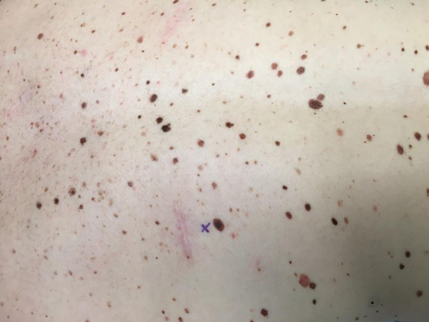
Convolutional Neural Networks
    Multilayer neural networks: involve local connections, shared weights, pooling

• Capture relationship of pixels to each other using filter as a matrix
• Run algorithms over and over until errors are minimized across all images
• Allows for profound level of pattern recognition beyond human brain capability

                   https://ujjwalkarn.me/2016/08/11/intuitive-explanation-convnets/
10
     Advances in AI/Machine Learning/Computer Vision

     Breakthroughs in artificial
       intelligence over past 7
       years

     Convolutional neural
      networks + large
      databases + processing
      power = deep learning

              Krizhevsky, Sutskever, Hinton et al. ImageNet Classification with Deep Convolutional Neural
              Networks, NIPS 2012
11 Algorithms train themselves to recognize what is important and
what is not, without human intervention
- ImageNet project: CNNs
trained to classify 1.4
million high-resolution
images into 1000 different
classes with low error rates
- With sufficient examples
of huskies, chihuahuas,
basal cell carcinomas or
melanomas, algorithms
learn the relevant patterns
of these categories
Hsu et al, 2013Clinical and Dermoscopic Images Spanning Breadth of Disease Constructing the dataset: Nearly 130,000 images: - clinician-labeled and/or biopsy- proven from 18 different, open- access online repositories - clinical data from Stanford Dermatology clinics Biopsy proven images: - University of Edinburgh library - ISDIS ISBI challenge (dermoscopic) Images comprised more than 2,000 dermatologic diseases
Visual Taxonomy – Disease Partitioning Algorithm

      129K images
      2K diseases
Our Objectives Evaluate performance of deep learning algorithms (namely, a single CNN) on classification of cutaneous malignancies Compare to dermatologists 2 main (binary) questions: ▪ Lesion benign or malignant? ▪ Biopsy/treat or reassure?
Methods
 Recruited 21 board-certified dermatologists
 ▪   Stanford University
 ▪   University of Pennsylvania
 ▪   Massachusetts General Hospital/Harvard
 ▪   University of Iowa
 Three 100+ question tests
      a.   Epidermal tumors (BCC/SCC vs SKs)
      b.   Clinical images of melanocytic lesions (melanoma vs benign
           nevi, excluded SKs)
      c.   Dermoscopic images of melanocytic lesions (separate dataset)
Training Objective: build a system that could accommodate significant variation inherent in photographic images (e.g. lighting, zoom, angle) with no pre- processing or lesion segmentation Goal: feed any captured skin image directly into the system and output a classification
Deep Convolutional Neural Network (CNN)

         Inception v3 CNN architecture reprinted from https://research.googleblog.com/2016/03/train-your-own-
         image-classifier-with.html

          - Used GoogleNet Inception v3 CNN architecture pretrained on ImageNet dataset
          - Trained and fine-tuned our CNN using transfer learning
          - Resulted in probability distribution over clinical classes of skin
          - Applied a partitioning algorithm to our taxonomy to define training classes
Sample Test Images Used new dermatologist-labeled dataset of >129K clinical images, including >3300 separate dermoscopic images
Algorithm ready for testing within 1 year

  CNN outputs a malignancy probability for each image
Results for CNN vs Dermatologists

       CNN performed at least as well as dermatologists as a whole
         (Limitation of dermoscopy test as most dermatologists were not experts in
                            pigmented lesions or dermoscopy)

                   Esteva A et a. Nature. 2017;542:115-118.
Limitations of our study
▪ Retrospective
▪ Potential spectrum bias
› Study design→ 2 disease categories
▪ Differences between in-person exam and telederm (“in silico”)
› We “blink, think, and compare” and use dermoscopy to aid diagnosis
▪ Transparency on pathology labels/ dermatopathology accuracy/ wide
variability for melanocytic neoplasms (Elmore JG et al. BMJ 2017)
▪ Opportunities for bias/confounding
› Need to ensure extensive representation by varied skin types
› Extensive additional work is needed
• Prospective clinical validation studies needed23
Limitations of Deep Learning
• System is opaque; we don’t
know why it calls an image
benign or malignant
• Dots, rulers, marks, etc. may
introduce bias into dataset
• Investigators may not be aware
of them or extent
• Impact on MD cognition/learning?Strengths Computer can assess image data imperceptible to human eye ▪ Only looking at specific lesions, not whole-body or sequential change detection Broadly applicable across disciplines ▪ Algorithms will improve with transfer learning ▪ Edges, shapes remain important ▪ Show a skin cancer app cat pictures and it will improve at classifying skin cancer Capable of running on a smartphone device ▪ App created to study this, prospective “real-time” validation in clinical setting
What’s happened with AI in Dermatology since then?
  ▪ At least 8 publications pertaining to AI (CNNs) for:
    › dermoscopy (with or without clinical images), dermatopathology (nod BCC, dermal
      nevi, SK), melanocytic lesions, melanoma, acral melanoma dermoscopy,
      nonmelanoma skin cancer (BCC/SCC), onychomycosis
  ▪ CNN consistently performs as well if not better than “expert”
    clinicians
  ▪ Limitations: datasets lack full spectrum of skin phenotypes and
    lesions (particularly less common banal lesions/disorders)
  ▪ No real-world prospective use/validation as yet

       Gilmore SF. PLoS One 2018; Han SS. PLoS One 2018; Marchetti MA. J Am Acad Dermatol
       2018; Yu C. PLoS One 2018; Haenssle HA. Ann Oncol 2018; Brinker TJ. Eur J Cancer
       2019; Fujisawa Y. Brit J Dermatol 2019; Marka A. BMC Med Imaging 2019
Can AI be used to quantify and monitor skin disease severity?
Context matters
   Dermatologist’s clinical impression based on factors beyond visual and
   dermoscopic examination of a lesion in isolation
      - Sequential imaging, change detection, incorporation of clinical metadata?
Are we comfortable with AI as black box?
A trained neural net does not necessarily
mimic the decision-making approach of
humans
Identifies its own criteria for informative
patterns associated with a diseaseIs AI a Pandora’s box?
• Probably not, and it won’t replace dermatologists!
• Human decisions made based on machine output of probability and other factors!
• Real vs imaginary dangers:
• “AI is a fundamental existential risk for human civilization.” (Elon Musk, 2017)
• “AI software will help us understand biology, understand how to intervene and improve lives very dramatically.”
(Bill Gates, 2018)
• Roles in health care and education
• Regulation and liability need to be addressed proactivelyReal-world, clinical validation of app in progress
“Smart dermoscope”
“Augmented
clinician”
54.8Can AI help dermatologists? -35 Y female with atypical mole syndrome, seven cutaneous melanomas (3 melanomas in situ, 3 T1a invasive melanomas and 1 T2a SLNB-positive melanoma) and ten severely dysplastic nevi diagnosed since 2016 -negative for p16/CDKN2A mutation -followed q 3 mos with total body photography and digital dermoscopy
A tool for surveillance? – NOT YET

                                     New moderately
                                     dysplastic nevi
                                     with focal severe
                                     atypia
Can AI enhance clinical decision-making?
Janda M, Soyer HP. Brit J Dermatol. 2019:180: 247-248.Future of melanoma early detection
Targeted
surveillance
of high risk
individuals
with high
resolution
3D imaging
and
integration
of AI
Mar VJ, Soyer HP. Ann Oncol. 2018;29:1625-1628. (Schematic from Smithers BM, Dunn J and
Soyer HP. Whither melanoma in Australia? Med J Aust 2017;207:330–331.)AI can augment, but not replace decision-making and
human interactions in medicineYou can also read