Case Report Metastatic Papillary Thyroid Carcinoma in a Paediatric Patient
←
→
Page content transcription
If your browser does not render page correctly, please read the page content below
Hindawi
Case Reports in Endocrinology
Volume 2021, Article ID 6655491, 4 pages
https://doi.org/10.1155/2021/6655491

Case Report
Metastatic Papillary Thyroid Carcinoma in a Paediatric Patient

          Nthabiseng Ellen Mothata ,1 Takalani Gidion Morulana ,2 Nyaweleni Tshifularo ,2
          Phumudzo Bridgett Nemutaduni ,3 Nozipho Elizabeth Nyakale ,3
          and Moshawa Calvin Khaba 1
          1
            Department of Anatomical Pathology, Dr George Mukhari Academic Laboratory, National Health Laboratory Services,
            Sefako Makgatho Health Sciences University, Ga-Rankuwa, South Africa
          2
            Department of Paediatric Surgery, Dr George Mukhari Academic Hospital, Sefako Makgatho Health Sciences University,
            Ga-Rankuwa, South Africa
          3
            Department of Nuclear Medicine, Dr George Mukhari Academic Hospital, Sefako Makgatho Health Sciences University,
            Ga-Rankuwa, South Africa

          Correspondence should be addressed to Moshawa Calvin Khaba; ckhaba@yahoo.co.uk

          Received 13 October 2020; Revised 11 December 2020; Accepted 19 December 2020; Published 6 January 2021

          Academic Editor: Mihail A. Boyanov

          Copyright © 2021 Nthabiseng Ellen Mothata et al. This is an open access article distributed under the Creative Commons
          Attribution License, which permits unrestricted use, distribution, and reproduction in any medium, provided the original work is
          properly cited.
          Papillary thyroid carcinoma is the most common endocrine cancer in the paediatric population. Although the disease is diagnosed
          at a later stage, the prognosis is favourable. When these patients present with lymph nodal and/or pulmonary metastases, they may
          be initially confused for infectious diseases such as tuberculosis. Therefore, thorough clinical assessment including radiology and
          microbiological and histopathological assessment is important for early and correct diagnosis. We report an 11-year-old female
          patient who presented with cervical lymphadenopathy and the histopathological assessment confirmed metastatic papillary
          thyroid carcinoma. Subsequent radiological investigation revealed further metastasis to the lung. This manuscript highlights the
          difficulties that might be encountered in the initial management of paediatric PTC which present atypically.

1. Introduction                                                         Although they present with metastatic disease, the prognosis
                                                                        still remains good [2]. We present a paediatric patient who
Paediatric thyroid carcinomas are rare endocrine cancers,               presented atypically with metastatic PTC to the cervical
majority of which are comprised of papillary thyroid car-               lymph nodes and lungs.
cinoma (PTC) followed by follicular carcinoma [1]. They are
associated with radiation exposure, thyroid disease such as             2. Case Presentation
Hashimoto’s thyroiditis, and inherited condition such as
familial adenomatous polyposis. They are more common in                 An 11-year-old African female patient presented to paedi-
older children and adolescence with a female-to-male ratio              atric surgery department with a 4 months’ history of lump
of 5 : 1 [2, 3]. Approximately less than 3% of the patients have        on the left side of the neck. She gave a history of a painless
thyroid nodules which should raise a high index of suspicion            and slow-growing lump on the left side of the neck that was
for malignancy [1, 4]. A definite diagnosis is usually delayed           treated with antibiotics by several general practitioners with
due to the subtle presentation of the primary malignancy,               no improvement. She did not have any comorbidities;
and therefore, they may present late with metastatic disease            however, her mother has a thyroidectomy for nodular goiter.
[1]. The gold standard of diagnosis is histopathology. Sur-             She appeared chronically ill without pallor, jaundice, or
gery and radioactive iodine are the treatment of choice in              proptosis. Her vital signs were within normal limits. She had
these patients and are dependent on the risk groups [5].                a lateral neck mass that measured 50 mm × 40 mm which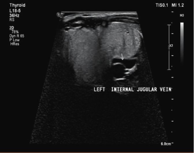
2 Case Reports in Endocrinology
was mobile, nontender, and hard with attachment to the involving both lobes and 50% of children present with
skin. Other systemic examinations were unremarkable. cervical lymph node metastasis and lung metastasis. The
Blood investigation was within normal limits including initial evaluation of thyroid nodule or neck mass is serum
thyroid function tests. A neck ultrasound showed an en- TSH and T4 which in most cases is normal at presentation,
larged left cervical lymph node with a normal blood flow in followed by neck ultrasound. This is followed by a confir-
the right and left thyroid lobes. There was a macrolobulated matory fine-needle aspiration (FNA) [6]. Our patient pre-
lesion (TIRADS 5) with increased flow and mass effect to the sented with atypical feature, an asymptomatic neck mass on
internal jugular vein (Figure 1(a)). A lymph node biopsy was the left side of the neck; therefore, a normal workup for
done and confirmed a metastatic papillary thyroid carci- thyroid disease was not followed. She did not have risk
noma. A subsequent staging computed tomography (CT) factors for thyroid malignancy; hence, a clinical diagnosis of
scan showed a distinct focal peripheral mass lesion on the thyroid malignancy was not entertained. Whilst Hashimo-
inferior pole of the left thyroid lobe with calcifications, mass to’s thyroiditis (HT) is the commonest risk factor associated
effect on the left internal jugular vein (Figure 1(b)), and with paediatric PTC [7], the patient did not report any
bilateral upper lung lobe nodules. An uneventful total history of autoimmune disease. Furthermore, histopatho-
thyroidectomy with a modified neck dissection was done logical assessment of thyroid did not reveal background HT.
and sent for histopathological assessment. While thyroid function tests would not have helped as they
Gross pathology showed a thyroid tumour that mea- are usually normal, FNA of the mass should still be done.
sured 55 × 35 × 10 mm and matted lymph nodes. The tumour This helps to exclude nonmalignant and malignant disease.
was unencapsulated, hemorrhagic, and firm without Clinician should always have a high index of suspicion for
necrosis. malignancy in children who presents with asymptomatic
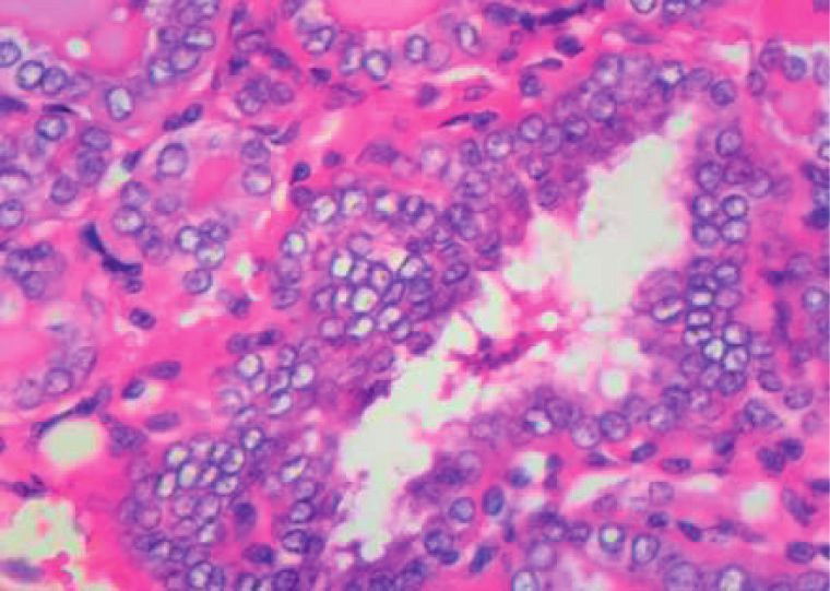
Microscopic examination confirmed papillary variant of neck mass.
PTC evidenced by tumour arranged in papillae, nests, and The histomorphological features of PTC in children are
follicles (Figures 2(a) and 2(b)). These structures were lined the same as in adults. Although it may present with varied
by follicular cells with nuclei exhibiting grooves, clearing, growth patterns including papillary, follicular, and solid,
pseudoinclusions, overlapping, and crowding. In addition, amongst others, the diagnosis is based on typical nuclear
five lymph nodes that were sampled showed metastatic features. These features include Orphan Annie eye nuclei
disease. which are optically clear. They also exhibit pseudoinclusion
An iodine-123 single photon emission computed to- and grooves [8]. The diagnosis on the index patient was done
mography (SPECT) scan done postsurgery, under high based on these features.
thyrotropin stimulation (>30 mU/L), showed increased The management of PTC involves extensive planning
nonuniform uptake in level II, III, and IV cervical lymph from staging preoperatively and deciding on approach that
nodes, mild residual uptake in the thyroid bed, and increased will benefit both the patient and family. Given the very high
uptake in the lungs bilaterally (Figure 3(a)). These features rate of metastatic disease in this population, preoperative
confirmed metastatic disease to the lymph nodes and lungs. neck ultrasound to identify suspicious lymph nodes and
A left lateral lymph node clearance was done which chest X-ray and/or chest CT should be considered in these
further confirmed metastatic papillary thyroid carcinoma. patients as it will guide on the type of surgical treatment to be
Four weeks postsurgery, the patient received 100 mCi of followed.
I-131 therapy. This was under TSH stimulation with the The latest American Thyroid Association (ATA) chil-
withdrawal of eltroxin for 4 weeks and given I131 under dren’s guidelines, published in 2015, recommend radioactive
maximally stimulated TSH. Prophylactic dose of prednisone iodine therapy (RAI) after total thyroidectomy for inter-
was given in conjunction with this therapy to minimize mediate- and high-risk groups who have extensive central
inflammatory response that may result from the radiation. neck lymph node involvement or locally invasive or ex-
The posttherapeutic iodine-131 images (Figure 3(b)) tensive lateral neck cervical lymph nodes. The aim of RAI is
done prior to discharge were congruent in pattern to the to increase the response rate to surgical management. It has
staging iodine-123 scan images. Thyroid function tests were been reported that when radioactive iodine therapy is used
low (thyroxine: 10.4 pmol/L, tri-iodo thyronine: 5.6 pmol/L, in conjunction with thyroid-stimulating hormone (TSH)
and thyroid-stimulating hormone: 0.54 mIU/L). The patient suppression, the rate for complete response is 47.32% and
was discharged on eltroxin daily. for partial response is 38.39%. Postsurgical RAI therapy is
At the 6th month follow-up visit, she tolerated the RAI indicated in paediatric PTC due to its increased risk of
well with improved nutritional status. She was euthyroid bilateral (30%) or multifocal involvement (65%) [6, 8].
with TSH: 4.13 mIU/L, T4: 9.4 pmol/L, T3: 6.0 pmol/L, and Lymph node dissection is recommended to reduce the
thyroglobulin: >300 μg/L. She was put on lifetime thyroid risk of recurrence and increase the efficacy of RAI therapy in
hormone replacement therapy. patients with extrathyroidal invasion or locoregional me-
tastasis [8–10].
3. Discussion The index patient was in the high-risk group, and hence,
a total thyroidectomy with radical neck dissection and ra-
Papillary thyroid carcinoma is the leading cause of paediatric dioactive therapy was the preferred choice of treatment.
endocrine cancers with an incidence of 6% of all cancers [1]. The follow-up guidelines include serum thyroglobulin
In typical cases of PTC, patients present as multifocal on levothyroxine every 3–6 months for 2–3 years then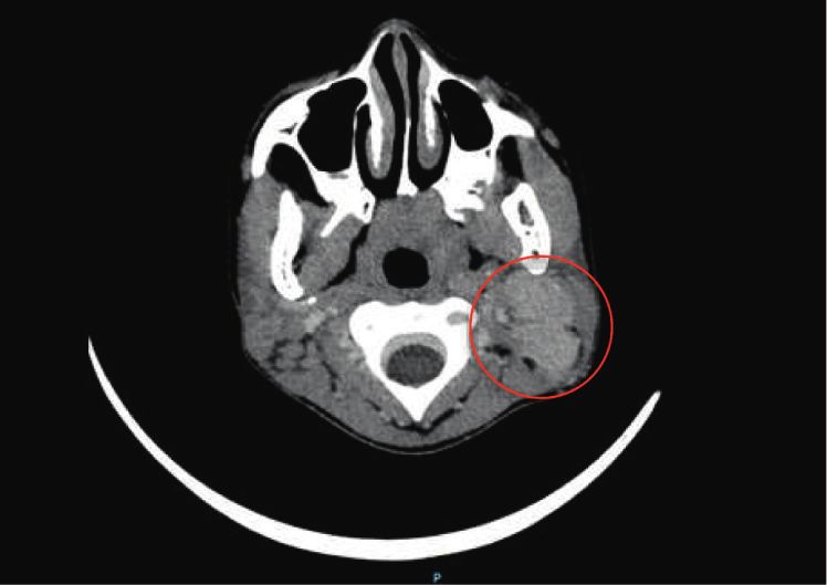
Case Reports in Endocrinology                                                                                                            3

                                         (a)                                                (b)

Figure 1: (a) Neck ultrasound shows enlarged left cervical lymph node compressing on the left internal jugular vein; (b) CT scan shows
mattered left cervical lymphadenopathy ( ).

                                   (a)                                                               (b)

Figure 2: (a) I123 and (b) I131 show increased nonuniform uptake in level II, III, and IV cervical lymph nodes, mild residual uptake in the
thyroid bed, and increased uptake in the lungs bilaterally.

                                         (a)                                               (b)

Figure 3: (a) Thyroid tumour with papillary and follicular pattern; (b) typical nuclear features of PTC: nuclear overlapping, clearing, and
occasional grooves.

annually and thyroid ultrasound including cervical lymph               thyroglobulin level is elevated, and the thyroid ultrasound is
nodes for 6–12 months for intermediate- and high-risk                  negative, then a CT scan of the chest and neck is recom-
groups and annually for low-risk patients. In case the                 mended. In children, more than 98% of cases have a good
4 Case Reports in Endocrinology
survival rate at 10 years while few patients develop pro- overdiagnosed condition? a literature review,” Diagnostics,
gressive or refractory disease not amenable to surgical in- vol. 10, no. 2, p. 112, 2020.
tervention or resistant to radioactive therapy. [5] P. K. Prasad, P. Mahajan, D. S. Hawkins, S. Mostoufi-Moab,
The prognosis for PTC is excellent with low mortality and R. Venkatramani, “Management of pediatric differenti-
rate. The survival rate for different risk groups is as follows: ated thyroid cancer: an overview for the pediatric oncologist,”
Pediatric Blood and Cancer, vol. 67, no. 6, p. e28141, 2020.
99% for low-risk groups, 83% for intermediate-risk groups,
[6] J. D. Prescott and M. A. Zeiger, “The RET oncogene in
and 43% for high-risk groups [11]. papillary thyroid carcinoma,” Cancer, vol. 121, no. 13,
The survivors of childhood ductal thyroid carcinoma pp. 2137–2146, 2015.
need a follow-up for life since recurrence has been reported [7] M. L. Sur, R. Gaga, C. Lazǎr, C. Lazea, C. Aldea, and D. Sur,
at 40 years after initial therapy [8]. “Papillary thyroid carcinoma in children with Hashimoto’s
In conclusion, fine-needle aspiration may provide early thyroiditis-a review of the literature between 2000 and 2020,”
diagnosis in children with neck mass and avoid delayed Journal of Pediatric Endocrinology and Metabolism, vol. 33,
management. A high index of suspicion for PTC should be no. 12, pp. 1511–1517, 2020.
maintained, regardless of risk factors. This allows for early [8] G. L. Francis, S. G. Waguespack, A. J. Bauer et al., “Man-
targeted treatment with reduction in morbidity and mor- agement guidelines for children with thyroid nodules and
differentiated thyroid cancer,” Thyroid, vol. 25, no. 7,
tality in this population. Surgical treatment with or without
pp. 716–759, 2015.
lymph node dissection and radiotherapy remains the cor- [9] H. D. Baumgarten, A. J. Bauer, A. Isaza, S. Mostoufi-Moab,
nerstone of management in paediatric PTC. K. Kazahaya, and N. S. Adzick, “Surgical management of
pediatric thyroid disease: complication rates after thyroid-
Data Availability ectomy at the children’s hospital of philadelphia high-volume
pediatric thyroid center,” Journal of Pediatric Surgery, vol. 54,
The relevant data and materials are available from the no. 10, pp. 1969–1975, 2019.
corresponding author upon request. [10] L. Massimo, D. Zarri, and D. Caprino, “Psychosocial aspects
of survivors of childhood cancer or leukemia,” Minerva
pediatrica, vol. 57, no. 6, pp. 389–397, 2005.
Ethical Approval [11] I. D. Hay, T. Gonzalez-Losada, M. S. Reinalda,
J. A. Honetschlager, M. L. Richards, and G. B. Thompson,
Sefako Makgatho University Research Ethics Committee “Long-term outcome in 215 children and adolescents with
(SMUREC) approved the publication of this case report. papillary thyroid cancer treated during 1940 through 2008,”
World Journal of Surgery, vol. 34, no. 6, pp. 1192–1202, 2010.
Consent
Written, informed consent was obtained from the parent of
the patient for publication of this case report. A copy of the
consent form is available for review.
Conflicts of Interest
The authors declare that they have no conflicts of interest.
Acknowledgments
The authors all express their gratitude to the patient’s
parents who gave consent for this case to be presented in this
paper.
References
[1] V. A. Paulson, E. R. Rudzinski, and D. S. Hawkins, “Thyroid
cancer in the pediatric population,” Genes (Basel), vol. 10,
no. 9, p. 723, 2019.
[2] K. A. Lee, M. T. A. Sharabiani, D. Tumino et al., “Differen-
tiated thyroid cancer in children: a UK multicentre review and
review of the literature,” Clinical Oncology, vol. 31, no. 6,
pp. 385–390, 2019.
[3] J. Chen, N. Huang, Q. Ji, Y. Wang, Y. Zhu, and D. Li,
“Multifocal papillary thyroid cancer in children and adoles-
cents: 12-year experience in a single center,” Gland Surgery,
vol. 8, no. 5, pp. 507–515, 2019.
[4] A. I. Stefan, A. Piciu, A. Mester, D. Apostu, M. Badan, and
C. I. Badulescu, “Pediatric thyroid cancer in Europe: anYou can also read