Cervico-Facial Necrotizing Fasciitis: A Ten-Year Clinical Evaluation of 80 Cases in Enugu, Eastern Nigeria - Remedy Publications
←
→
Page content transcription
If your browser does not render page correctly, please read the page content below
World Journal of Oral and Maxillofacial Surgery                                                                                                    Research Article
                                                                                                                                             Published: 11 Jan, 2019

    Cervico-Facial Necrotizing Fasciitis: A Ten-Year Clinical
       Evaluation of 80 Cases in Enugu, Eastern Nigeria
                                             Chukwuneke FN1*, Okechi UC1, Nwosu JN2, Onyeka TC3, Okoroafor IJ4 and Akpeh JO4
                                             1
                                                 Department of Oral and Maxillofacial Surgery, University of Nigeria Enugu Campus, Nigeria
                                             2
                                                 Department of Otolaryngology, University of Nigeria Teaching Hospital, Nigeria
                                             3
                                                 Department of Anesthesia, University of Nigeria Enugu Campus, Nigeria
                                             4
                                                 Department of Otorhinolaryngology Surgery, University of Nigeria Enugu Campus, Nigeria

                                             Abstract
                                             Introduction: The purpose of this article was to evaluate the 80 patients with Cervico-Facial
                                             Necrotizing Fasciitis (CNF) amongst 1109 oral and maxillofacial patients seen at the Oral &
                                             Maxillofacial surgery department of a Nigerian Teaching hospital.
                                             Patients and Methods: We carried out a 10-year retrospective evaluation of 80 patients with
                                             Cervico-facial necrotizing fasciitis seen at the Oral and Maxillofacial Department of the University
                                             of Nigeria Teaching Hospital, Enugu, Nigeria from 2004 to 2013. We reviewed all patients who had
                                             a diagnosis of cervico-facial necrotizing fasciitis during this period from notes kept in the Records
                                             Department. We identified the trends in the number of cases categorized by oral hygiene status,
                                             yearly occurrence, age, and sex and treatment outcome.
                                             Result: Eighty cases were recorded, accounting for 7.2% of all oral and maxillofacial cases reviewed.
                                             Of the 80 cases, 31 (38.75%) were males, while 49 (61.25%) were females giving a male-to-female
                                             ratio of 1:1.6. The year 2013 presented more number of cases (26; 32.5%) and 2006 was the least (1;
                                             1.25%). Patients between the ages of 20 years to 29 years were more affected (18; 22.5%). They were
                    OPEN ACCESS              all emergencies and most 71(88.7%) came from rural areas. Poor oral hygiene was evident in all the
                                             cases. The treatment of choice was surgical debridement and hospitalization. Five patients (6.3%)
                     *Correspondence:
                                             were lost.
   Felix N Chukwuneke, Department of
 Oral and Maxillofacial Surgery, College     Conclusion: This study has shown an increasing trend in occurrence of Cervico-facial necrotizing
       of Medicine, University of Nigeria    fasciitis with morbidity and mortality surge in Enugu, Nigeria. While emphasis should be on
   Enugu Campus, Enugu, Nigeria, Tel:        individual oral health care and health-seeking behavior, there is a need also for health care policy
                      +2347064531609;        makers to re-focus on this morbidly increasing orofacial infection.
       E-mail: ichiefn2002@yahoo.com         Keywords: Necrotizing fasciitis; Clinical evaluation; Enugu nigeria
           Received Date: 21 Dec 2018
           Accepted Date: 09 Jan 2019        Introduction
          Published Date: 11 Jan 2019
                                                  Cervicofacial Necrotizing Fasciitis (CNF) remains a health challenging burden in the developing
                              Citation:      and underdeveloped nations, mainly due to neglected or untreated and severe dentofacial infections.
  Chukwuneke FN, Okechi UC, Nwosu            It was first detected and described in 1871 by Joseph Jones who referenced more than 2600 during
   JN, Onyeka TC, Okoroafor IJ, Akpeh        the American civil war [1]. The disease was named “Streptococcal gangrene” by Meleny in 1924
JO. Cervico-Facial Necrotizing Fasciitis:    and described the condition as a more generalized lesion when he isolated Hemolytic streptococci
    A Ten-Year Clinical Evaluation of 80     in 20 cases studied [2]. However, recent studies supported the polymicrobial etiology of the disease
Cases in Enugu, Eastern Nigeria. World       and ascribed various names to the lesion which include hospital gangrene, necrotizing erysipelas,
     J Oral Maxillofac Surg. 2019; 2(1):     Streptococcal gangrene, gangrenous erysipelas, Hemolytic streptococcal gangrene, and suppurative
                                  1018.      fasciitis [3]. Poverty, immune-compromised infections, lack of adequate awareness on sound
       Copyright © 2019 Chukwuneke           oral health care and other poor health conditions are also the major factors contributing to the
      FN. This is an open access article     prevalence of the disease. According to Vinod, the probability of infection occurring at the lower
          distributed under the Creative     head (face) and neck region is increased as the teeth act as the focus of the infection [4]. It can be
   Commons Attribution License, which        said that 90% of the head and neck pyogenic infection have tooth as the causative factor, while the
   permits unrestricted use, distribution,   rest 10% may have different aetiology. Thus in all head and neck pyogenic infection, tooth should be
       and reproduction in any medium,
                                             considered as the causative factor, unless otherwise proved [5]. The teeth in the jaws have classical
   provided the original work is properly
                                             location and anatomical criteria and so the clinical presentation is so specific that depending on the
                                             character of the infection, one can trace the source and find out which tooth is the culprit. Authors
                                    cited.

Remedy Publications LLC.                                                        1                                          2019 | Volume 2 | Issue 1 | Article 1018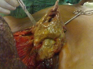
Chukwuneke FN, et al., World Journal of Oral and Maxillofacial Surgery
Materials and Methods
We carried out a 10-year retrospective review of 1109 oral and
maxillofacial patients seen at the Oral and Maxillofacial Department
of the University of Nigeria Teaching Hospital (UNTH), Enugu,
Nigeria. The focus was on the 80 cases diagnosed with Cervicofacial
Nectrotizing Faciitis. We recorded and reviewed all the patients
during this period from notes kept in the Records department of
the Hospital. Relevant information retrieved included patients’ oral
habits, past medical and drug history, socio-demographic data, onset
of symptoms and the treatment outcome. Oral cavities were properly
assessed to identify the affected tooth as well as the periodontal tissues
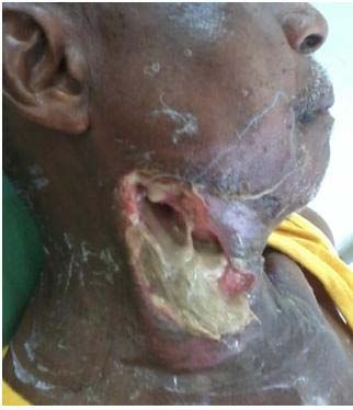
Figure 1: One of the female patients with necrotizing fasciitis.
using Sillness-Loe plaque index and Sulcus Bleeding Index (SBI). All
the 80 patients had the same spectrum of presentation: life threatening
cervico-facial infection characterized by aggressive spread of
inflammation and necrosis of the surrounding tissues (Figure 1,2 and
3). Clinical and laboratory findings carried out were also noted. Some
of the patients also presented with oral lesion characterized by the
presence of patchy white plaques and confluent pseudo-membranous
mucosa suggesting opportunistic infection with oral candidiasis. We
identified the trends in the number of cases categorized by the yearly
occurrence and time of presentation, age, and sex and treatment
outcome. To study changes in the yearly occurrence, we grouped
the cases by the year of presentation and diagnosis while treatment
outcome was assessed based on total remission and abatement of the
disease.
Figure 2: An elderly patient with necrotizing fasciitis. Results
Most of the patients came from remote rural areas, lived at
a subsistence level with little or no formal knowledge and sound
oral health care education. Consequently, they were ignorant of
the availability of orthodox treatment for their cases and resulted
to the use of local herbs and patronizing quacks. Poor oral hygiene
was evident in all the cases treated (Table 1). We lost some of the
patients due to complications of the spread of the infection to the
vital organs such as the brain and the mediasternal region, late
presentation and co-morbidity from other systemic diseases such as
diabetes and hypertension. Of the 1109 oral & maxillofacial patients
recorded within the ten-year study period, 80 (7.21%) cases of CNF
were observed (Table 2). Patients between the ages of 20 years to 29
Figure 3: A debridement procedure in one of the patients. years were more affected (18; 22.5%) while 70 years to 79 years were
least (1; 1.25%). The total number CNF patients were 80 while that of
the unaffected was 1029. Of the 80 recorded cases, 31 (38.75%) were
have expressed different opinion on the role of the co-morbidities
males; and 49 (61.25%) were females giving a male-to female ratio
as a risk factor in the pathogenesis and progression of the disease.
of 1:1.6. The trends in yearly occurrence shows that more cases of
While some are of the opinion that pre existing ill health, co morbid
states like Diabetes mellitus, alcoholism, retroviral disease, vascular
insufficiency, neutropenia play a vital role in the pathogenesis of the
disease, others think otherwise [6-9].The prevalence and management
of CNF as a potentially fatal fulminant disease condition in our
environment Nigeria is presently sub-optimal and largely under-
reported in dental literature [1]. In recent time, there has been an
increasing trend in the occurrence of CNF in our environment. We
postulate that Poverty, ignorance, superstitious beliefs in addition to
improper health seeking behavior and poor oral health care habits
may be a chain reactions causing CNF in our environment. The fact
that these aetiological factors and causes of CNF are treatable and
modifiable risk factors emphasizes the need for increasing oral health
care awareness among the general public by the health policy makers
as a first step in the prevention of this ailment. Figure 4: Patient after debridement and progression of secondary healing.
Remedy Publications LLC. 2 2019 | Volume 2 | Issue 1 | Article 1018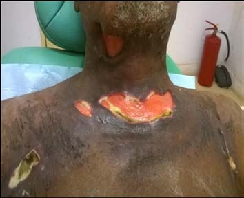
Chukwuneke FN, et al.,                                                                                                        World Journal of Oral and Maxillofacial Surgery

                                                                                                neck region it is known as cervicofacial necrotizing fascitis [6].
                                                                                                Cervicofacial necrotizing fascitis is uncommon, possibly due to
                                                                                                highly vascular nature of this area [9,10]. The most common reported
                                                                                                etiological causes include dental caries, periodontal disease and
                                                                                                pericoronitis with the mandibular second and third molar teeth
                                                                                                being the most implicated [10-13]. Poor oral hygiene is therefore a
                                                                                                major contributing risk factor to developing CNF as evidenced in our
                                                                                                findings. Most of the patients were from lower economic class of the
                                                                                                society and live at a substance level with little or no formal education.
                                                                                                Some were ignorant of the availability of orthodox management
                                                                                                of their cases and resulted to the use of local herbs. Authors have
                                                                                                expressed divergent views regarding age and sex distributions. While
                                                                                                some findings suggest the rate of occurrence to be more in males others
  Figure 5: Patient after total healing and abatement of the disease.                           observed that females are more affected [1,9,13-15]. We observed
                                                                                                in our study that females were more affected than the males to the
Table 1: Distribution of the oral health status of the patients.                                ratio of 1:1.6 male-to-females. These findings may not be unrelated
 Oral health status     Male      (%)       Female        (%)         Frequency     (%)         to the geographical variations and place of study. The mean age of the
          Good            -           -         -          -              -           -         patient seen was 59.3 years (29 to 76 years) which is similar to other
                                                                                                studies [10,13,14]. Patients between the ages of 20 years to 29 years
          Fair            -           -         -          -              -           -
                                                                                                were more affected (18; 22.5%) while 70 years to 79 years were least
          Poor            2         2.5        1          1.25           3          3.75
                                                                                                (1; 1.25%). This may better be explained in terms of poor oral health
      Very poor          29      36.25         48         60             77         96.25       care awareness and improper health seeking behaviour regarding oral
          Total          31      38.75         49     61.25              80         100         health amongst the youths. There seems to be an increasing trend in
                                                                                                the rate of yearly occurrence of CNF in recent time as observed in this
CNF were observed in the year 2013, followed by 2012, 2011, 2010,                               study. The trends in yearly occurrence shows that more cases of CNF
2005 with 2006 having the least frequency. The prevalence of CNF                                were observed in the year 2013, followed by 2012, 2011, 2010, 2005
among the two sex groups were equal in 2004 and 2007 while more                                 with 2006 having the least frequency. No satisfactory explanation
amongst females in 2008, 2009 and 2012. We carried out surgical                                 could better exist with this observation other than the dwindling
debridement on 60 (75%) of the patients; 23 (28.75%) males and 37                               emphasis on oral health care awareness in Nigerian environment.
(46.25%) females. Seventeen patients (21.25%); six (7.5%) males and                             Poor oral hygiene was evidence in all the cases. This is in line with
11 (13.75%) females were mainly treated with antibiotics in addition                            other observations by some authors [16,17]. Although other sources
to tooth extraction (Figure 4). Seventy-one of the patients came from                           such as trauma, tonsilar and pharyngeal infections, cervical adenitis,
the rural areas with the number of male 25 (31.25%) to female 46                                tumour infections, mastoid and salivary gland infections as well as
(57.5%) while 9 (11.25%) of the patients came from the urban areas                              postauricular lymphadenitis may be among the aetiological factor, the
with the number of males 6 (7.5%) to females 3 (3.75%). All the                                 major causes include periapical infections of the mandibular molars,
patients presented late and were hospitalized. Five patients (6.3%)                             pericoronitis and periodontal disease suggesting poor oral hygiene as
were lost due to complication arising from the spread of infection and                          a modifiable risk factor [16]. Diagnosis of necrotizing fascitis could be
poorly controlled co-morbidities while the rest had total remission                             quite challenging, it may be misdiagnosed as it presents with features
and abatement of the diseases (Figure 5).                                                       similar to other odontogenic infections [10,16,18]. The main stay in
                                                                                                its diagnosis is a thorough history, adequate knowledge of the clinical
Discussion                                                                                      presentation, accompanied by advanced radiographic imaging as
   Necrotizing Faciitis commonly affect the extremities, but                                    Computed Tomography (CT) scan and Magnetic Resonance Imaging
can affect any part of the body and when it involves the head and                               (MRI) [10,19,20].

Table 2: Trends in the prevalence number of cases seen amongst the 1109 oral & maxillofacial cases within the study period.
                                                                                                                                                  Frequency of
 Years     Unaffected male      %         Affected male         %      Unaffected female        %   Affected female    %        Affected %                              Total %
                                                                                                                                                   Unaffected
  2004            39           3.75            2               2.5            41            3.98           2           2.5          5                   80               7.77

  2005            45           4.37            1               1.25           45            4.37           4           5           6.25                 90               8.75

  2006            30           2.92             -               0             30            2.92           1          1.25         1.25                 60               5.83

  2007            34            3.3            1               1.25           34             3.3           1          1.25         2.5                  68                6.61

  2008            60           5.83            3               3.75           59            5.73           1          1.25          5                  119               11.56

  2009            43           4.18            2               2.5            47            4.57           0           0           2.5                  90                8.75

  2010            48           4.66            3               3.75           53            5.15           5          6.25          10                 101                9.82

  2011            59           5.73            1               1.25           51            4.95           8           10         11.25                110               10.69

  2012            68           6.61            10              12.5           79            7.68           9          11.25       23.75                147               14.28

  2013            76           7.39            8               10             88            8.55          18          22.5         32.5                164               15.94

  Total           502          48.74           31          38.75              527           51.26         49          61.25        100                 1029               100

Remedy Publications LLC.                                                                    3                                             2019 | Volume 2 | Issue 1 | Article 1018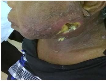
Chukwuneke FN, et al.,                                                                                            World Journal of Oral and Maxillofacial Surgery

Table 3: Recorded treatment methods adjusted by sex.                                 6. Fomete B, Ononiwu CN, Agbara R, Okeke UA, Idehen KO. Cervicofacial
Recorded Treatment Method              Male (%)    Females (%)    Freq. (%)             necrotizing fasciitis: Case series and review of the Literature. Case Study
                                                                                        Case Rep. 2013;3(1):26-33.
Surgical debridement                  23 (28.75)    37 (46.25)     60 (75)
                                                                                     7. Fliss DM, Tovi F, Zirkin HJ. Necrotizing soft-tissue infections of dental
Antibiotics medications/Extraction      6 (7.5)     11(13.75)     17 (21.25)
                                                                                        origin. J Oral Maxillofac Surg. 1990;48(10):1104-8.
Management of co-morbidity              2 (2.5)      1 (1.25)      3 (3.75)
                                                                                     8. Ndukwe KC, Fatusi OA, Ugboko VI. Craniocervical necrotizing fasciitis in
Total                                 31 (38.75)    49 (61.25)     80 (100)             Ile-Ife, Nigeria. Br J Oral Maxillofac Surg. 2002;40(1):64-7.
                                                                                     9. Obiechina AE, Arotiba JT, Fasola AO. Necrotizing fasciitis of odontogenic
    Treatment outcome is often hampered by chain reactions of                           origin in Ibadan, Nigeria. Br J Oral Maxillofac Surg. 2001;39(2):122-6.
events and they include; presence of co-morbid conditions, late
surgical intervention and delayed hospitalization, aggressive spread                 10. Subhashraj K, Jayakumar N, Ravindran C. Cervical necrotizing fascitis: An
                                                                                         unusual sequel of Odontogenic Infection. Med Oral Patol Oral Cir Bucal.
of infection to the meditational and thoracic region, polymicrobial
                                                                                         2008;13(12):E788-91.
nature of the infection and inappropriate diagnosis and treatment
procedure [1,17]. Consequently we lost 5 patients. There is increasing               11. Fliss DM, Tovi F, Zirkin HJ. Necrotizing soft-tissue infections of dental
yearly occurrence of CNF in our environment due to odontogenic                           origin. J Oral Maxillofac Surg. 1990;48(10):1104-8.
infection caused by poor oral hygiene. In our environment patients                   12. Edwards JD, Sadeghi N, Najam F, Margolis M. Craniocervical necrotizing
presents late when management challenges and limited treatment                           fascitis of odontogenic origin with mediastinal exntension. Ear Nose
options results in high mortality rate. The fact that this is a modifiable               Throat J. 2004;83(8):579-82.
and preventable risk factor emphasizes the need for oral health care                 13. Obiechina AE, Arotiba JT, Fasola AO. Necrotizing fasciitis of odontogenic
awareness campaign and programme of oral health education among                          origin in Ibadan, Nigeria. Br J Oral Maxillofac Surg. 2001;39(2):122-6.
the populace as a first step to prevention and early presentation of
                                                                                     14. Iynen I, San I, Bozkus F, Beklen H. Life threatening necrotizing fascitis of
dental infections for proper treatment.                                                  the neck: a case report. J Curr Surg. 2011;1(1):35-7.
Acknowledgement                                                                      15. Maria A, Rajnikanth K. Cervical necrotizing fascitis caused by dental
                                                                                         infection: A review and case report. Natl J Maxillofac Surg. 2010;1(2):135-
    We would like to acknowledge Rev. Christian Okoye dental                             8.
technologist with the Federal College of Dental Technology and
Therapy for his assistance in the collection of data at the department               16. Lambade PN, Dolas RS, Virani N, Lambade DP. Cervicofacial necrotizing
                                                                                         fasciitis of odontogenic origin: A review. Open Access Sci Rep.
of Oral & Maxillofacial Surgery, University of Nigeria Teaching
                                                                                         2012;1(8):414.
Hospital Ituku-Ozalla.
                                                                                     17. Sikkerimath SB, Sikkerimath BC. Cervical Necrotizing fasciitis – The flesh
References                                                                               eating disease: Review of literature and Report of five cases. J Indian Aca
1. Obimakinde OS, Okoje VN, Akinmoladum VI, Fasola AO, Arotiba JT.                       Oral Med Radiol. 2013;25(2):131-6.
   Retrospective evaluation of necrotizing fascitis in University college            18. Lin C, Yeh FL, Lin JT, Ma H, Hwang CH, Shen BH, et al. Necrotizing
   hospital Ibadan. Niger J Clin pract. 2012;15(3):344-8.                                fasciitis of the head and neck: an analysis of 47 cases. Plast Reconstr Surg.
2. Umeanuka TO, Okechi CU, Chukwuneke NF. Challenges In The                              2001;107(7):1684-93.
   Management Of Cervicofacial Necrotizing Fasciitis In A Developing                 19. Shaariyah MM, Marina MB, Mohd Razif MY, Mazita A, Primuharsa Putra
   Economy: Our Experience. Niger J Med. 2016;25(44):368-73.                             SH. Necrotizing fasciitis of the head and neck: Surgical outcomes in three
3. Edlich RF, Cross CL, Dahlstrom JJ, Long WB 3rd. Modern concepts                       cases. Malays J Med Sci. 2010;17(2):51-5.
   of the diagnosis and treatment of necrotizing fasciitis. J Emerg Med.             20. Mastronikolis NS, Stathas T, Naxakis SS, Mallis A, Papadas T,
   2010;39(2):261-5.                                                                     Kalogeropoulou C, et al. Necrotizing fasciitis of the head and neck:
4. Vinod K. A Textbook of Oral and Maxillofacial Surgery. Arya (Medi)                    Report of 5 cases and review of the literature. Eur Rev Med Pharmacol Sci.
   Publishing House. 2nd Ed. 2011;35.                                                    2010;14(2):123-34.

5. Anisha M, Rajnikanth K. Cervical necrotizing fascitis caused by dental
   infection: A review and case report. Natl J Maxillofac Surg. 2010;1(2):135-
   8.

Remedy Publications LLC.                                                         4                                             2019 | Volume 2 | Issue 1 | Article 1018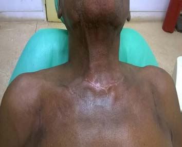
You can also read