Central retinal artery occlusion and optic neuropathy secondary to platelet rich plasma injection: a case report
←
→
Page content transcription
If your browser does not render page correctly, please read the page content below
Int J Ophthalmol, Vol. 14, No. 6, Jun.18, 2021 www.ijo.cn
Tel: 8629-82245172 8629-82210956 Email: ijopress@163.com
·Letter to the Editor·
Central retinal artery occlusion and optic neuropathy
secondary to platelet rich plasma injection: a case report
Norashikin Maslan, Wan Haslina Wan Abdul Halim, Norshamsiah Md Din, Seng Fai Tang
Department of Ophthalmology, Universiti Kebangsaan note that her previous PRP injection, however, was uneventful
Malaysia Medical Centre (UKMMC), Cheras 56000, Kuala and patient denies skin filler injections.
Lumpur, Malaysia Patient was referred to a nearby ophthalmology clinic within
Correspondence to: Seng Fai Tang. Department of 3h after the initial complaint. Ocular massage was performed
Ophthalmology UKMMC, Jalan Yaacob Latif, Bandar Tun for 10-15min before patient was hyperventilated. Oral
Razak, Cheras 56000, Kuala Lumpur, Malaysia. drtangsf@ medication was given to lower her intraocular pressure (IOP).
ukm.my Her vision did not improve regardless, so she decided to seek
Received: 2020-07-04 Accepted: 2020-08-26 treatment at our center.
She arrived in 9h after the loss of vision. Her vision was 20/20
DOI:10.18240/ijo.2021.06.23 in right eye (OD) and 20/60 OS. There was a grade 3 relative
afferent pupillary defect (RAPD) OS. Anterior segment
Citation: Maslan N, Wan Abdul Halim WH, Din NM, Tang SF. Central examination OS was unremarkable with an IOP of 16 mm Hg.
retinal artery occlusion and optic neuropathy secondary to platelet rich A fundus examination revealed generalized edematous retina
plasma injection: a case report. Int J Ophthalmol 2021;14(6):945-947 with dull foveal reflex but no cherry red spot present (Figure
1A). Ocular examination OD was unremarkable. Her blood
Dear Editor, pressure was 150/90 mm Hg, with a pulse rate of 90 beats
I am Dr. Norashikin Maslan, from the Department of
Ophthalmology of University Kebangsaan Malaysia
Medical Centre in Kuala Lumpur, Malaysia. I am writing to
per minute and regular rhythm. A diagnosis of left CRAO
secondary to PRP injection was then made. She was started on
topical timolol 0.5%, topical dexamethasone 0.1%, and oral
present a case report on central retina artery occlusion (CRAO) Acetazolamide. Optical coherence tomography angiography
and optic neuropathy secondary to platelet rich plasma (PRP) (OCTA) OS showed significant reduction in both vessel and
injection. perfusion density of the superficial capillary plexus (SCP)
In recent times, PRP injection is increasingly being used compared with the fellow eye (Figures 2 and 3). Humphrey
in medical specialties especially in aesthetic industry. PRP visual field (HVF) 24-2 test showed-generalized constricted
injection delays ageing with the presence of cytokines and visual field OS with normal finding OD (Figure 4A).
growth factors in it[1-2]. It is administered either topically, Four days after the PRP injection, the patient developed left
superficially or through deep dermal injections[3]. The injection periorbital hematoma associated with worsening of vision,
promotes the production of type 1 procollagen (skin fibroblast) occasional headache, and numbness over the glabella site.
and hyaluronic acid which in turn improves the skin texture, Her visual acuity dropped to 10/200 in OS. There was
firmness and color homogeneity[4]. There are, however, some subconjunctival hemorrhage with anterior chamber cells of
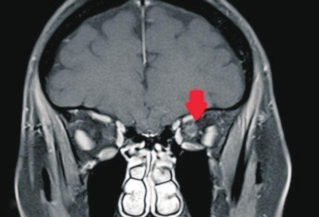
minor adverse effects of PRP such as discomfort, small prick 4+. The fundus was hazy but no vitritis was seen. MRI orbit
bleeding and swelling that have been reported[4]. Although showed perineural enhancement of the optic nerve (Figure 5).
many have noted its efficacy[4-5], we would like to highlight a The magnetic resonance venography (MRV) was normal and
visually threatening complication caused by pure PRP injection there was no retro-orbital hemorrhage. She was therefore
in this report. treated for optic neuropathy and covered with intravenous
CASE PRESENTATION methylprednisolone 250 mg q.i.d. for 3d followed by an oral
A 48-year-old woman with no known medical illness arrived prednisolone (1 mg/kg·d) for 11d.
with a sudden onset loss of vision in left eye (OS) immediately Fortunately, her vision improved slowly. Three months after
after receiving a PRP injection on the glabellae region by the event, visual acuity has improved to 20/20 OS, but RAPD
an aesthetic practitioner. The monocular loss of vision was remained positive (grade 2) and red color saturation was 8/10
accompanied by an ocular discomfort. It is important to also OS. Her periorbital hematoma resolved. Anterior segment
945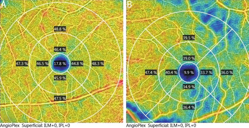
CRAO and ON secondary to platelet rich plasma injection
Figure 1 Fundus photos of the left eye at first presentation (A)
and after 2mo showing pale and cupped optic disc (B). Figure 5 MRI of the orbit showing perineural enhancement of the
left optic nerve.
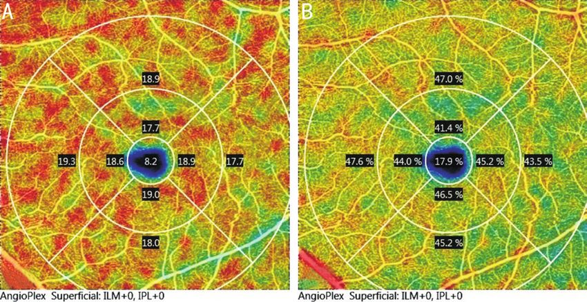
Figure 2 OCTA images of the patient Reduced vessel density in the
superficial capillary plexus of the left eye (B) compared to the right Figure 6 OCTA images of the patient Improved vessel (A) and
eye (A) at initial presentation. perfusion density (B) on heat map of the superficial capillary plexus
of the left eye after 2mo of follow up.
examination was unremarkable, but optic disc OS became
pale with an increase cupping to 0.9 (Figure 1B). Repeated
OCTA showed improvement in perfusion and vessel density
of the SCP (Figure 6). Her latest HVF also showed significant
improvement (Figure 4B).
DISCUSSION
Treatment with PRP is generally safe and effective. While
it appears simple to administer, devastating intravascular
Figure 3 OCTA images of the patient Reduced perfusion density complications can still occur. Periocular vascular events
in the superficial capillary plexus of the left eye (B) compared to the are particularly important to be aware of as immediate
right eye (A) at initial presentation. intervention may restore the ocular circulation and prevent
permanent blindness. Inadvertent intravascular injection of
materials, either fillers, PRP or local anesthetic medications
can cause retrograde flow to the original artery resulting in the
obstruction of the retinal vascular lumen[1,6-7]. The inner retina
layer is supplied by central retinal artery and occlusion will
result in ischemia of the inner retina layer, leading to edema
and subsequently atrophy. In CRAO, some vision may be
preserved since the choroidal circulation also supplies part of
the inner retinal tissue. Experiments on animal models have
demonstrated irreparable retinal damage occurs after 105min
of vascular occlusion[1,8].
Visual loss following filler injections has been reported
Figure 4 HVF 24-2 showing generalized constricted visual field in although its occurrence is very rare. Facial injections can
the left eye at presentation (A) and 3mo after treatment showing be performed at various locations such as at the glabella,
significant improvement (B). nasolabial fold, or periorbital region. These regions are rich in
946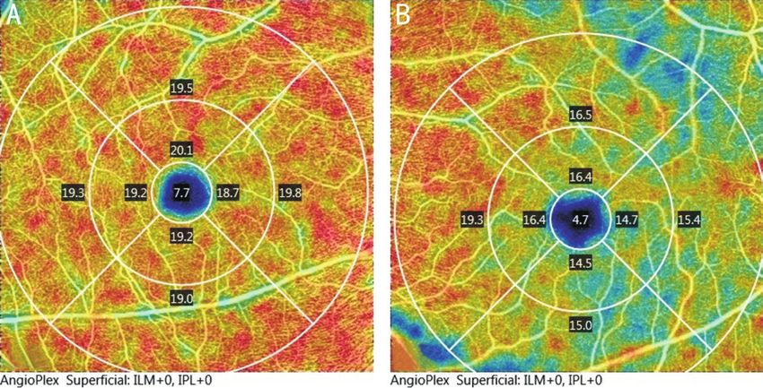
Int J Ophthalmol, Vol. 14, No. 6, Jun.18, 2021 www.ijo.cn
                                                                     Tel: 8629-82245172 8629-82210956 Email: ijopress@163.com

blood supply. Injections at the glabella is commonly associated      recognition and appropriate intervention may help prevent
with compression or obstruction of blood vessel because the          irreversible visual loss.
supratrochlear artery which supplies this region does not have       ACKNOWLEDGEMENTS
a strong collateral circulation[9].                                  Authors’ contributions: Maslan N analyzed, interpreted
Blood supply to the face is from branches of the external            investigation result and major contributor in the writing
and internal carotid arteries. Collaterals between the anterior      manuscript, Tang SF analysed, interpreted, managed the patient
and posterior ethmoidal arteries medial to the orbit, between        and contribute in the writing manuscript, Wan Abdul Halim
the supraorbital and infraorbital artery anteriorly and the          WH contributed in the writing of the manuscript, Din NM
zygomaticotemporal branches laterally provides a rich supply         analyzed and contributed in the writing of the manuscript”.
to the periorbital and orbital region. Angular artery supplies the   Conflicts of Interest: Maslan N, None; Wan Abdul Halim
dorsal part of the nose and inferomedial fossa of the orbit[8].      WH, None; Din NM, None; Tang SF, None.
Connections between the terminal branches of angular artery          REFERENCES
with supraorbital/supratrochlear artery plays an important role      1 Hu XZ, Hu JY, Wu PS, Yu SB, Kikkawa DO, Lu W. Posterior ciliary
in establishing anastomosis[8].                                        artery occlusion caused by hyaluronic acid injections into the forehead:
In our case, the patient received PRP injections at the glabella       a case report. Medicine (Baltimore) 2016;95(11):e3124.
area which is an area known to have arterial anastomosis.            2 Charles-de-Sá L, Gontijo-de-Amorim NF, Takiya CM, Borojevic R,
Hu et al[1] highlighted three essential concomitant factors that       Benati D, Bernardi P, Sbarbati A, Rigotti G. Effect of use of platelet-
eventually lead to spread into the ophthalmic circulation which        rich plasma (PRP) in skin with intrinsic aging process. Aesthet Surg J
are retrograde movement of material, forceful injection with           2018;38(3):321-328.
high pressure and adequate amount of injectable material.            3 Banihashemi M, Nakhaeizadeh S. An introduction to application of
Following that, the embolus can travel along the central retinal       platelet rich plasma (PRP) in skin rejuvenation. Reviews in Clinical
artery causing occlusion of the retinal vessels.                       Medicine 2014;1(2):38-43.
Currently, limited literature suggested a correlation between        4 Aust M, Pototschnig H, Jamchi S, Busch KH. Platelet-rich plasma for
CRAO and optic neuropathy and to our knowledge, this                   skin rejuvenation and treatment of actinic elastosis in the lower eyelid
is the first case of unilateral CRAO with optic neuropathy             area. Cureus 2018;10(7):e2999.
after a PRP injection. In this patient, optic neuropathy was         5 Amini F, Abiri F, Ramasamy TS, Tan ESS. Efficacy of platelet rich
diagnosed based on perineural optic nerve enhancement seen             plasma (PRP) on skin rejuvenation: a systematic review. Iranian
on MRI. Optic neuropathy may be caused by either ischemia              Journal of Dermatology 2015;18(3):119-127.
or inflammation, leading to disturbance of the blood-optic           6 DeLorenzi C. Complications of injectable fillers, part 2: vascular
nerve barrier. We postulate that the retrograde flow of the            complications. Aesthet Surg J 2014;34(4):584-600.
PRP material may have affected both the central retinal artery       7 Park KH, Kim YK, Woo SJ, Kang SW, Lee WK, Choi KS, Kwak
and the posterior ciliary artery that mainly supplies the intra-       HW, Yoon IH, Huh K, Kim JW, Korean Retina Society. Iatrogenic
orbital and intraocular portion of the optic nerve, resulting          occlusion of the ophthalmic artery after cosmetic facial filler injections:
in impairment of small nutrient vessels feeding the nerve by           a national survey by the Korean Retina Society. JAMA Ophthalmol
ischemic or inflammatory process and consequently causing              2014;132(6):714-723.
optic neuropathy.                                                    8 Loh KT, Chua JJ, Lee HM, Lim JT, Chuah G, Yim B, Puah BK.
Our case study shows that OCTA is a useful tool to establish           Prevention and management of vision loss relating to facial filler
reduced perfusion and vessel density, conforming to the study          injections. Singapore Med J 2016;57(8):438-443.
by Yang et al[10] that vessel density of the SCP and a 300 μm        9 Lafaille P, Benedetto A. Fillers: contraindications, side effects and
area around the foveal avascular zone is significantly reduced         precautions. J Cutan Aesthet Surg 2010;3(1):16-19.
in retinal artery occlusion. OCTA is also a non-invasive             10 Yang S, Liu XQ, Li H, Xu J, Wang F. Optical coherence tomography
alternative diagnostic tool to monitor progression of patients         angiography characteristics of acute retinal arterial occlusion. BMC
with vascular occlusions[11].                                          Ophthalmol 2019;19(1):147.
To conclude, our case highlights a potentially blinding              11 Nakayama LF, Bergamo VC, Silva LSC, Moraes NSB. Optical
complication of PRP injection around the ocular region. Even           coherence tomography (OCT) angiography of central retinal artery
though it is a rare complication, ophthalmologist need to be           occlusion in the patent cilioretinal artery: a case report. Arq Bras
aware of this due to its devastating complication. Prompt              Oftalmol 2018;81(3):242-246.

                                                                                                                                      947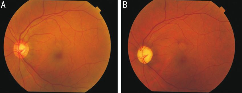
You can also read