Case Report Acute Myocardial Infarction due to External Compression of the Left Main Coronary Artery by a Large Pulmonary Artery Aneurysm
←
→
Page content transcription
If your browser does not render page correctly, please read the page content below
Hindawi
Case Reports in Cardiology
Volume 2021, Article ID 8850044, 4 pages
https://doi.org/10.1155/2021/8850044
Case Report
Acute Myocardial Infarction due to External Compression of the
Left Main Coronary Artery by a Large Pulmonary
Artery Aneurysm
H. Sharma ,1 S. N. Doshi,2 and M. A. Nadir2
1
Institute of Cardiovascular Sciences, University of Birmingham, Birmingham, UK
2
Cardiology Department, Queen Elizabeth Hospital, Birmingham, UK
Correspondence should be addressed to H. Sharma; harish.sharma@nhs.net
Received 30 September 2020; Revised 23 December 2020; Accepted 27 January 2021; Published 23 February 2021
Academic Editor: Manabu Shirotani
Copyright © 2021 H. Sharma et al. This is an open access article distributed under the Creative Commons Attribution License,
which permits unrestricted use, distribution, and reproduction in any medium, provided the original work is properly cited.
Background. Although rare, external compression of the left main coronary artery (LMCA) by a pulmonary arterial aneurysm
(PAA) as a consequence of pulmonary arterial hypertension causing stable angina pectoris is well described. However, acute
myocardial infarction is extremely rare, particularly with a full array of electrocardiographic, biochemical, and
echocardiographic features, as in this scenario. Case. In this case, a 62-year-old man with a past history of severe fibrotic lung
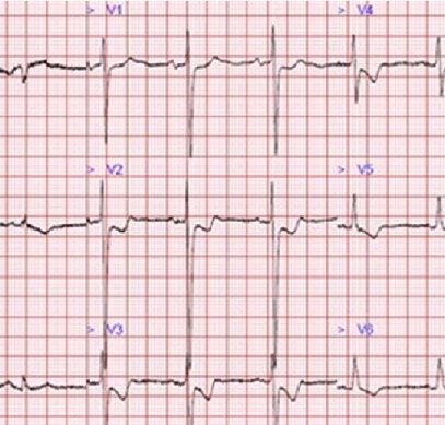
disease was hospitalised with chest pain. The patient had dynamic anterolateral ischaemic changes on electrocardiography and
serially elevated high-sensitivity troponin I. Transthoracic echocardiography revealed impaired left ventricular ejection fraction
with anterolateral hypokinesis. Coronary angiography with intracoronary imaging revealed external compression of the LMCA.
Computer tomography (CT) scans confirmed new PAA, compared to previous scans. The patient was successfully treated by
percutaneous coronary stent implantation. Conclusion. Progressive dilatation of the pulmonary artery due to pulmonary arterial
hypertension can result in acute MI secondary to external compression of the LMCA. Clinicians should be mindful of acute
coronary syndromes in patients with long-standing pulmonary hypertension presenting with chest pain.
1. Introduction ponin, and a corresponding regional wall motion abnormal-
ity on echocardiography.
Pulmonary arterial hypertension (PAH) is associated with
aneurysm of the main pulmonary artery (PA). In normal anat- 2. Case
omy, the PA lies adjacent to the left coronary sinus of the
aorta. As the left main coronary artery (LMCA) arises from A 62-year-old man with a past history of severe fibrotic lung
the left coronary sinus, it may be compressed by a PA aneu- disease requiring long-term oxygen therapy was hospitalised
rysm (PAA). Although rare, a small number of case reports with typical angina pain with no resolution despite sublin-
have described this phenomenon in the presence [1, 2] and gual glyceryl trinitrate use. ECG demonstrated dynamic
absence of PAH [3]. The most commonly described clinical anterolateral ST depression with T wave inversion in anterior
sequelae of this scenario is stable angina pectoris [4–7]. and lateral leads (Figure 1). High-sensitivity troponin I assays
Myocardial infarction (MI) as a result of external com- (normal: < 5 ng/L) measured 99 and 250 ng/L. Transthoracic
pression of a coronary artery has been previously docu- echocardiography revealed a left ventricle ejection fraction of
mented in patients with anomalous coronary origin or 52% and anterolateral hypokinesis (video 1) with a high like-
course [8] and normal coronary anatomy [9], attributed to lihood of pulmonary hypertension (estimated systolic PA
PAA (with or without PAH). However, it is rare to see the full pressure 110 mmHg), indicating likely group III pulmonary
gamut of evidence suggesting acute MI, including dynamic hypertension (secondary to hypoxic lung disease). The main
electrogram (ECG) changes, elevation of high-sensitivity tro- PA diameter was measured at 5.0 cm (Figure 2). Cardiac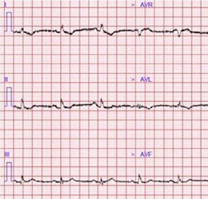
2                                                                                                     Case Reports in Cardiology

                                 (a)                                                           (b)

Figure 1: Electrocardiogram demonstrating (a) anterolateral ischaemic changes (ST depression and T wave inversion); (b) older ECG 6
months earlier.

                                                                       The patient was not felt to be a suitable candidate for sur-
                                                                   gery and underwent IVUS-guided percutaneous coronary
                                                                   intervention with a 5 × 20 mm everolimus drug-eluting stent
                                                                   deployed directly without any predilatation. Postdeploy-
                                                                   ment, IVUS confirmed a well-apposed stent (video 3) with
                                                                   resolution of extrinsic compression and restoration of the
                                       5.0 cm
                                                                   LMCA lumen (Figures 4(b) and 4(d)). The patient was ren-
                                                                   dered pain free and discharged home after a short period of
                                                                   observation and remained angina free on subsequent fol-
                                                                   low-up.

                                                                   3. Discussion
                                                                   MI due to external compression of the left main stem can
Figure 2: Transthoracic echocardiogram demonstrating significant
                                                                   occur by 2 main mechanisms. In normal coronary anatomy,
dilatation of the main pulmonary artery (diameter: 5 cm).
                                                                   severe PAA can develop following longstanding PAH, result-
                                                                   ing in acute compression of the LMCA. In patients with
                                                                   anomalous coronary origin or course interruption of coro-
catheterisation assessment as part of a lung transplant work-      nary flow, it occurs due to compression of the LMCA
up 7 years earlier had shown no significant coronary disease        between the PA and the high-pressured aorta and can occur
(Figure 3(c)) with invasive measurements demonstrating             without PAA.
only mild pulmonary hypertension (mean systolic PA pres-                In PAH patients who experience LMCA compression by
sure 30 mmHg). The differential diagnosis included pulmo-           the PA, stable angina pectoris has been the dominant syn-
nary thromboembolism and pulmonary/aortic dissection,              drome reported by numerous case reports, but this case dem-
although the clinical picture was most consistent with acute       onstrates that acute MI can also occur, and is supported by
MI. The patient was commenced on pharmacological treat-            electrocardiographic, biochemical, and echocardiographic
ment for non-ST elevation myocardial infarction. In view of        parameters. The LMCA is the vessel most at risk of compres-
the significant lung disease, an initial conservative approach      sion, and due to the significant myocardial territory supplied,
was taken, but after continual chest pain despite optimal          occlusion can result in significant or even life-threatening
medical therapy, invasive coronary angiography was necessi-        myocardial injury.
tated. The transradial coronary angiogram showed a tight                Even if the acute MI event is survivable, major infarction of
narrowing of the LMCA ostium (Figure 3(d)). The smooth             the LMCA territory in PAH patients could significantly impair
tapering appearance (Figure 4(a)) raised the possibility of        long-term prognosis. Such patients are likely to have coexist-
external compression. This was subsequently confirmed with          ing right ventricular pressure or volume overload, and acute
the use of intravascular ultrasound (IVUS) which showed            left ventricular infarction may result in biventricular failure.
dynamic external compression of LMCA with a slit-like              This has the potential to decompensate pulmonary function
lumen and absence of atheroma (Figure 4(c), video 2)—sug-          due to the combination of pre- and postcapillary pulmonary
gesting that the patient had developed a type 2 NSTEMI due         hypertension. Clinicians should therefore be mindful of acute
to demand ischaemia. A gated CT scan confirmed normal               coronary syndromes occurring in PAH patients presenting
origin and course of the LMCA; however, it was compressed          with chest pain. As demonstrated in this case, such patients
by a grossly dilated PA measuring 58 mm (normal ~30 mm)            can be easily assessed by CT and intracoronary imaging
at its maximal diameter (Figure 3(b)), compared to 33 mm 7         and successfully treated with prompt percutaneous implanta-
years ago (Figure 3(a)).                                           tion of an intracoronary stent. It is therefore suggested that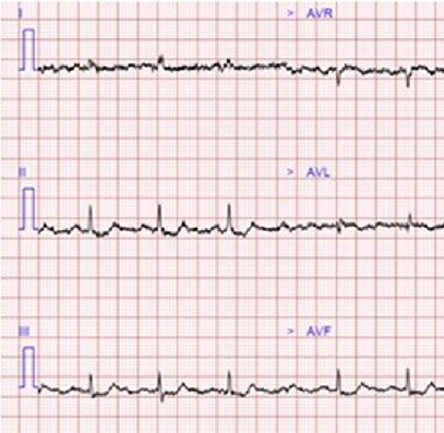
Case Reports in Cardiology                                                                                                          3

                                                                                       Ao     MPA
                                               MPA
                                          Ao

                                          RPA                                          RPA
                                                     LPA                                          LPA

                                               (a)                                          (b)

                                        LMS
                                                                                 LMS

                           Guide
                                                                      Guide
                           catheter
                                                                      catheter

                                               (c)                                          (d)

Figure 3: (a) CT scan showing main pulmonary artery with a diameter of 33 mm in 2013; (b) CT scan showing main pulmonary artery with a
diameter of 58 mm in 2020; (c) normal appearance of LMCA in 2013; (d) severe tapering stenosis of LMCA in 2020. MPA = main pulmonary
artery; RPA = right pulmonary artery; LPA = left pulmonary artery; Ao = ascending aorta; LMS = left main stem.

                                               (a)                                          (b)

                                               (c)                                          (d)

Figure 4: (a) Angiography demonstrating severe ostial LMCA stenosis despite intracoronary nitrate and disengaged guide catheter; (b)
angiography following deployment of 5 × 20 mm stent; (c) baseline IVUS with a slit-like lumen of LMCA and distinct lack of atheroma;
(d) well-apposed coronary stent and near-circular lumen of LMCA.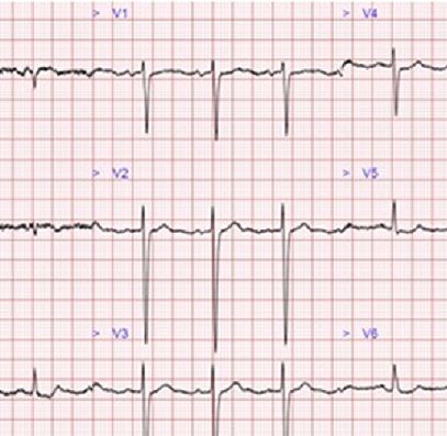
4                                                                                                       Case Reports in Cardiology

extrinsic compression by an aneurysmally dilated PA should          [2] K. Albadri, J. M. Jensen, E. H. Christiansen, S. Mellemkjær,
be considered in the differential diagnosis of chest pain in             and J. E. Nielsen-Kudsk, “Left main coronary artery compres-
patients with severe PAH. Coronary angiography alone can                sion in pulmonary arterial hypertension,” Pulmonary Circula-
misdiagnose extrinsic LMCA compression, but IVUS can                    tion, vol. 5, no. 4, pp. 734–736, 2016.
more accurately determine the extent of LMCA stenosis and           [3] B. Miranda-Barrio, E. Garcia-Romero, H. Cuellar-Calabria,
is associated with lower rates of major clinical events [10].           and L. Dos-Subira, “Left main coronary artery compression
     It is worth noting that because of the slit-like nature of         by a large pulmonary artery aneurysm in the absence of pul-
the narrowing, 45° left anterior oblique (LAO) or 30° LAO               monary hypertension: a case report,” European Heart Journal
                                                                        - Case Reports, vol. 2, no. 4, 2018https://academic.oup.com/
cranial angulation coronary angiographic planes cross-
                                                                        ehjcr/article/2/4/yty105/5126422.
sectioning the narrow axis of the compressed LMCA best
visualise this pathology, whereas other planes crossing the         [4] N. Galiè, F. Saia, M. Palazzini et al., “Journal of the American
                                                                        College of Cardiology,” vol. 69, no. 23, pp. 2808–2817, 2017.
wide axis of the narrowing frequently miss the compression
[11]. Shorter stents are often necessary to eliminate the risk      [5] V. A. de Jesus Perez, F. Haddad, R. H. Vagelos, W. Fearon,
of circumflex artery obliteration. Moreover, displacement of             J. Feinstein, and R. T. Zamanian, “Angina associated with left
                                                                        main coronary artery compression in pulmonary hyperten-
the aortic lumina is an important risk during ostial LMCA
                                                                        sion,” The Journal of Heart and Lung Transplantation,
stenting. Intravascular ultrasound imaging can aid diagnosis            vol. 28, no. 5, pp. 527–530, 2009.
(absence of plaque and dynamic external compression) and
                                                                    [6] M. Bijl, J. G. F. Bronzwaer, A. C. van Rossum, and F. W. A.
determine stent size and optimal position of deployment.
                                                                        Verheugt, “Angina pectoris due to left main coronary artery
                                                                        compression in Eisenmenger ductus arteriosus,” American
Data Availability                                                       Heart Journal, vol. 125, no. 6, pp. 1767–1771, 1993, https://
                                                                        pubmed.ncbi.nlm.nih.gov/8498323/.
Data are available on request.                                      [7] J. F. Patrat, G. Jondeau, O. Dubourg et al., “Left main coronary
                                                                        artery compression during primary pulmonary hypertension,”
Additional Points                                                       Chest, vol. 112, no. 3, pp. 842-843, 1997, https://pubmed.ncbi
                                                                        .nlm.nih.gov/9315824/.
Learning Objectives. Longstanding PAH may result in PAA.            [8] K. Shishido, N. Moriyama, T. Shimizu, and S. Saito, “Anoma-
Patients with known PAA and chest pain should be consid-                lous acute left main myocardial infarction due to compression
ered for urgent coronary angiography with IVUS imaging                  between pulmonary artery and aorta by acute pulmonary
to diagnose and treat possible extrinsic coronary compres-              thromboembolism,” JACC: Cardiovascular Interventions,
sion. 45° left anterior oblique (LAO) or 30° LAO cranial cor-           vol. 9, no. 23, pp. e227–e228, 2016, https://interventions
onary views are best to visualise the narrowing (other views            .onlinejacc.org/content/9/23/e227.
frequently miss the compression). IVUS can aid diagnosis            [9] M. Vaseghi, J. S. Lee, and J. W. Currier, “Acute myocardial
and determine stent size and optimal position of deployment.            infarction secondary to left main coronary artery compression
                                                                        by pulmonary artery aneurysm in pulmonary arterial hyper-
                                                                        tension,” Journal of Invasive Cardiology, vol. 19, pp. 375–377,
Conflicts of Interest                                                   2009, https://www.invasivecardiology.com/articles/Acute-
                                                                        Myocardial-Infarction-Secondary-Left-Main-Coronary-
The authors declare that they have no conflicts of interest.             Artery-Compression-Pulmonary-Arter.
                                                                   [10] M. Velázquez Martín, J. M. Montero Cabezas, S. Huertas et al.,
Authors’ Contributions                                                  “Clinical relevance of adding intravascular ultrasound to coro-
                                                                        nary angiography for the diagnosis of extrinsic left main coro-
All authors contributed to and approved this manuscript.                nary artery compression by a pulmonary artery aneurysm in
                                                                        pulmonary hypertension,” Catheterization and cardiovascular
                                                                        diagnosis, pp. 1–10, 2020.
Supplementary Materials
                                                                   [11] O. Y. Akbal, C. Kaymaz, I. H. Tanboga et al., “Extrinsic com-
Video 1: transthoracic echocardiography demonstrating                   pression of left main coronary artery by aneurysmal pulmo-
anterolateral left ventricular hypokinesis. Video 2: intravas-          nary artery in severe pulmonary hypertension: its correlates,
cular ultrasound examination of the left main coronary                  clinical impact, and management strategies,” European Heart
artery, demonstrating external compression with a slit-like             Journal-Cardiovascular Imaging, vol. 19, no. 11, pp. 1302–
                                                                        1308, 2018, https://academic.oup.com/ehjcimaging/article/
lumen and absence of atheroma. Video 3: intravascular ultra-
                                                                        19/11/1302/4717459.
sound examination of the left main coronary artery, demon-
strating resolution of external compression and well-apposed
stent. (Supplementary Materials)

References
 [1] W. Karrowni, G. Sigurdsson, and P. A. Horwitz, “Left main
     coronary artery compression by an enlarged pulmonary
     artery,” JACC. Cardiovascular Interventions, vol. 6, no. 1,
     pp. e3–e4, 2013.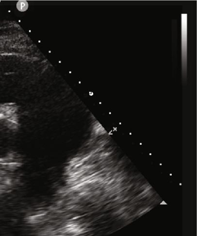
You can also read