GSJ: Volume 9, Issue 1, January 2021, Online: ISSN 2320-9186 - Global Scientific Journal
←
→
Page content transcription
If your browser does not render page correctly, please read the page content below
GSJ: Volume 9, Issue 1, January 2021
ISSN 2320-9186                                                                  2393

               GSJ: Volume 9, Issue 1, January 2021, Online: ISSN 2320-9186
                                       www.globalscientificjournal.com
Chest CT findings in COVID-19- Retrospective Study –Our
Experience in KAAH
Authors:
Dr. Malak H. Alawi MD1
Dr. Wael Hamza Mutair MD2
Dr. Nadir Hamza Mutair MD3
Dr. Azra Begum DMRD, DNB4
                                                    5
Dr. Hatim Abdul Aziz Khogeer MD
                                                6
Dr. Asim Abdul Aziz Khogeer MD
Dr. Mohammed Harraz MD7
Dr. Lubna Abdullrahman Hafiz MD8
                                                           9
Mr. Fawzi Mohammed Ali Jeninah B.Sc.
                                                                 10
Dr. Hannan AbdulFattah Bin Saddaq MBBS                                .

1
    Head of Department of Radiology and Consultant Radiologist, King Abdul
Aziz Hospital, Makkah, Saudi Arabia. Email-m6alawi@hotmail.com.
2
    General Director of Health Affairs, General Directorate of Health Affairs
Makkah, Ministry of Health, Makkah, Saudi Arabia. Email-
wmotair@moh.gov.sa.
3
    Director of Planning and Research in the health of Makkah region, Ministry
of Health, Makkah, Saudi Arabia. Email-nmetair@moh.gov.sa.
4
    Radiology Specialist, King Abdul Aziz Hospital, Makkah. Email-
drazrabegum@gmail.com.
5
    Surgical specialist, KFH, Ministry of Health, Makkah, Saudi Arabia.
    Email-dr_foryou@yahoo.com.
6
    Ministry of Health, Makkah, Saudi Arabia. Email-asimkhogeer@yahoo.uk.
7
    Consultant Radiologist, King Abdul Aziz Hospital, Makkah, Saudi Arabia.

                                                  GSJ© 2021
                                          www.globalscientificjournal.com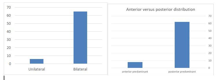
GSJ: Volume 9, Issue 1, January 2021
ISSN 2320-9186                                                              2394

     Email- harrazharraz@live.com.
8
    Consultant Family Medicine, Al Zaher PHC, Ministry of Health, Makkah,
    Saudi Arabia. Email-drlubnahafiz@yahoo.com.
9
    Lab specialist, King Abdul Aziz Hospital, Makkah, Saudi Arabia.
    Email-Fawzimohd_ali@yahoo.com
10
     Radiology Resident, King Abdul Aziz Hospital, Makkah.
     Email-hananbinsadeq@gmail.com.

Corresponding author:
Dr. Azra Begum
Dept of Diagnostic Imaging
King Abdul Aziz Hospital,
Zaher,
Makkah, Saudi Arabia
Phone-+966543575319
Email-drazrabegum@gmail.com

Key words- computed tomography, coronavirus, COVID-19, crazy pavement
pattern, ground glass opacities , pneumonia , sub-pleural line.

Abstract-
The coronavirus 2019 (COVID-19) pandemic has emerged as a severe
healthcare crisis. Thoracic imaging with Chest radiography and CT are crucial
for identifying the disease early and help in excluding the differentials and
thereby help in management of the disease and its progression. Hence it is
crucial that we are familiar with the imaging pattern of COVID-19. Our
research is aimed at outlining the common patterns of presentation of
COVID-19 on CT chest.

Objective-
 Our objective was to analyse the chest computed tomography findings of
corona virus disease of COVID-19 pneumonia during the treatment of 71
patients presenting with symptoms such as cough, fever and shortness of
breath and to integrate the most frequent imaging features and distribution
patterns of this disease.

                                               GSJ© 2021
                                       www.globalscientificjournal.com
GSJ: Volume 9, Issue 1, January 2021
ISSN 2320-9186 2395
Introduction-
The novel Coronavirus, currently named as SARS-COV-2, was first identified
in Wuhan, in December 2019. Within a few months, the virus has spread
from China to worldwide. On March 11, 2020, the World Health Organization
(WHO) declared this global outbreak as a pandemic (1). In Saudi Arabia, the
first case was reported on 2 March, 2020 (2). There have been 21,756,357
confirmed cases of COVID-19 worldwide, including 771,635 deaths, reported
to WHO upto 18 August ,2020 (3). In Saudi Arabia, there have been a total
of 301,323 confirmed cases, including 3,470 deaths uptodate (4).
As this disease spreads rapidly through close contact, droplets and aerosol
transmission from person to person, there is need for early diagnosis and
isolation to control it(5).
The standard confirmatory test is the real-time reverse transcription-
polymerase chain reaction(RT-PCR) test(6).
Computed tomography can detect lesions in the lungs at a very early stage,
specially in those patients who have symptoms but show negative RT-PCR
test initially(7-9).
Materials and methods-
A retrospective study as conducted with the approval of the institutional
review board, and written informed consent was waived. Totally 71 patients
(50men, 21women, mean age 54.2years), with initial positive RT-PCR test or
on followup, confirming the diagnosis of COVID -19 disease, who were
treated in our hospital between 1st March till 31st July were included in our
study . These patients underwent CT chest for respiratory symptoms, chest
xray findings or to rule out pulmonary embolism.
A comprehensive analysis was done of the most common findings on CT
chest (eg. Types of opacities, distribution pattern, additional findings) and
also the atypical presentations in patients diagnosed with COVID-19
pneumonia.
CT imaging and evaluation
Images were taken via a 64-slice CT scanner (GE). The axial images were
acquired craniocaudally at shallow inspiration and included the body part
from the thoracic inlet to the diaphragm. Multiplanar reconstruction was
done. The CT images were evaluated for the following charecterisctics-
distribution pattern, laterality of the lesions, lobe involvement, density of the
lesions –ground glass opacities or consolidation or both, peribronchovascular
thickening, nodules, halo sign, reverse halo sign, air bronchogram,
subpleural line, bronchiectasis, cavitation, tree in bud opacities, pleural
effusion, pericardial effusion, mediastinal lymphadenopathy, pneumothorax.
GSJ© 2021
www.globalscientificjournal.comGSJ: Volume 9, Issue 1, January 2021
ISSN 2320-9186 2396
Statistical analysis
Continuous data were expressed as mean and standard deviation, while cat-
egorical data were expressed as counts (n) and percentages (%). The
statistical analysis was carried out on SSPS software(version25.0, IBM).
RESULTS
We evaluated 71 patients who tested positive for RT-PCR on initial testing or
on follow up test. Out of 71 patients , 50 were male and 21 were females,
age range between 19-86 years.
Most of the patients belonged to the age groups 41-60(40%) and over 60
(38%) (Figure1).
Distribution according to age groups
Figure 1- Distribution of study population according to
age groups.
The most common distribution pattern was bilateral (91.5%), peripheral
(67.6%) and posterior predominant (87.3%) (Figure 2),(Figure 3). Among
the lobar distribution, most commonly involved lobe was the right lower lobe
(90.1%), followed by the left lower lobe (84.5%), indicating the tendency for
basal predominance (Figure 4).
GSJ© 2021
www.globalscientificjournal.comGSJ: Volume 9, Issue 1, January 2021
ISSN 2320-9186 2397
Distribution Pattern
Figure 2- distribution pattern
Central vs peripheral Distribution according to age groups
Figure 3- Central vs peripheral distribution pattern according to age groups.
GSJ© 2021
www.globalscientificjournal.comGSJ: Volume 9, Issue 1, January 2021
ISSN 2320-9186 2398
Figure 4- Distribution according to the lobes involved.
Most common detected lesions were GGO (ground glass opacities) with
multifocal pattern (69%) while pure consolidation was seen in 28.1% of
patients and a mixed GGO with consolidation pattern seen in 32.3% of
patients and associated with air bronchograms in (43.6%). Septal thickening
(67.6%), crazy pavement pattern (35.2%) , subpleural line (33.8%) ,
traction bronchiectasis (39.4%) and peribronchovascular wall thickening
(33.8%) were a common occurrence. Halo sign (7%) was more commonly
seen than the reverse halo sign (1.4%) . Less commonly seen were the
reticulonodular opacities, cavitation, pleural thickening and pleural
effusion(5-16%), (Table1), (Figure 5).
We did not detect any patients with pneumothorax, tree in bud opacities,
mediastinal lymphadenopathy and pericardial effusion.
GSJ© 2021
www.globalscientificjournal.comGSJ: Volume 9, Issue 1, January 2021
ISSN 2320-9186 2399
Table 1- CT imaging findings in COVID-19 pneumonia (n-72)
TYPES OF DENSITIES n %
Ground glass opacification –unifocal 2 2.8
Ground glass opacification –multifocal 49 69.0
Consolidation 20 28.1
Ground glass opacification with 23 32.3
consolidation
Halo sign 5 7.0
reverse halo sign 1 1.4
Plerual effusion 4 5.6
Pleural thickening 11 15.4
Airbronchogram 31 43.6
Reticulonodular 11 15.4
Septal thickening 48 67.6
Crazy pavement pattern 25 35.2
Subplerual line 24 33.8
Cavitation 4 5.6
Bronchiectasis 28 39.4
Peribronchovascular wall thickening 24 33.8
Micronodular opacities 1 1.4
Not found in our study
Mediastinal lymphadenopathy 0 0
Pericardial effusion 0 0
Pneumothorax 0 0
Tree in bud 0 0
GSJ© 2021
www.globalscientificjournal.comGSJ: Volume 9, Issue 1, January 2021
ISSN 2320-9186 2400
Types of Densities on CT chest imaging
60
50
40
30
20
10
0
Figure 5- Distribution according to the types of densities seen on CT
chest.
Discussion-
Based on our study, the typical CT chest findings of COVID -19 infections are
ground glass opacities GGO in multifocal, peripheral and basal distribution
bilaterally. Along with these, there is increased incidence of consolidation
with air bronchogram, traction bronchiectasis, crazy pavement pattern and
sub pleural line (Figure 6). However, these findings are non specific and
could be seen in other infectious diseases, For eg. Viral pneumonias
(influenza pneumonia, SARS, RSV, rhino virus and adenovirus pneumonias,
Non viral infections eg mycoplasma pneumonia, or non-infectious pneumonia
eg hypersensitivity pneumonitis, interstitial pneumonia etc.(10-18).
Therefore, it is essential to take thorough relevant clinical history in order to
narrow the differentiatial.
GSJ© 2021
www.globalscientificjournal.comGSJ: Volume 9, Issue 1, January 2021
ISSN 2320-9186 2401
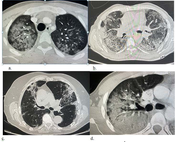
Figure6a.GGO ground glass opacities. b.subpleural line c.traction
bronchiectasis, d.crazy pavement pattern.
CT is more sensitive for early parenchymal lung disease, disease
progression, and alternative diagnoses including acute heart failure,
myocardial injury and, when performed with intravenous contrast material,
pulmonary thromboembolism.(19)
The characteristic lesions and their distribution pattern can help narrow
down the differentials and help save live by initiating early treatment.
According to research study and guidelines set by Fleischner Society, the
recommendations for imaging are for those patients who are symptomatic,
age over 65 years and those with co morbities eg. Diabetes, cardiovascular
disease, hypertension etc.(19)
Patients with functional impairment after recovery from COVID-19 should
undergo imaging to differentiate between expected morphologic
abnormalities as sequelae of infection, mechanical ventilation, or both versus
a different and potentially treatable process.(19)
GSJ© 2021
www.globalscientificjournal.comGSJ: Volume 9, Issue 1, January 2021
ISSN 2320-9186 2402
Conclusion-
The most common findings in CT chest imaging of COVID-19 patients are
bilateral, peripheral distribution, septal thickening and multifocal GGO with
basal and posterior predominance and crazy pavement pattern.
Therefore CT proves to a very reliable, easily accessable and effective tool in
the early diagnosis and management of symptomatic patient’s who initially
test negative for COVID-19 and also for the assessment of the progression
of the disease.
References-
1.World Health Organization. 2020. Coronavirus disease 2019 (COV-ID- 19)
situation report-51. World Health Organization, Geneva. Avail-able from:
https://www.who.int/docs/default-source/coronaviruse/situa-
tionreports/20200311-sitrep-51-covid-19.pdf?sfvrsn=1ba62e57_10.
2.Saudi Arabia government. Ministry of health. Available
fromhttps://www.moh.gov.sa/en/Ministry/MediaCenter/News/Pages/News-
2020-03-02-002.aspx
3. World Health Organisation Coronavirus Disease Dashboard. Available from
https://covid19.who.int/
4. Saudi Arabia government. Ministry of health. Available from
https://covid19.moh.gov.sa/
5. Riou J, Althaus CL. Pattern of early human-to-human transmission of
Wuhan 2019 novel coronavirus (2019-nCoV), December 2019 to January
2020. Euro Surveill 2020; 25:2000058. [Crossref]
6. Kim H, Hong H, Yoon SH. Diagnostic performance of CT and reverse
transcriptase-polymerase chain reaction for coronavirus disease 2019: A
meta-analysis. Radiology 2020; 201343. [Crossref]
GSJ© 2021
www.globalscientificjournal.comGSJ: Volume 9, Issue 1, January 2021
ISSN 2320-9186 2403
7. Ai T, Yang Z, Hou H, et al. Correlation of chest CT and RT-PCR testing in
coronavirus disease 2019 (COVID-19) in China: A report of 1014 cases. Ra-
diology 2020; 200642. [Crossref]
8. Fang Y, Zhang H, Xie J, et al. Sensitivity of chest CT for COVID-19:
comparison to RT-PCR. Radiology 2020; 200432. [Crossref]
9. Dai WC, Zhang HW, Yu J, et al. CT imaging and differential diagnosis of
COVID-19. Can Assoc Radiol J 2020; 71:195–200. [Crossref]
10. Oikonomou A, Müller NL, Nantel S.. Radiographic and high-Resolution CT
findings of influenza virus pneumonia in patients with Hematologic
Malignancies. Am J Roentgenol. 2003/08/01;181(2):507–511. doi:
10.2214/ajr.181.2.1810507 [PubMed] [CrossRef] [Google Scholar]
11. Franquet T. Imaging of pulmonary viral pneumonia. Radiology.
2011/07/01;260(1):18–39. doi: 10.1148/radiol.11092149 [PubMed]
[CrossRef] [Google Scholar]
12. Choi S-H, Hong S-B, Ko G-B, et al. . Viral infection in patients with
severe pneumonia requiring intensive care unit admission. Am J Respir Crit
Care Med. 2012;186(4):325–332. doi: 10.1164/rccm.201112-2240OC
[PubMed] [CrossRef] [Google Scholar]
13. Choi S-H, Huh JW, Hong S-B, et al. . Clinical characteristics and
outcomes of severe rhinovirus-associated pneumonia identified by
bronchoscopic bronchoalveolar lavage in adults: comparison with severe
influenza virus-associated pneumonia. J Clin Virol. 2015/01/01/;62:41–47.
doi: 10.1016/j.jcv.2014.11.010 [PMC free article] [PubMed]
[CrossRef] [Google Scholar]
14. Ooi GC, Khong PL, Müller NL, et al. . Severe acute respiratory
Syndrome: Temporal lung changes at Thin-Section CT in 30
patients. Radiology. 2004/03/01;230(3):836–844. doi:
10.1148/radiol.2303030853 [PubMed] [CrossRef] [Google Scholar]
GSJ© 2021
www.globalscientificjournal.comGSJ: Volume 9, Issue 1, January 2021
ISSN 2320-9186 2404
15. Mackay IM, Arden KE.. MERS coronavirus: diagnostics, epidemiology and
transmission. Virol J. 2015/12/22;12(1):222. doi: 10.1186/s12985-015-
0439-5 [PMC free article] [PubMed] [CrossRef] [Google Scholar]
16. Akira M, Inoue Y, Arai T, et al. . Pulmonary Fibrosis on high-Resolution
CT of patients With pulmonary alveolar proteinosis. Am J Roentgenol.
2016/09/01;207(3):544–551. doi: 10.2214/AJR.15.14982 [PubMed]
[CrossRef] [Google Scholar]
17. Silva CIS, Churg A, Müller NL.. Hypersensitivity Pneumonitis: Spectrum
of high-Resolution CT and Pathologic findings. Am J Roentgenol.
2007/02/01;188(2):334–344. doi: 10.2214/AJR.05.1826 [PubMed]
[CrossRef] [Google Scholar]
18. Ichikado K, Johkoh T, Ikezoe J, et al. . Acute interstitial pneumonia:
high-resolution CT findings correlated with pathology. Am J Roentgenol.
1997/02/01;168(2):333–338. doi: 10.2214/ajr.168.2.9016201 [PubMed]
[CrossRef] [Google Scholar]
19. Geoffrey D. Rubin, MD, MBA • Christopher J. Ryerson, MD, MAS et al. .
The Role of Chest Imaging in Patient Management during the COVID-19
Pandemic: A Multinational Consensus
Statement from the Fleischner Society. Radiology.2020/07/01;296(1).
doi/10.1148/radiol.2020201365[PubMed] [CrossRef] [Google Scholar]
GSJ© 2021
www.globalscientificjournal.comYou can also read