Scale and Pustule on Dermoscopy of Rosacea: A Diagnostic Clue for Demodex Species - Dermatology: Practical and ...
←
→
Page content transcription
If your browser does not render page correctly, please read the page content below
Dermatology Practical & Conceptual

        Scale and Pustule on Dermoscopy of Rosacea:
           A Diagnostic Clue for Demodex Species
                               Gamze Serarslan1, Özlem Makbule Kaya2, Emre Dirican3

1 Department of Dermatology, Mustafa Kemal University, Hatay, Turkey
2 Department of Parasitology, Mustafa Kemal University Hatay, Turkey
3 Department of Biostatistics, Mustafa Kemal University Hatay, Turkey

Key words: Demodex folliculorum, Demodex brevis, rosacea, dermoscopy
Citation: Serarslan G, Makbule Ö, Dirican E. Scale and pustule on dermoscopy of rosacea: a diagnostic clue for Demodex species.
Dermatol Pract Concept. 2021;11(1):e2021139. DOI: https://doi.org/10.5826/dpc.1101a139
Accepted: September 8, 2020; Published: January 29, 2021
Copyright: ©2021 Serarslan et al. This is an open-access article distributed under the terms of the Creative Commons Attribution License
BY-NC-4.0, which permits unrestricted noncommercial use, distribution, and reproduction in any medium, provided the original author
and source are credited.
Funding: None.
Competing interests: The authors have no conflicts of interest to disclose.
Authorship: All authors have contributed significantly to this publication.
Corresponding Author: Gamze Serarslan, MD, Mustafa Kemal University, Faculty of Medicine, Department of Dermatology, 31100 Hatay,
Turkey. Email: gserarslan@hotmail.com

  Abstract              Background: Demodex mites are highly found in the skin of patients with rosacea.The diagnosis of
                        Demodex can be made by standardized skin surface biopsy. Dermoscopy is a tool used in the nonin-
                        vasive diagnosis of various dermatological diseases.
                        Objectives: To determine whether dermoscopic features of demodicosis are associated with the result
                        of standardized skin surface biopsy in patients with rosacea and to compare dermoscopic features of
                        rosacea in Demodex-positive and negative samples and Demodex type.
                        Methods: A total of 30 patients (7 male, 23 female) were included in the study. Dermoscopic exami-
                        nation was performed on both the clinically most severely affected areas and adjacent healthy skin.
                        The skin surface biopsy sample was taken from the same place from where the dermoscopic image
                        was taken.
                        Results: A total of 83 (lesion n = 60, non-lesion n = 23) areas were evaluated. Demodex was detected
                        in 60.2% (n = 50) of the samples. Half of these samples revealed only Demodex folliculorum, and the
                        remaining half revealed D folliculorum and Demodex brevis. Of theDemodex-positive samples, 88%
                        had Demodex tails (P =0.001) and68% Demodex follicular openings (P = 0.002) on dermoscopy. In
                        D folliculorum+D brevis-positive samples, the rate of scale and pustule was higher than D folliculo-
                        rum-positive samples (P = 0.017 and P = 0032,respectively).
                        Conclusions: The sensitivity and specificity of Demodex tail are higher than Demodex follicular open-
                        ing and scale and pustule detection with dermoscopy and may indicate the coexistence of both D
                        folliculorum and D brevis.

Research | Dermatol Pract Concept 2021;11(1):e2021139                                                                                      1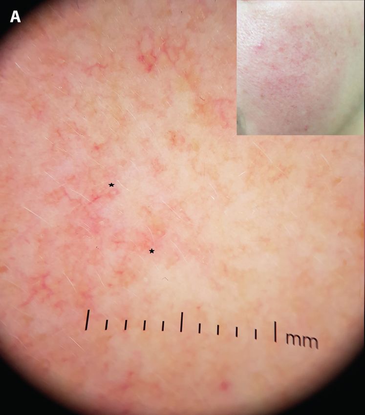
Introduction                                                         Dermoscopic Evaluation
                                                                     The dermoscopic evaluation was performed by the same clini-
Rosacea is a chronic inflammatory disease that affects the           cian (G.S.) by using a handheld dermoscope (DermLite DL4;
face, including cheeks, chin, nose, and forehead. There is no        3Gen, Inc., San Juan Capistrano, USA) at ×10 magnification
diagnostic laboratory test for rosacea. The diagnosis and clas-      (cross-polarized light). Images were recorded directly by the
sification of rosacea are based on the clinical characteristics      smartphones attached magnetically to the dermoscope. Der-
of the patient. Although the pathogenesis of rosacea is not          moscopic examination was performed on both the clinically
fully understood, genetics, immune factors, neurovascular            most severely affected areas and adjacent healthy skin.
dysregulation, microorganisms, and environmental factors
are thought to play a role. There are differences in skin flora      Dermoscopic Definitions
composition, such as increased commensal organisms of skin           • Demodex tail; a gelatinous, whitish creamy thread, 1-3
in rosacea patients. Demodex species (D folliculorum and D             mm in length [7].
brevis) are known commensals of facial skin. D folliculorum          • Demodex follicular opening; containing round, amorphic,
is mostly located in the hair follicle, and D brevis is frequently     grayish/light brown plugs surrounded by an erythematous
found in sebaceous and Meiboman glands [1]. D folliculorum             halo [7].
is the largest member of its genus and can reach a length of         • Dermoscopic features of rosacea; vascular structures,
0.3-0.4 mm. D brevis is shorter and is 0.2-0.3mm long. The             follicular plug, white or yellowish scale, orange yellowish
opisthosomal tip of D folliculorum is round, and in D brevis           areas, dilated follicles, and follicular pustules [6,9].
is pointed. In addition, D folliculorum has spurs on the legs,
but D brevis does not. The mouthparts of D folliculorum are          Standardized Skin Surface Biopsy
more developed than those of D brevis [2-4]. The number of           An SSSB sample was taken from the same place from which the
Demodex mites is higher on the skin in patients with rosacea         dermoscopic image was taken. An area of 1 cm2 was marked on
[1]. The diagnosis of Demodex can be made by a method                a microscope slide. A drop of cyanoacrylate was placed on the
called standardized skin surface biopsy (SSSB), by which it is       other side of the slide in the middle of this area. The sample was
possible to collect the superficial part of the horny layer and      gently pressed onto the surface and removed slowly after about
the complete follicle contents [5].                                  30-45 seconds. A few drops of glycerin were dropped onto
    Dermoscopy is a tool used in the noninvasive diagnosis           the biopsy specimens and covered with coverslip. The sam-
of various dermatological diseases such as scalp and hair dis-       ples were examined with a light microscope (Leica DM750,
eases, nail and nail fold anomalies, and cutaneous infections        Switzerland) at ×4, ×10, and ×40 magnifications by an expert
(infestations and inflammatory dermatoses) [6]. Demodex              parasitologist. The diagnosis of 5 or more Demodex mites in
tails (DT) and Demodex follicular openings (DFO) have been           1 cm2 was evaluated as positive. Species identification of mites
reported to be demodicosis-specific dermoscopic features [7].        was made in accordance with the relevant literature [2].
    In this study, we aimed to determine whether dermoscopic
features specific to demodicosis are associated with the results     Statistical Analysis
of SSSB obtained from the same localization in patients              Data was analyzed with 95% confidence using the SPSS for
with rosacea. We aimed to compare dermoscopic features               Windows (version 21; IBM Corp, Armonk, New York, USA).
of rosacea in Demodex-positive and negative samples and              Frequency percent was used in the expression of clinical
Demodex type.                                                        data and mean ± standard deviation was used in continuous
                                                                     variables. Chi-square test and Kappa coefficient were used to
Methods                                                              analyze the relationship of categorical variables. In addition,
                                                                     sensitivity and specificity values, which are among the basic
Patients                                                             measures, were included to evaluate the diagnostic perfor-
This prospective study was conducted in a tertiary hospital.         mance of the developed tests.
It was accepted by the local ethics committee. A total of 30
patients (7 male, 23 female), who were seen in the dermatol-         Results
ogy outpatient clinic and were diagnosed with rosacea, were
included in the study. The diagnosis of rosacea was made             Demographic and Clinical Data
according to National Rosacea Society criteria [8]. Individuals      A total of 83 lesion areas and non-lesion areas from 30
who had received any topical or systemic rosacea treatment           (7 male, 23 female) patients were evaluated. The mean age
within the previous 2 months of enrollment were excluded             of the patients was 42.50 ± 12.74 years (range, 18-72 years),
from the study. Information such as age, gender, duration            and the duration of the disease was 5.41 ± 6.90 years (range,
of disease, and clinical subtype of the disease, was recorded.       0.125-30 years). The patients had erythematotelangiectatic

2                                                                             Research | Dermatol Pract Concept 2021;11(1):e2021139
Table 1. Clinical and Demographic significant differences between the D folliculorum-positive
Characteristics of the Patients and Lesions and D folliculorum + D brevis-positive samples in terms of
DT (P = 1.00) and DFO (P = 0.363).
Characteristics n (%)
Dermoscopic features of the D folliculorum-positive
Sex
lesion samples and D folliculorum + D brevis-positive lesion
Female 23 (76.6)
samples were compared. In the D folliculorum + D bre-
Male 7 (23.4)
vis-positive samples, the rate of scale and pustule was higher,
Age (years); mean ± SD 42.50 ± 12.74 compared to theD folliculorum-positive samples (P = 0.017
Duration of disease(years); mean ± SD 5.41 ± 6.90 and P = 0.032, respectively). The details of dermoscopic fea-
Rosacea subytpe tures of the lesion areas according to the Demodex type are
Erythematotelangiectatic 17 (56.6)
Papulopustular 13 (43.4)
Total number of samples
Lesions 60 (72.2)
Controls 23 (27.8)
Location of lesions
Cheek 38 (63.3)
Chin 11 (18.3)
Forehead 7 (11.6)
Nose 4 (6.8)
(ET) (n = 17) and papulopustular (PP) (n = 13) rosacea sub-
types. SSSB and dermoscopy of 83 samples were evaluated.
Sixty of these samples were from the lesion area [cheek (n =
38), chin (n = 11), forehead (n = 7), nose (n = 4)] and 23 were
from the normal skin area. Thirty-three of the samples taken
from the lesion sites were from patients with ET rosacea, and
27 were from patients with PP rosacea. Fourteen of the sam-
ples taken from normal skin areas were from patients with ET
rosacea, and 9 were from patients with PP rosacea (Table 1).
SSSB Findings
Demodex was detected by SSSB in 60.2% (n = 50) of the sam-
ples. Half of these samples revealed only D folliculorum, and
the remaining half D folliculorum and D brevis (Figure 1).
All but 2 of the samples detected in Demodex belonged to
the lesion areas.
Dermoscopic Findings
Dermoscopy revealed that DT was present in 88% (n = 44)
of the samples that were positive with SSSB (P = 0.001).
DFO was present in 68% (n = 34) of SSSB-positive samples
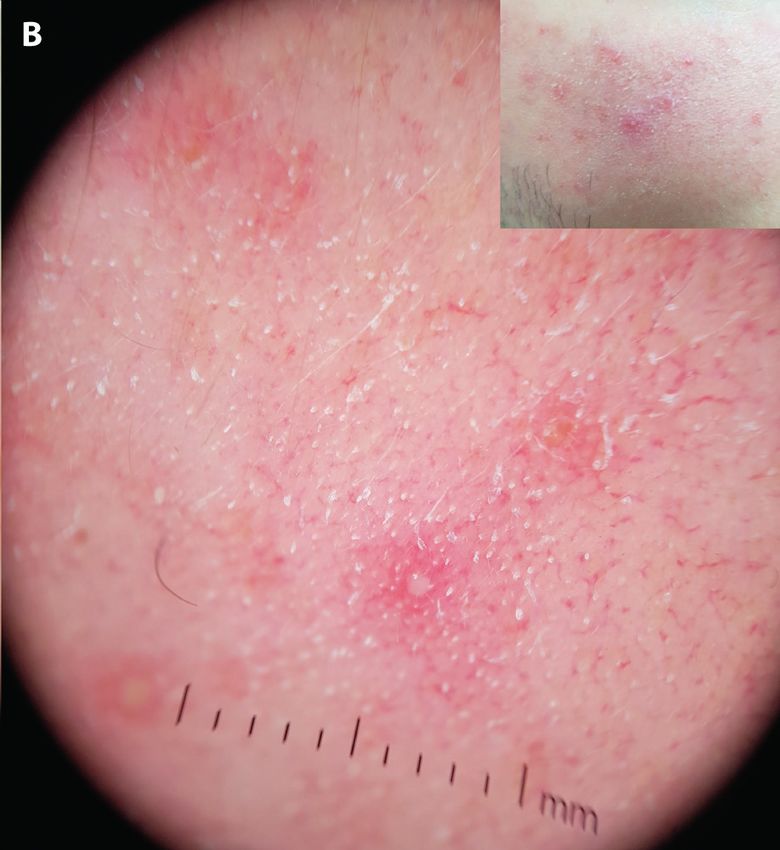
(P = 0,002) on dermoscopy. Examples of DT and DFO are
shown in Figure 2. Kappa value, sensitivity, and specificity of
DT and DFO are shown in Table 2. There were no statistically
Figure 1. (A) Demodex folliculorum. (B) Demodex brevis (original Figure 2. Examples of (A) Demodex follicular openings (stars) and
magnification ×400). (B) Demodex tails.
Research | Dermatol Pract Concept 2021;11(1):e2021139 3Table 2. P Value, Kappa Value, Sensitivity and Specifity of
Demodex Tail and Demodex Follicular Opening
Demodex (–) Demodex (+) P value Kappa Sensitivity Specifity
Demodex tail (–) 24 (72.7) 6 (12.0) 0.001* 0.617 0.88 0.73
(+) 9 (27.3) 44 (88.0)
Dilated (–) 22 (66.7) 16 (32.0) 0.002* 0.338 0.68 0.67
follicular (+) 11 (33.3) 34 (68.0)
opening
Table 3. Dermoscopic Features of Lesion Areas According to the Demodex Type
Demodex type
D folliculorum +
D folliculorum Total P value
D brevis
n (%)
n (%)
Scale (–) 13 (54.2) 5 (20.8) 18 0.017
(+) 11 (45.8) 19 (79.2) 30
Pustule (–) 20 (83.3) 12 (50.0) 32 0.032
(+) 4 (16.7) 12 (50.0) 16
Follicular plug (–) 17 (70.8) 13 (54.2) 30 0.371
(+) 7 (29.2) 11 (45.8) 18
Vascular structures (–) 4 (16.7) 8 (33.3) 12 0.317
(+) 20 (83.3) 16 (66.7) 36
Orangeyellowish area (–) 13 (54.2) 16 (66.7) 29 0.555
(+) 11 (45.8) 8 (33.3) 19
shown in Table 3. As reported by Lallas et al. some pustules (P = 0.001 and P = 0.002, respectively). The sensitivity and
that were not clinically noticeable could be detected in the specificity of DT and DFO were 0.88/0.73 and 0.68/0.67,
dermoscope [8]. Dilated follicles were not included in the respectively. There are a few studies on the relationship
statistical analysis because of low rate. between DT and DFO findings and Demodex in dermoscopy.
Lesion areas were analyzed according to the rosacea Segal et al. were the first to describe these 2dermoscopic fea-
subtype. The most common dermoscopic features in both ET tures associated with Demodex. The authors reported that
rosacea and PP rosacea were vascular structure (59.6% and the dermoscopy findings showed excellent agreement with
40.4%, respectively) and scale (54.1% and 45.9%, respec- the microscopic findings [7]. It was also reported that the
tively). The dermoscopic features of Demodex-positive and tails are less abundant in the inflammatory forms of demod-
negative lesion samples were compared and the results were icosis including rosacea-like demodicosis [7]. In a study con-
as follows: follicular plugging (87.5%), vascular structures ducted in patients with demodicosis including rosacea, it was
(71.4%), and orange-yellow areas (70.6%) were common in reported that DT was the only specific and sensitive criterion
Demodex-positive samples of ET rosacea. Although dilated in the diagnosis [11]. In another study, the sensitivity and
follicles were present in 100% of these samples, the number specificity of the DT were reported as 66.7% and 100%; the
of this dermoscopic feature was low (n=3). Scale (100%), sensitivity and specificity of the DFO were reported as 54.8%
orange-yellow areas (87.5%), and follicular plugging (84.6%) and 97%, respectively, in patients with Demodex-associated
were frequently detected dermoscopic findings in the Demo- folliculitis [12]. In the studies mentioned above, some of the
dex-positive samples of PP rosacea (Figure 3). results are not compatible with each other—including our
study. The reason for this may be the difference in the patient
groups studied.
Conclusions
The superficial layer of the horn layer and the piloseba-
We found that the dermoscopic findings of DT and DFO were ceous follicle content can be collected by SSSB. However, not
statistically significant in terms of the presence of Demodex all biotopes of D folliculorum can be collected with SSSB,
4 Research | Dermatol Pract Concept 2021;11(1):e202113920
15
DILATED FOLLICLES
10
5
FOLLICULAR PLUGS
0
VASCULAR STRUCTURES
VALUE
WHITE/YELLOWISH SCALES 20
PUSTULES 15
10
ORANGE-YELLOWISH AREAS
5
0
–)
–)
+)
+)
D(
D(
D(
D(
ET
PP
PP
ET
Figure 3. The frequency of dermoscopic features of the lesion samples with and without Demodex.
and it can cause false-negative results. Forton et al. offered Microsc Res Tech. 2005;68(5):284-289. DOI: 10.1002/
to perform a second SSSB at the same place [13]. Although jemt.20253. PMID: 16315233.
5. Forton F, Seys B. Density of Demodex folliculorum in rosacea:
DT was detected on dermoscopy, SSSB was negative in 9 of
a case-control study using standardized skin-surface biposy.
83 samples in our study. This may be because no sample was
Br J Dermatol. 1993;128(6):650-659. DOI: 10.1111/j.1365-
taken from the same place more than once. In addition, the 2133.1993.tb00261.x. PMID: 8338749.
number of samples taken from the control area was less than 6. Errichetti E, Stinco G. Dermoscopy in general dermatology: a
the number of samples taken from the lesion area. This is the practical overview. Dermatol Ther (Heidelb). 2016;6(4):471-507.
limitation of our study. DOI: 10.1007/s13555-016-0141-6.PMID: 27613297.
7. Segal R, Mimouni D, Feuerman H, Pagovitz O, David M.
In the D folliculorum + D brevis-positive samples, the
Dermoscopy as a diagnostic tool in demodicidosis. Int
rate of scale and pustule was higher compared to the D
J Dermatol. 2010;49(9):1018-1023. DOI: 10.1111/j.1365-
folliculorum-positive samples. Karadağ Köse et al. reported 4632.2010.04495.x. PMID: 20931672.
similarly that demodicosis might be suspected in the presence 8. Tan J, Almeida LMC, Bewley A, et al. Updating the diagnosis,
of epidermal scale [11]. classification and assessment of rosacea: recommendations from
The results of our study can generally be evaluated as the global ROSacea COnsensus (ROSCO) panel. Br J Dermatol.
2017;176(2):431-438. DOI: 10.1111/bjd.15122.PMID: 27718519.
follows: (1) Although DT and DFO indicate Demodex on der-
9. Lallas A, Argenziano G, Apalla Z, et al. Dermoscopic patterns of
moscopy, the sensitivity and specificity of DT are higher than
common facial inflammatory skin diseases. J Eur Acad Dermatol
the DFO. (2) Scale and pustule detection on dermoscopy may Venereol. 2014;28(5):609-614. DOI: 10.1111/jdv.12146. PMID:
indicate the coexistence of both D folliculorum and D brevis. 23489377.
10. Lallas A, Argenziano G, Longo C, et al. Polygonal vessels of rosacea
References are highlighted by dermoscopy. Int J Dermatol. 2014;53(5):e325-
327. DOI: 10.1111/ijd.12270.PMID: 23879349.
1. Ahn CS, Huang WW. Rosacea pathogenesis. Dermatol Clin. 11. Karadağ Köse Ö, Borlu M. Definition of videodermoscopic fea-
2018;36(2):81-86. DOI: 10.1016/j.det.2017.11.001.PMID: tures of demodicosis. Int J Dermatol. 2019;58(10):1153-1159.
29499802. DOI: 10.1111/ijd.14547. PMID: 31198996.
2. Desch C, Nutting WB. Demodex folliculorum (Simon) and D. bre- 12. Durdu M, Errichetti E, Eskiocak AH, Ilkit M. High accuracy of
vis akbulatova of man: redescription and reevaluation. J Parasitol. recognition of common forms of folliculitis by dermoscopy: An
1972;58(1):169-177. DOI: 10.2307/3278267. PMID: 5062457. observational study. J Am Acad Dermatol. 2019;81(2):463-471.
3. Rufli T, Mumcuoglu Y. The hair follicle mites D. folliculorum and D. DOI: 10.1016/j.jaad.2019.03.054.PMID: 30914342.
brevis: biology and medical importance. A review. Dermatologica. 13. Forton F, Song M. Limitations of standardized skin surface
1981;162(1):1-11. DOI: 10.1159/000250228. PMID: 6453029. biopsy in measurement of the density of Demodex folliculo-
4. Jing X, Shuling G, Ying L. Environmental scanning electron rum. A case report. Br J Dermatol. 1998;139(4):697-700. DOI:
microscopy observation of the ultrastructure of Demaodex. 10.1046/j.1365-2133.1998.02471.x.PMID: 9892917.
Research | Dermatol Pract Concept 2021;11(1):e2021139 5You can also read