The poor prognosis and influencing factors of high D dimer levels for COVID 19 patients - Nature
←
→
Page content transcription
If your browser does not render page correctly, please read the page content below
www.nature.com/scientificreports
OPEN The poor prognosis and influencing
factors of high D‑dimer levels
for COVID‑19 patients
Xiaokang He1,5, Fei Yao2,5, Jie Chen1,5, Yan Wang1, Xiangming Fang3, Xuan Lin4, Hui Long1*,
Qiang Wang2* & Qingming Wu1,2*
To explore the value, and influencing factors, of D-dimer on the prognosis of patients with COVID-19.
A total of 1,114 patients with confirmed COVID-19 who were admitted to three designated COVID-
19 hospitals in Wuhan, China from January 18, 2020, to March 24, 2020, were included in this study.
We examined the relationship between peripheral blood levels of D-dimer, and clinical classification
and prognosis, as well as its related influencing factors. D-dimer levels were found to be related to
the clinical classification and the prognosis of clinical outcome. D-dimer levels were more likely to be
abnormal in severely and critically ill patients compared with mild and ordinary cases, while D-dimer
levels of patients who had died were significantly higher than those of surviving patients according to
the results of the first and last lab tests. The results from ROC analyses for mortality risk showed that
the AUCs of D-dimer were 0.909, YI was 0.765 at the last lab test, and a D-dimer value of 2.025 mg/L
was regarded to be the optimal probability cutoff for a prognosis of death. In addition, we found that
patients with advanced age, male gender, dyspnea symptoms, and some underlying diseases have a
higher D-dimer value (p < 0.05). In short, D-dimer is related to the clinical classification and can be used
to evaluate the prognosis of COVID-19 patients. The D-dimer value of 2.025 mg/L was the optimal
probability cutoff for judging an outcome of death. Advanced age, male gender, dyspnea symptoms,
and some underlying diseases are influencing factors for D-dimer levels, which impacts the prognosis
of patients.
Corona Virus Disease 2019 (COVID-19), caused by the new coronavirus (SARS-CoV-2), is spread mainly via
respiratory droplets and close contact with highly infectious p eople1. COVID-19 primarily causes lung injury,
and most patients have a good prognosis, but the condition of some patients with severe infections and who are
critically ill, worsens rapidly, resulting in coagulation dysfunction and even d eath2,3. Therefore, an early iden-
tification of the possible prognosis is very important for the clinical diagnosis and treatment of the COVID-19
patients. In a previous single-center, prospective study of COVID-19 patients, we observed the dynamic changes
of in the peripheral blood coagulation function indices: D-dimer (DD); prothrombin time (PT); activated partial
thromboplastin time (APTT); and fibrinogen (Fg). We found that D-dimer levels in particular, could be used
to predict the severity and prognosis of COVID-194. Based on this, we expanded the scope of our sample col-
lection, conducted a multi-center and retrospective analysis, and further systematically studied the value and
influencing factors of using D-dimer levels in the clinical classification and prognosis of COVID-19 patients.
Materials and methods
Source of patients and diagnosis criteria. We conducted a retrospective study focusing on the signifi-
cance of D-dimer in evaluating the severity and prognosis of COVID-19. A total of 1,114 COVID-19 patients
with a positive nucleic acid test for SARS-CoV-2 were collected from the COVID-19 designated hospitals in
Wuhan, Hubei Province, China, including Tianyou Hospital Affiliated to Wuhan University of Science and
Technology, Puren Hospital Affiliated to Wuhan University of Science and Technology, and China Resources
& WISCO General Hospital between January 18, 2020, and March 24, 2020. Among them, 115 patients with
1
Tianyou Hospital, Wuhan University of Science and Technology, Wuhan 430064, China. 2Institute of Infection,
Immunology and Tumor Microenvironment, Hubei Province Key Laboratory of Occupational Hazard Identification
and Control, Medical College, Wuhan University of Science and Technology, Wuhan 430065, China. 3Department
of Gastroenterology, Puren Hospital, Wuhan University of Science and Technology, Wuhan 430081, China. 4China
Resources & WISCO General Hospital, Wuhan 430081, China. 5These authors contributed equally: Xiaokang He, Fei
Yao, and Jie Chen. *email: longhui@wust.edu.cn; wangqiang@wust.edu.cn; wuhe9224@sina.com
Scientific Reports | (2021) 11:1830 | https://doi.org/10.1038/s41598-021-81300-w 1
Vol.:(0123456789)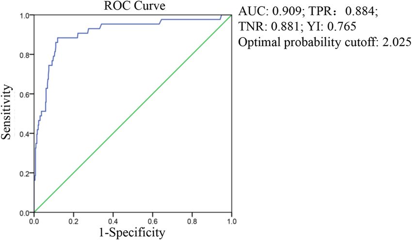
www.nature.com/scientificreports/

                                            COVID-19 confirmed in Tianyou Hospital have been reported in our previous study. Pregnancy patients were
                                            excluded from the study. The study was approved by the Medical Ethics Review Board of Wuhan University of
                                            Science and Technology (No. 202009), all patients involved in this study were fully informed and each provided
                                            a written informed consent. In addition, written informed consent was obtained from the legally authorized
                                            representatives or next of kin of the patients who had died. The patient data used in this study contained no
                                            personal or identifying information. All methods were performed in accordance with the relevant guidelines
                                            and regulations.

                                            Case classification. According to the COVID-19 diagnosis and treatment guidelines (seventh version)
                                            issued by the National Health Commission of China, COVID-19 patients are divided into four categories: mild;
                                            ordinary; severe; and critical. For our research purposes, we combined the mild and ordinary categories into a
                                            single mild or ordinary type.

                                            Outcome of illness. Illness outcomes were divided into: hospital discharge; improved; exacerbation; and
                                            death, according to clinical progression. We combined hospital discharge, improved and exacerbation into sur-
                                            vival for the purpose of this study.

                                            Experimental data collection. A retrospective analysis of the COVID-19 patients admitted from between
                                            January 18, 2020 to and March 24, 2020 was undertaken. The D-dimer values were determined using D-dimer
                                            Assay kits (Long Island Biotech, Shanghai, China) by automatic blood coagulation analyzer mindray ExC810
                                            (Shenzhen, China) according to the operating manual. The first D-dimer test results were obtained at admission
                                            or within 1–3 days of hospitalization, and the last D-dimer test results were from before discharge or death, or
                                            from the last test during hospitalization.

                                            Statistical methods. SPSS software (SPSS 24.0 for Windows, IBM, Chicago, IL, USA) was used for statisti-
                                            cal analysis, and quantitative variables were expressed as the mean plus standard deviation (SD). Comparison
                                            between groups were performed using a t test and Mann Whitney nonparametric U test or Kruskal–Wallis
                                            H-test. The differences between groups of enumeration data were analyzed using a chi-square test. The Receiver
                                            Operating Characteristic curve (ROC) was used to calculate the area under the D-dimer curve, to evaluate the
                                            sensitivity and specificity in predicting mortality and hospital discharge. p < 0.05 was considered statistically
                                            significant.

                                            Ethical approval. This study was approved by the Medical Ethics Review Board of Wuhan University of
                                            Science and Technology (No. 202009).

                                            Consent to participate. Informed consent was obtained from all patients to be included in the study.

                                            Consent for publication. All authors approved the manuscript and gave their consent for publication.

                                            Results
                                            A total of 1,114 cases were finally selected in this study. 885 cases and 471 cases were tested for D-dimer at the
                                            first and last lab test, respectively.

                                            Demographic characteristics and clinical symptoms of D‑dimer patients for the first detec‑
                                            tion.. Table 1 lists the general demographic characteristics and the clinical symptoms of the 885 COVID-19
                                            patients who received a D-dimer test as part of the initial lab test. Most patients with abnormal D-dimer values
                                            were over 60 years old and have a higher average age (p < 0.001). There was no significant difference in gender
                                            and length of hospital stay. The clinical symptoms presented by patients at admission were mainly fever (73.4%)
                                            and cough (67.0%). Patients with abnormal D-dimer values were more likely to have clinical manifestations of
                                            dyspnea, gastrointestinal and mental symptoms (p < 0.05), and to have some previous underlying condition,
                                            such as hypertension, renal insufficiency, cerebrovascular disease, or traumatic fracture (p < 0.05).

                                            The relationship between D‑dimer level and clinical classification. There were significant differ-
                                            ences in the first and last D-dimer test results for the different clinical classifications (p < 0.001). As shown in
                                            Table 2, D-dimer values were more likely to be abnormal in patients with severe and critical cases, than for those
                                            with mild or ordinary cases.

                                            The relationship between D‑dimer level and disseminated intravascular coagulation. We
                                            explored the relationship between D-dimer levels and patients that meet ISTH criteria for disseminated intra-
                                            vascular coagulopathy (DIC). As shown in Table 3, the D-dimer levels of COVID-19 patients with DIC who met
                                            the ISTH diagnostic criteria were higher than for those without DIC at the first and last lab test, and the differ-
                                            ences were significant.

                                            The relationship between D‑dimer levels and prognosis for COVID‑19 patients. The final out-
                                            comes for COVID-19 patients were divided into either survival or death. Significant differences were found
                                            between D-dimer levels and outcomes at endpoints at the first and last test (p < 0.001). In addition, D-dimer

          Scientific Reports |   (2021) 11:1830 |                https://doi.org/10.1038/s41598-021-81300-w                                                   2

Vol:.(1234567890)
www.nature.com/scientificreports/
Demographic N (%) D-dimer < 0.5 mg/L D-dimer ≥ 0.5 mg/L P
Total 885 (100.0%) 556 329
Age (years)
≤ 60 427 (48.2%) 326 101 < 0.001
> 60 458 (51.8%) 230 228
Average age ( x ± s) 58.83 ± 14.77 55.73 ± 14.48 64.08 ± 13.77 < 0.001
Sex
Male 431 (48.7%) 272 159 0.87
Female 454 (51.3%) 284 170
Average hospital stay (days) 16.16 ± 7.89 16.00 ± 7.71 16.47 ± 8.19 0.37
Clinical manifestations
Fever 650 (73.4%) 409 241 0.92
Cough 593 (67.0%) 381 212 0.21
Chest tightness 235 (26.6%) 150 85 0.71
Difficulty breathing 233 (26.3%) 131 102 < 0.05
Gastrointestinal symptoms 64 (7.2%) 49 15 < 0.05
Spiritual consciousness 8 (0.9%) 1 7 < 0.05
Past medical history
Hypertension 288 (32.5%) 160 128 < 0.05
Coronary heart disease 72 (8.1%) 39 33 0.11
Diabetes 97 (11.0%) 56 41 0.27
Renal insufficiency 20 (2.3%) 6 14 < 0.05
Cerebrovascular disease 53 (6.0%) 18 35 < 0.001
Malignant tumor 23 (2.6%) 16 7 0.50
Surgical history 86 (9.7%) 60 26 0.16
Traumatic fracture 16 (1.8%) 6 10 < 0.05
Table 1. General demographic characteristics and clinical manifestations of 885 COVID-19 patients with
D-dimer (first lab test). Normal range of D-dimer value:www.nature.com/scientificreports/
D-dimer x ± s (n)
N Survival (n) Death (n) t/p H(p)
The first test 885 0.80 ± 2.08(820) 3.92 ± 8.29(65) − 3.02(< 0.05) < 0.001
The last test 471 1.17 ± 2.67(428) 10.74 ± 11.74(43) − 5.33(< 0.001) < 0.001
Table 4. Relationship between D-dimer levels and the progression of disease.
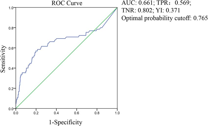
Figure 1. ROC curve of D-dimer from the first test, in predicting hospital discharge and mortality.
Figure 2. ROC curve of D-dimer from the last test, in predicting hospital discharge and mortality.
Influencing factors of D‑dimer levels. The patients whose D-dimer levels were measured at the last lab
test were divided into two groups with 2.025 mg/L as the critical value. The 382 patients with D-dimer levels
less than the critical value were called the former group, and the 89 patients with levels greater than or equal
to the critical value were called the latter group. The average age, male ratio, and proportion of patients with
dyspnea or spiritual consciousness in the latter group were significantly higher. However, the number of people
with spiritual consciousness in these groups was relatively small, so a larger sample is still needed to support
this conclusion. Additionally, hypertension, coronary heart disease, diabetes, and cerebrovascular disease were
statistically significant in both groups (Table 5). Overall, advanced age, male gender, and underlying diseases
such as hypertension, coronary heart disease, diabetes, and cerebrovascular disease were influencing factors of
D-dimer levels which affects the prognosis for COVID-19 patients.
Scientific Reports | (2021) 11:1830 | https://doi.org/10.1038/s41598-021-81300-w 4
Vol:.(1234567890)www.nature.com/scientificreports/
D-dimer < 2.025 (mg/L) D-dimer ≥ 2.025 (mg/L)
n = 382 n = 89
Influencing factors N M ± SD N (%) M ± SD N (%) t/χ2 (p) H (p)
Age (years) 471 59.53 ± 14.23 382 (100%) 68.55 ± 11.76 89 (100%) − 6.25 (< 0.001) < 0.001
Sex
Male 242 176 (46.1%) 66 (74.2%) 22.79 (< 0.001)
Female 229 206 (53.9%) 23 (25.8%)
Average hospital stay (days) 471 18.13 ± 7.61 382 (100%) 18.21 ± 10.08 89 (100%) − 0.75 (0.94) 0.65
Clinical manifestations
Fever 361 290 (75.9%) 71 (79.8%) 0.60 (0.44)
Cough 322 263 (68.8%) 59 (66.3%) 0.23 (0.64)
Chest tightness 140 118 (30.9%) 22 (24.7%) 1.32 (0.25)
Difficulty breathing 144 101 (26.4%) 43 (48.3%) 14.7 (< 0.001)
Gastrointestinal symptoms 38 31 (8.1%) 7 (7.9%) 0.06 (0.94)
Spiritual consciousness 5 2 (0.5%) 3 (3.4%) (< 0.05)
Past medical history
Hypertension 159 114 (29.8%) 45 (50.6%) 13.86 (< 0.001)
Coronary heart disease 46 30 (7.9%) 16 (18.0%) 8.40 (< 0.01)
Diabetes 62 44 (11.5%) 18 (20.2%) 4.79 (< 0.05)
Renal insufficiency 7 6 (1.6%) 1 (1.1%) (1.00)
Cerebrovascular disease 34 21 (5.5%) 13 (14.6%) 8.94 (< 0.01)
Malignant tumor 13 8 (2.1%) 5 (5.6%) 3.34 (0.07)
Surgical history 42 34 (8.9%) 8 (9.0%) 0.00 (0.98)
Traumatic fracture 10 8 (2.1%) 2 (2.2%) (1.00)
Total 225 165(43.2%) 60(67.4) 16.97 (< 0.001)
Table 5. Influencing factors of D-dimer.
Discussion
The clinical symptoms of COVID-19 patients were mainly fever, most being mild cases, and a few severe cases.
The condition and prognosis of COVID-19 patients were complicated due to the variety of symptoms and imag-
ing findings, and the varying degree of disease p rogression4. Notably, some severe and critical cases, as well as
patients who had died, had differing degrees of coagulation dysfunction. Autopsy, and puncture histopathological
observations revealed thrombus or microthrombus in the lungs, heart and liver5–7. In our previous study, the
dynamic changes in the peripheral blood coagulation function indices (D-dimer, PT, APTT and Fg) of COVID-
19 patients in a single-center and prospective study were observed, which indicated that D-dimer levels could
be used for predicting the severity and prognosis of COVID-194.
D-dimer is the product of fibrinolytic degradation of fibrin, and elevated levels indicate that there is a hyper-
coagulable state and secondary fibrinolysis in the body, which is extremely useful for the diagnosis of thrombotic
diseases. Patients with COVID-19 were reported to have a hypercoagulable s tate8, with 71% of patients who died
from COVID-19 were found to have met the DIC standard, this ratio among surviving patients was only 0.6%2.
In addition, the incidence of venous thromboembolism (VTE) in patients with severe COVID-19 was 25%,
and 30% of COVID-19 patients were diagnosed with pulmonary e mbolism9,10. D-dimer levels in the blood of
COVID-19 patients with ischemic stroke were also i ncreased11.
The results of this study showed that there were 329 (37.2%) and 256 (54.4%) patients with abnormal D-dimer
values detected at the first and last test, respectively, indicating the presence of hypercoagulability in COVID-19
patients, which is consistent with the results reported above. The patients were divided into two groups based
on a D-dimer value of 0.5 mg/L being the cut-off value. D-dimer values were significantly correlated with the
clinical classification of the patients at the first and last test. The patients were then divided into survival and
death groups according to the final outcome, and the significant differences between D-dimer and final outcomes
were observed (p < 0.001). The D-dimer levels of the dead patients were significantly higher than those of surviv-
ing cases, and the D-dimer of COVID-19 patients with DIC who met the ISTH diagnostic criteria were higher
than those without DIC at the first and last lab test, indicating that D-dimer levels have predictive value for the
prognosis of patients. A higher D-dimer value indicated that the condition of the patient may be more serious,
and these patients even combined with other serious complication. Possible reasons for increased D-dimer val-
ues for COVID-19 patients are: (1) infection can cause the release of pro-inflammatory cytokines, thus causing
an inflammatory s torm12. The levels of pro-inflammatory cytokines, such as IL-2, IL-7, G-CSF, IP-10, MCP-1,
MIP-1A and TNF-α in plasma were higher especially in severe COVID-19 patients, and T cells, macrophages
and natural killer cells rapidly proliferate and are highly activated, accompanied by overproduction of immune or
non-immune defense cells and the release of more than 150 inflammatory cytokines and chemical m ediators13–16.
These may induce endothelial cell dysfunction, resulting in damage to the microvascular system, and abnormal
activation of the coagulation system, pathological manifestations of systemic small vessel vasculitis and extensive
microthrombosis17. (2) Some patients with COVID-19 have different degrees of hypoxia, and inflammation can
Scientific Reports | (2021) 11:1830 | https://doi.org/10.1038/s41598-021-81300-w 5
Vol.:(0123456789)www.nature.com/scientificreports/
lead to thrombosis or increased oxygen c onsumption18. Absolute oxygen demand increases during abnormal
hemodynamics, which triggers molecular and cellular pathways and leads to t hrombosis19. (3) Severe infection,
or acute inflammation caused by sepsis, can also affect blood coagulation, such as increased levels of plasminogen
activator inhibitor 1 (PAI-1), and excessive inhabited fibrinolysis20, which will eventually activate the coagulation
cascade, and inhibit fibrinolysis as well as promoting thrombosis.
After grouping patients according to a D-dimer value of 2.025 mg/L as the critical value, we found that
advanced age, male gender, dyspnea, hypertension, coronary heart disease, diabetes, and cerebrovascular dis-
ease can affect D-dimer levels and prognosis (p < 0.05). In addition, gastrointestinal and mental consciousness
symptoms, renal insufficiency, and traumatic fractures were also related to D-dimer levels. Patients with mental
consciousness symptoms indicated critical illness; over 50% of patients with chronic renal insufficiency already
had uremia hemodialysis status. Both of these groups of patients had poor prognosis and high mortality. The
COVID-19 patients with gastrointestinal symptoms and trauma fractures may have abnormal D-dimer values.
D-dimer levels change with age. Male patients were more likely to have a history of smoking, which might be an
influential factor. The number of patients with dyspnea was 47.2%, considering the combination of acute respira-
tory distress (ARDS). COVID-19 patients with hypertension and coronary heart disease might have a higher
risk of death. Hypertension and COVID-19 may be related to the role of A CE221. In a recent study, it was found
that pericytes express high levels of ACE2 in the heart. The damage of pericytes caused by viral infections can
lead to capillary vascular cell dysfunction and microvascular dysfunction22, which seems to explain the possible
causes of acute coronary syndrome (ACS) in COVID-19 patients. Diabetes and cerebrovascular disease are risk
factors affecting prognosis, COVID-19 patients with diabetes had higher D-dimer values, higher inflammatory
markers, and a worse prognosis than those without d iabetes23. In COVID-19 patients with a history of previ-
ous cerebrovascular disease (mostly cerebral infarction), there is an existing risk of atherosclerosis and emboli
falling off that can lead to a hypercoagulable state. These patients also had higher levels of D-dimer. The study
also showed that patients with additional underlying diseases had higher D-dimer levels, and a worse prognosis.
In summary, the results of this multi-center clinical study showed that D-dimer is related to the clinical
classification of COVID-19 patients and can be used to evaluate the prognosis of patients. The D-dimer value
of 2.025 mg/L is an optimal probability cutoff for judging the risk of death. After grouping according to this
value, advanced age, male gender, dyspnea symptoms, and underlying diseases such as hypertension, coronary
heart disease, diabetes, cerebrovascular disease became the influencing factors of D-dimer value, impacting the
prognosis of patients. COVID-19 patients with the above-mentioned influencing factors have a higher risk of
death. It is important to dynamically monitor D-dimer levels, to detect thrombotic complications as soon as
possible, and take corresponding preventive measures to reduce thromboembolism and the risk of hemorrhage
in DIC secondary fibrinolysis, thus reducing the mortality rate of COVID-19.
Data availability
The datasets generated and analyzed during the study are available from the corresponding author upon reason-
able request.
Received: 16 August 2020; Accepted: 5 January 2021
References
1. Guan, W. J. et al. Clinical characteristics of coronavirus disease 2019 in China. N. Engl. J. Med. 382(1708–1720), 2020. https://doi.
org/10.1056/NEJMoa2002032 (2019).
2. Tang, N., Li, D., Wang, X. & Sun, Z. Abnormal coagulation parameters are associated with poor prognosis in patients with novel
coronavirus pneumonia. J. Thromb. Haemost.: JTH 18, 844–847. https://doi.org/10.1111/jth.14768 (2020).
3. Zhu, J. et al. Clinical characteristics of 3,062 COVID-19 patients: a meta-analysis. J. Med. Virol. https://doi.org/10.1002/jmv.25884
(2020).
4. Long, H. et al. D-Dimer and prothrombin time are the significant indicators of severe COVID-19 and poor prognosis. Biomed.
Res. Int. 2020, 6159720. https://doi.org/10.1155/2020/6159720 (2020).
5. Fox, S. E. et al. Pulmonary and cardiac pathology in African American patients with COVID-19: an autopsy series from New
Orleans. Lancet Respir. Med. https://doi.org/10.1016/s2213-2600(20)30243-5 (2020).
6. Edler, C. et al. Dying with SARS-CoV-2 infection—an autopsy study of the first consecutive 80 cases in Hamburg, Germany. Int.
J. Legal Med. 134, 1275–1284. https://doi.org/10.1007/s00414-020-02317-w (2020).
7. Lax, S. F. et al. Pulmonary arterial thrombosis in COVID-19 with fatal outcome: results from a prospective, single-center, clinico-
pathologic case series. Ann. Intern. Med. https://doi.org/10.7326/m20-2566 (2020).
8. Spiezia, L. et al. COVID-19-related severe hypercoagulability in patients admitted to intensive care unit for acute respiratory failure.
Thromb. Haemost. 120, 998–1000. https://doi.org/10.1055/s-0040-1710018 (2020).
9. Cui, S., Chen, S., Li, X., Liu, S. & Wang, F. Prevalence of venous thromboembolism in patients with severe novel coronavirus
pneumonia. J. Thromb. Haemost.: JTH 18, 1421–1424. https://doi.org/10.1111/jth.14830 (2020).
10. Leonard-Lorant, I. et al. Acute pulmonary embolism in COVID-19 patients on CT angiography and relationship to D-Dimer
levels. Radiology https://doi.org/10.1148/radiol.2020201561 (2020).
11. Beyrouti, R. et al. Characteristics of ischaemic stroke associated with COVID-19. J. Neurol. Neurosurg. Psychiatry https://doi.
org/10.1136/jnnp-2020-323586 (2020).
12. Chousterman, B. G., Swirski, F. K. & Weber, G. F. Cytokine storm and sepsis disease pathogenesis. Semin. Immunopathol. 39,
517–528. https://doi.org/10.1007/s00281-017-0639-8 (2017).
13. Chen, G. et al. Clinical and immunological features of severe and moderate coronavirus disease 2019. J. Clin. Investig. 130,
2620–2629. https://doi.org/10.1172/jci137244 (2020).
14. Mehta, P. et al. COVID-19: consider cytokine storm syndromes and immunosuppression. Lancet (London, England) 395, 1033–
1034. https://doi.org/10.1016/s0140-6736(20)30628-0 (2020).
15. Wong, J. P. et al. Current and future developments in the treatment of virus-induced hypercytokinemia. Future Med. Chem. 9,
169–178. https://doi.org/10.4155/fmc-2016-0181 (2017).
Scientific Reports | (2021) 11:1830 | https://doi.org/10.1038/s41598-021-81300-w 6
Vol:.(1234567890)www.nature.com/scientificreports/
16. Li, H. et al. Serum Amyloid A is a biomarker of severe coronavirus disease and poor prognosis. J. Infect. 80, 646–655. https://doi.
org/10.1016/j.jinf.2020.03.035 (2020).
17. Tian, S. et al. Pulmonary pathology of early-phase 2019 novel coronavirus (COVID-19) pneumonia in two patients with lung
cancer. J. Thoracic Oncol.: Off. Publ. Int. Assoc. Study Lung Cancer 15, 700–704. https://doi.org/10.1016/j.jtho.2020.02.010 (2020).
18. Gupta, N., Zhao, Y. Y. & Evans, C. E. The stimulation of thrombosis by hypoxia. Thromb. Res. 181, 77–83. https: //doi.org/10.1016/j.
thromres.2019.07.013 (2019).
19. Pugh, C. W. & Ratcliffe, P. J. New horizons in hypoxia signaling pathways. Exp. Cell Res. 356, 116–121. https://doi.org/10.1016/j.
yexcr.2017.03.008 (2017).
20. Iba, T. et al. Diagnosis and management of sepsis-induced coagulopathy and disseminated intravascular coagulation. J. Thromb.
Haemost.: JTH 17, 1989–1994. https://doi.org/10.1111/jth.14578 (2019).
21. Guzik, T. J. et al. COVID-19 and the cardiovascular system: implications for risk assessment, diagnosis, and treatment options.
Cardiovasc. Res. https://doi.org/10.1093/cvr/cvaa106 (2020).
22. Chen, L., Li, X., Chen, M., Feng, Y. & Xiong, C. The ACE2 expression in human heart indicates new potential mechanism of heart
injury among patients infected with SARS-CoV-2. Cardiovasc. Res. 116, 1097–1100. https://doi.org/10.1093/cvr/cvaa078 (2020).
23. Guo, W. et al. Diabetes is a risk factor for the progression and prognosis of COVID-19. Diabetes/Metab. Res. Rev. https://doi.
org/10.1002/dmrr.3319 (2020).
Author contributions
Q.M.W., Q.W. and H.L. designed the study. X.K.H. and F.Y. wrote the main manuscript text. J.C., Y.W., X.M.F.
and X.L. collected data. X.K.H. and F.Y. performed statistical analysis. All authors reviewed the manuscript, and
agreed with its submission.
Competing interests
The authors declare no competing interests.
Additional information
Correspondence and requests for materials should be addressed to H.L., Q.W. or Q.W.
Reprints and permissions information is available at www.nature.com/reprints.
Publisher’s note Springer Nature remains neutral with regard to jurisdictional claims in published maps and
institutional affiliations.
Open Access This article is licensed under a Creative Commons Attribution 4.0 International
License, which permits use, sharing, adaptation, distribution and reproduction in any medium or
format, as long as you give appropriate credit to the original author(s) and the source, provide a link to the
Creative Commons licence, and indicate if changes were made. The images or other third party material in this
article are included in the article’s Creative Commons licence, unless indicated otherwise in a credit line to the
material. If material is not included in the article’s Creative Commons licence and your intended use is not
permitted by statutory regulation or exceeds the permitted use, you will need to obtain permission directly from
the copyright holder. To view a copy of this licence, visit http://creativecommons.org/licenses/by/4.0/.
© The Author(s) 2021
Scientific Reports | (2021) 11:1830 | https://doi.org/10.1038/s41598-021-81300-w 7
Vol.:(0123456789)You can also read