Pure Talocrural Dislocation without Associated Malleolar Fracture
←
→
Page content transcription
If your browser does not render page correctly, please read the page content below
Open Journal of Orthopedics, 2020, 10, 13-20
                                                                                                     https://www.scirp.org/journal/ojo
                                                                                                                ISSN Online: 2164-3016
                                                                                                                  ISSN Print: 2164-3008

Pure Talocrural Dislocation without
Associated Malleolar Fracture

Souleymane Diao, Amadou Ndiassé Kasse*, Jean Claude Sane, Joseph Davy Diouf,
Abdoulaye Keita, Pape Matar Fall, Pape Alkaly Diouf, Ndiaga Dieye, Mouhamadou Habib Sy

Hôpital Général Idrissa Pouye, University Cheikh Anta Diop, Dakar, Senegal

How to cite this paper: Diao, S., Kasse,            Abstract
A.N., Sane, J.C., Diouf, J.D., Keita, A., Fall,
P.M., Diouf, P.A., Dieye, N. and Sy, M.H.           Pure talocrural dislocation is an uncommon injury of the ankle. Malleolar
(2020) Pure Talocrural Dislocation without
                                                    fracture is usually associated. We report two cases of pure talocrural disloca-
Associated Malleolar Fracture. Open Jour-
nal of Orthopedics, 10, 13-20.                      tion, to describe its therapeutic and prognostic clinical aspects through a re-
https://doi.org/10.4236/ojo.2020.101003             view of the literature.

Received: November 12, 2019
                                                    Keywords
Accepted: January 6, 2020
Published: January 9, 2020
                                                    Talocrural, Pure Dislocation, Closed Reduction
Copyright © 2020 by author(s) and
Scientific Research Publishing Inc.
This work is licensed under the Creative
Commons Attribution International                 1. Introduction
License (CC BY 4.0).
http://creativecommons.org/licenses/by/4.0/       Pure talocrural dislocation is the dislocation of the ankle. The talocalcaneal block
                 Open Access                      is expelled out of the mortise which is intact both with regard to malleoli and
                                                  syndesmosis [1].
                                                     It is a simple dislocation compared to subtalar dislocation, which is a double
                                                  dislocation, and enucleation of the talus which is a triple dislocation.
                                                     This is a very rare lesion that occurs in a context of violent trauma. Histori-
                                                  cally, the first case was published in 1912 by Auvray [2]. It is an emergency
                                                  whose management, apart from complications (cutaneous opening, vascular and
                                                  nerve lesions), is most often orthopedic: reduction under general anesthesia fol-
                                                  lowed by a restraint by plaster cast.
                                                     Pure talocrural dislocation can compromise the functional development of the
                                                  ankle due to late complications such as stiffness of the ankle, osteoarthritis or
                                                  necrosis of the talus.
                                                     The goal of this work was to report two cases of pure talocrural dislocation, to
                                                  describe its therapeutic and prognostic clinical aspects through a review of the
                                                  literature.

DOI: 10.4236/ojo.2020.101003 Jan. 9, 2020                              13                                  Open Journal of Orthopedics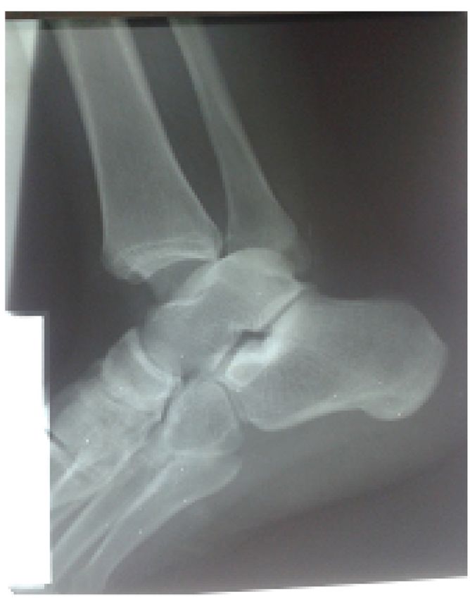
S. Diao et al.
2. Our Observations
Observation 1:
Mr. H. S. D. 32 years old, with no significant pathological history, has been
received at H1 from a sports-related life-style accident (football). He would have
done a faux pas with the foot in inversion. Physical examination at admission
noted deformity and swelling of the left ankle without cutaneous opening or
vasculoneural involvement.
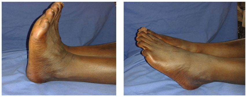
The palpation and the mobilization of the ankle caused a sharp pain. The
X-ray of the ankle showed pure posterior talocrural dislocation without asso-
ciated bone lesion (Figure 1).
The reduction was carried out in an emergency by maneuvering a boot jack.
The control X-ray confirmed the reduction of dislocation (Figure 2).
(a) (b)
Figure 1. X-rays of the ankle showing pure posterior Talocrural dislocation ((a) face; (b)
profile).
(a) (b)
Figure 2. Post reduction control X-ray ((a) face; (b) profile).
DOI: 10.4236/ojo.2020.101003 14 Open Journal of Orthopedics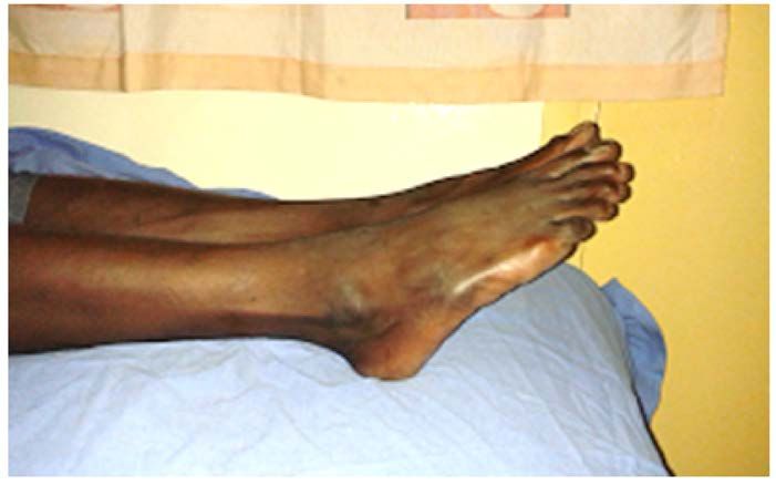
S. Diao et al.

                                 The arterial duplex scan of the limb was normal. The restraint has been en-
                               sured by a plastered boot for 6 weeks. Rehabilitation of the ankle has been
                               started as soon as the cast was removed.
                                 After 2 years of hindsight, the patient had no pain, and he had resumed his
                               sporting and professional activities. The functional results (Figure 3), judged on
                               the Gay and Evrard score (Table 1), were excellent with a score of 15 points. The
                               X-rays of the ankle were normal.

                               Figure 3. Pictures showing normal mobility of the ankle in hindsight of 2 years.

                               Table 1. Score of gay and Evrard.

                                                                   Criteria of Gay and Evrard
                                                                 Subjective Criteria                                      Points
                                                                                 Absence of pain                            3
                                                                             Pain on uneven ground                          2
                                           Pain
                                                                          Function pain limiting activity                   1
                                                                           Pain preventing any activity                     0
                                                                              Absence of instability                        3
                                                                           Instability on rough ground                      2
                                        Instability
                                                                        Awkward and insecurity Instability                  1
                                                                            Instability requiring a stick                   0
                                                                  Objective Criteria
                                                                                 Normal mobility                            3
                                                                 Mobility equal to or greater than 50% of normal            2
                                         Mobility
                                                                         Mobility less than 50% of normal                   1
                                                                           Ankylosis or foot deflection                     0
                                                                                Absence of edema                            3

                                       Edema and                           Mild or intermittent edema                       2
                                    trophic disorders                      Important edema to fatigue                       1
                                                                         Important and permanent edema                      0
                                                                          Identical activity or profession                  3
                                                                Retained profession with adjustment of the position         2
                                   Professional activity
                                                                         Change of profession or activity                   1
                                                                         Impossible professional activity                   0

DOI: 10.4236/ojo.2020.101003                               15                                       Open Journal of Orthopedics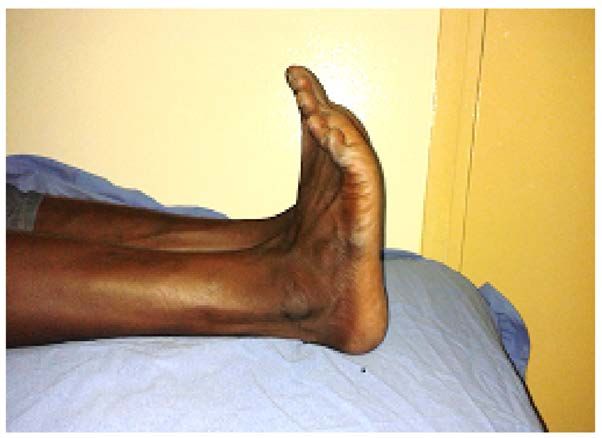
S. Diao et al.
− Disappearance of tibiotalar spacing and decrease in height of external mal-
leolus (a);
− Displacement of the talo-calcaneo-pedal block behind the tibia (b);
− The malleoluses are intact on both pictures.
Observation 2:
Mr. B. M. S., 29 years old, with no pathological history reported, was received
at H1 from a home-related accident; he would have done a faux pas when com-
ing down the stairs with foot in inversion. Physical examination at admission
noted edema and deformity of the left ankle. The palpation of the ankle was
painful. There was no cutaneous opening or vasculoneural lesion.
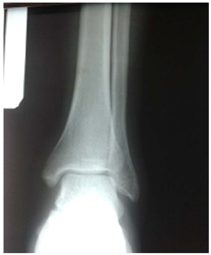
The X-ray of the ankle showed posterior pure tibiotalar dislocation with talo-
navicular sprain (Figure 4).
The orthopedic reduction was performed as an emergency, confirmed by an
ankle control X-ray (Figure 5).
The restraint has been ensured by a plastered boot for 6 weeks followed by
reeducation of the ankle. The patient had resumed his activities. He had no pain
or limitation of ankle mobility (Figure 6). The functional results were consi-
dered very good according to the score of Gay and Evrard modified by Elisé
(score = 15), in hindsight of 2 years.
The X-rays of the ankle (face and profile) did not show calcification with re-
gard to ligaments or osteoarthritis.
− Disappearance of joint space;
− Displacement of the talo-calcaneo-pedal block behind the tibia;
− Removal of the talonavicular ligament;
− Two malleoli are intact.
The result was as the following:
(a) (b)
Figure 4. Ankle X-ray showing posterior pure tibiotalar dislocation with talonavicular
sprain ((a) face; (b) profile).
DOI: 10.4236/ojo.2020.101003 16 Open Journal of Orthopedics
S. Diao et al.
(a) (b)
Figure 5. Post reduction control X-ray ((a) face; (b) profile).
Figure 6. Pictures showing normal mobility of the ankle in hindsight of 2 years.
− bad (score ≤ 4);
− passable (score between 5 and 9);
− good (score between 10 and 14) ;
− and excellent (score = 15).
3. Comments
Epidemiology
We have only found two cases over a period of 15 years.
Pure talocrural dislocation is exceptional [3] [4]. The first case documented by
X-ray was described in 1913 by Peraire [1]. The work reported in the literature
on this lesion is few [5]. Until 1995, only 73 cases were reported [6]. Most stu-
dies are based on clinical facts that rarely exceed 2 cases.
Only two series of more than 10 cases [1] [7] have been reported.
The scarcity of this lesion is due to the mechanical stability of the tibiofibular
mortise and the resistance of the collateral ligaments, which are as strong as the
malleolus, hence the high frequency of the fractures [8].
Mechanisms and circumstances of occurrence
For the cases of our two patients, these were respectively real-life accidents of
DOI: 10.4236/ojo.2020.101003 17 Open Journal of Orthopedics
S. Diao et al.
sports-related and home-related. Talocrural dislocation without associated frac-
ture always occurs in a context of violent trauma [1] [5]. Two-wheeled crashes
are the main cause [5] [8] [9] [10], followed by real-life sports-related accidents
(football, basketball, volleyball) [4] [10] [11] [12] and falls.
We had two posterior dislocations. In both cases, the mechanism was a mis-
step associated with inversion of the foot.
According to Fernandes [13], the mechanism of the lesion usually consists of a
high energy trauma, which produces sufficient anteroposterior force applied to
the foot in maximal plantar flexion resulting in a posterior dislocation of the an-
kle. The Plantar flexion was considered to be the unstable position of the talo-
crural joint because the narrow part of the body of the talus lies in the mortise,
allowing dislocation.
Fahey and Murphy [5] classified this lesion into five types according to the
direction of dislocation: anterior, posterior, medial, lateral and superior. They
add the combined forms. The posteromedial variety is the most common [1] [6]
[14] [15] [16].
No risk factors were found in any of our patients. Predisposing factors for
pure talocrural dislocation are ligament laxity, shortness of the medial malleolus
[1] [9] [17] [18], lack of talus cover, history of sprained ankle, and weakness of
the peroneal muscles.
Immediate complications and associated lesions
None early complications have been observed with our patients.
In the literature, cutaneous opening is frequent. It would be of the order of
50% [1] [2] [6]. The other lesions observed are ligamentous [6] [19], vascular [1]
[6] [17] [20] [21] [22], nervous [1] [7] [12] and musculo-tendinous [1] [17] [21]
[23] [24].
Lower tibiofibular ligaments are intact in posterior dislocations [18] [20].
Treatment
The management of pure talocrural dislocation should not be delayed because
it is an extreme emergency. Closed dislocations must be treated orthopedically.
The reduction is done by maneuvering a boot jack in the posterior varieties, un-
der general anesthesia, knee flexed to release the triceps sural muscle [10] [12].
Most authors recommend emergency reduction followed by plaster cast restraint
whether the dislocation is open or closed [7] [25]. Orthopaedic reduction was
performed without anaesthesia in both our patients. Indeed our working condi-
tions in our hospitals in sub-Saharan Africa do not often allow us to have a fast
general anaesthesia in our patients.
This immobilization must last 6 to 8 weeks [1] [15] [16]. Open dislocations
require trimming with drainage, and repair of capsular, neurovascular and
musculotendinous lesions. The repair of collateral ligaments is controversial.
Finkmeier [26] recommends ligament repair only in case of instability. Other
authors advocate their repair in open dislocations [1] [6] [20].
Prognosis
The functional results were ecxellent for both of our patients in hindsight of 2
DOI: 10.4236/ojo.2020.101003 18 Open Journal of Orthopedics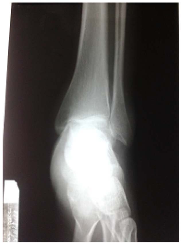
S. Diao et al.

                               years. Elisé [1] had had satisfactory results (6 very good, 5 good and 1 bad) in 11
                               cases in hindsight of 11 years. Its complications were stiffness, paresthesia and
                               trophic disorders.
                                 Other late complications are reported in the literature such as ankle os-
                               teoarthritis [6], avascular necrosis of the talus [7] and residual ligament laxity
                               [18].

                               4. Conclusion
                               The talocrural dislocation without malleolar fracture is an exceptional lesion. It
                               occurs in a context of high energy trauma. Its treatment is most often orthopedic
                               with excellent functional results.

                               Conflicts of Interest
                               The authors declare no conflicts of interest regarding the publication of this pa-
                               per.

                               References
                               [1]     Élisé, S., Maynou, C., Mestdagh, H., Forgeois, P. and Labourdette, P. (1998) Les
                                       luxations tibio-astragaliennes pures. A propos de 16 observations. Acta Orthopaedica
                                       Belgica, 64, 25-34.
                               [2]     Peraire, M. (1913) Luxation tibio-astragalienne avec issue à l’extérieur du péroné
                                       non fracturé à travers une boutonnière cutanée. Présentation de malade. Paris
                                       Chirurgical, 5, 959.
                               [3]     Lamraski, G. and Clegg, E. (2010) Unusual upward Closed Tibiotalar Dislocation
                                       without Fracture: A Case Report. Foot and Ankle Surgery, 16, 44-46.
                                       https://doi.org/10.1016/j.fas.2009.10.012
                               [4]     Wang, Y.T., Wu, X.T. and Chen, H. (2013) Pure Closed Posteromedial Dislocation
                                       of the Tibiotalar Joint without Fracture. Orthopaedic Surgery, 5, 214-218.
                                       https://doi.org/10.1111/os.12049
                               [5]     Fahey, J.J. and Murphy, J.L. (1965) Dislocations and Fractures of the Talus. Surgical
                                       Clinics of North America, 45, 79-102.
                                       https://doi.org/10.1016/S0039-6109(16)37485-0
                               [6]     Garbuio, P., Gérard, F. and Gagneux, E. (1995) Les luxations tibio-tarsiennes pures.
                                       À propos de 9 cas. Revue de Chirurgie Orthopédique et Réparatrice de l Appareil
                                       Moteur, 81, 601-608.
                               [7]     Toohey, J.S. and Worsing Jr., R.A. (1989) A Long-Term Follow-up Study of Tibio-
                                       talar Dislocations without Associated Fractures. Clinical Orthopaedics and Related
                                       Research, 239, 207-210. https://doi.org/10.1097/00003086-198902000-00023
                               [8]     Wilson, M.J., Michele, A.A. and Jacobson, E.W. (1939) Ankle Dislocations without
                                       Fracture. The Journal of Bone and Joint Surgery, 21, 198-204.
                               [9]     Thangarajah, T., Giotakis, N. and Matovu, E. (2008) Bilateral Ankle Dislocation
                                       without Malleolar Fracture. The Journal of Foot and Ankle Surgery, 47, 441-446.
                                       https://doi.org/10.1053/j.jfas.2008.05.004
                               [10] Uyar, M., Tan, A., Isler, M. and Cetinus, E. (2004) Closed Posteromedial Disloca-
                                    tion of the Tibiotalar Joint without Fracture. British Journal of Sports Medicine, 38,
                                    342-343.

DOI: 10.4236/ojo.2020.101003                            19                                    Open Journal of Orthopedics
S. Diao et al.

                               [11] Dahmani, O., Ouchrif, Y., Alarab, H., Blanc, S. and Elmrini, A. (2014) Luxation
                                    talo-crurale pure chez un footballeur (À propos d’un cas et revue de la littérature).
                                    Journal de Traumatologie du Sport, 31, 224-227.
                                    https://doi.org/10.1016/j.jts.2014.10.003
                               [12] Georgilas, I. and Mouzopoulos, G. (2008) Anterior Ankle Dislocation without As-
                                    sociated Fracture. A Case with an 11 Year Follow-up. Acta Orthopaedica Belgica,
                                    74, 266-269.
                               [13] Fernandes, T.J. (1976) The Mechanism of Talotibial Dislocation without Fracture.
                                    The Journal of Bone and Joint Surgery. British Volume, 58,364-365.
                               [14] Cyteval, C., Blin, D., Sarrrabère, M.P., Larroque, G. and Decoux, E. (2007) Imagerie
                                    des traumatismes de la cheville et du pied. Journal de Radiologie, 88, 789-800.
                                    https://doi.org/10.1016/S0221-0363(07)91347-5
                               [15] Lazarettos, I., Brilakis, E. and Efstathopoulos, N. (2013) Open Ankle Dislocation
                                    without Associated Malleolar Fracture. The Journal of Foot and Ankle Surgery, 52,
                                    508-512. https://doi.org/10.1053/j.jfas.2013.03.017
                               [16] Zizah, S., Benabid, M., Mezzani, A., Bennani, A., Lahrach, K., Marzouki, A. and
                                    Boutayeb, F. (2012) Un cas rare de luxation tibio-talienne pure. Journal de
                                    Traumatologie du Sport, 29, 212-214. https://doi.org/10.1016/j.jts.2012.09.008
                               [17] Prost à la Denise, J., Tabib, W. and Pauthier, F. (2009) Long-Term Result of a Pure
                                    Tibiotalar Dislocation in a Child. Orthopaedics & Traumatology: Surgery & Re-
                                    search, 95, 558-562. https://doi.org/10.1016/j.otsr.2009.06.007
                               [18] Colville, M.R., Colville, J.M. and Manoli, A. (1987) Posteromedial Dislocation of the
                                    Ankle without Fracture. The Journal of Bone & Joint Surgery, 69, 706-710.
                                    https://doi.org/10.2106/00004623-198769050-00011
                               [19] De Mourgues, G., Comtet, J.J. and Leclerc-Chalvet, F. (1969) La luxation tibio-tarsienne
                                    pure sans fracture associée. Revue de la littérature à propos d’un cas. Lyon Chirurgical,
                                    65, 661-667.
                               [20] Mohering, H.D., Tan, R.T., Marder, R.A. and Lian, G. (1994) Ankle Dislocation.
                                    Journal of Orthopaedic Trauma, 8, 167-172.
                                    https://doi.org/10.1097/00005131-199404000-00015
                               [21] Tsatedem, F.A., Tsiagadigui, J.G., Ndando, R.P., Arabo, M.S., Bayiha, A. and
                                    Kenfack, B. (2012) Décision d’amputation dans la prise en charge initiale d’une
                                    luxation ouverte grave de la cheville à propos d’un cas observé à l’Hôpital
                                    Laquintinie de Douala suite à un accident par moto-taxi. Pan African Medical
                                    Journal, 13, 73.
                               [22] Tondeur, G., Dufaz, J.P. and Nemry, C.H. (1964) Les luxations pures de la cheville.
                                    À propos de deux observations. Acta Orthopaedica Belgica, 30, 410-414.
                               [23] Kelly, P.J. and Peterson, L.F.A. (1962) Compound Dislocation of the Ankle without
                                    Fracture. American Journal of Surgery, 103, 170-172.
                                    https://doi.org/10.1016/0002-9610(62)90481-6
                               [24] Wehner, J. and Lorenz, M. (1990) Lateral Ankle Dislocation without Fracture.
                                    Journal of Orthopaedic Trauma, 4, 362-365.
                                    https://doi.org/10.1097/00005131-199004030-00022
                               [25] Wroble, R.R., Nepola, J.V. and Malvitz, T.A. (1988) Ankle Dislocation without
                                    Fracture. Foot & Ankle International, 9, 64-74.
                                    https://doi.org/10.1177/107110078800900202
                               [26] Finkemeier, C., Engebretsen, L. and Gannon, J. (1995) Tibial-Talar Dislocation
                                    without Fracture: Treatment Principles and Outcome. Knee Surgery, Sports Trau-
                                    matology, Arthroscopy, 3, 47-49. https://doi.org/10.1007/BF01553525

DOI: 10.4236/ojo.2020.101003                           20                                     Open Journal of Orthopedics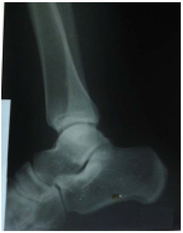
You can also read