Infective Endocarditis with Recurrent Epistaxis in a Young Patient: A Case Report
←
→
Page content transcription
If your browser does not render page correctly, please read the page content below
The Heart Surgery Forum 2021-3617                                                                 Online address: http://journal.hsforum.com
24 (2), 2021 [Epub April 2021]
doi: 10.1532/hsf.3617

Infective Endocarditis with Recurrent Epistaxis in a Young Patient: A Case Report

Liu Chunguang,1 Bhushan Sandeep, PhD,2 Long Mao, MD,2 Chen Jian,3 Shi Haipeng,3
Yuan Li, MD,2 Ke Gao, PhD2
1
  Graduate School of Zunyi Medical University, Zunyi, Guizhou 563003, Chengdu Second People’s Hospital;
2
  Department of Cardiothoracic Surgery, Chengdu Second People’s Hospital, Chengdu, Sichuan;
3
  Department of Surgery, Chengdu Second People’s Hospital, Chengdu, Sichuan

    ABSTRACT                                                          of congenital heart disease. In May 2020, he suffered from a
                                                                      moderate amount of epistaxis after nasal excavation, which
   Epistaxis is a common emergency, and its main causes are           was relieved after treatment in the clinic near his home (spe-
hypertensive crisis and trauma. Nasal packing is the primary          cific drugs are unknown) and self tamponade (toilet paper).
treatment. After active symptomatic treatment, the symptoms           After three times of intermittent epistaxis, the patient was
of epistaxis effectively can be controlled. In this case report,      relieved after self tamponade, and no rhinoscopy was per-
the patient was treated with epistaxis many times in the out-         formed. On September 30, he felt sore limbs, his body tem-
patient department. After nasal examination, there was a clear        perature was 380 C, and blood routine examination in other
bleeding point, and it was treated with gauze packing or silver       hospitals showed that WBC was 7.67 × 10⁹/L, RBC was 3.56 ×
nitrate cauterization. The symptoms of epistaxis gradually            1012/L, HGB was 98g/L, MCV was 85.5fl, MCH was 27.4pg,
got worse and was accompanied with fever and progressive              MCHC was 321g/L, PLT was 148 × 109/L. After symptom-
anemia. After blood culture and color Doppler ultrasound              atic treatment, he left the hospital. One week later, his epi-
examination, it was confirmed that it was endocarditis caused         staxis reoccurred. Nasal endoscopy showed active bleeding
by defective hypoxic bacterial infection. After active antibac-       points in the right and left nasal septum. The blood routine
terial and surgical treatment, the symptoms of epistaxis, fever       examination showed that WBC 12.79 × 10⁹/L, RBC 3.56
and anemia were relieved.                                             × 1012/L, Hgb 96g/L, MCV 82.9fl, MCH 27.1pg, MCHC
                                                                      327G/L, PLT 223 × 109/L. After the examination, Azithro-
                                                                      mycin tablets 0.5g was given orally 1 tablet per day (course of
    INTRODUCTION                                                      5 days) and furosemide nasal drops.
                                                                          On October 16, epistaxis with high fever reoccurred again.
   Infective endocarditis is caused by bacteria and other             The body temperature was 380C ~ 390C. Rhinoscopy exami-
microorganisms infecting the endocardium, especially the              nation in other hospitals showed that the nasal septum was
heart valve. Its characteristic lesion is the formation of vegeta-    deviated, the right side of the nasal septum has bleeding, left
tion, which often occurs in the heart valves, congenital car-         side of the patient's area had bleeding spots, and blood routine
diovascular malformation or after artificial valve replacement        examination showed that WBC was 8.67 × 10⁹/ L, RBC was
[Gungor 2012]. Fever and anemia are common clinical mani-             2.76 × 10¹²/L, HGB was 78g/L, MCV was 88fl, MCH was
festations. Atypical infective endocarditis often is covered by       28.3pg, MCHC was 321g/L, and PLT was 190 × 109/L. His
the clinical symptoms of complicated diseases. Mild cases only        chest CT showed that his spleen was enlarged, and the density
show bleeding from the nose, while severe cases can lead to           was not uniform. Direct antiglobulin test was positive, Ana
hemorrhagic shock [Wang 2014]. The causes also can include            1:100; anti dsDNA was weakly positive; bone marrow biopsy
local and systemic factors. In this case, the etiology of recur-      smear showed that the proliferation of granulocytic and eryth-
rent epistaxis is caused by two factors. After the control of         rocyte lineage was significantly active, with a slightly higher
infective endocarditis, the epistaxis was cured dramatically.         proportion of plasma cells. Some nucleated cells were degen-
                                                                      erated and mature red blood cells were dissolved. There was
                                                                      no evidence of abnormal immunophenotype associated with
    CASE REPORT                                                       acute leukemia, NHL and high-risk MDS.
                                                                          On October 28, the blood routine examination showed
  A 21-year-old male patient had the habit of blowing his             that WBC 12.58 × 10⁹/L, RBC 2.26 × 10¹²/L, Hgb 61G/L,
nose, digging at it, and staying up late; he had no clear history     MCV 89.8fl, MCH 27pg, MCHC 300g/L, PLT 203 × 109/L.
                                                                      Considering autoimmune hemolytic anemia, we mainly give
                                                                      prednisone acetate short-term shock therapy, assisted by nasal
Received January 16, 2021; accepted February 7, 2021
                                                                      packing and (compound paracetamol and amantadine cap-
                                                                      sules) cooling treatment, but the curative effect was not good.
Correspondence: Bhushan Sandeep, PhD, Department of Cardiothoracic    The frequency of epistaxis and high fever gradually increased,
Surgery, Chengdu Second People’s Hospital, Chengdu, Sichuan 610017,   mainly in the morning and at night, accompanied by cardiac
Telephone +8615891442217 (e-mail: dr.sandeep07@yahoo.com)             fatigue and chest tightness after activities.

© 2021 Forum Multimedia Publishing, LLC                                                                                              E317
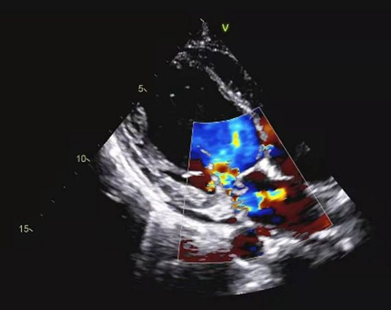
The Heart Surgery Forum #2021-3617

   Blood routine examination on November 6 in our hospi-
tal showed that WBC was 11.51 × 10⁹/L, RBC was 2.05 ×
1012/L, HGB was 53g/L, MCV was 86.3fl, MCH was 25.9pg,
MCHC was 299G/L, PLT was 219 × 109/L. Urine routine
occult blood + + +; our hospital outpatient find he had severe
anemia and he was admitted to our department. His exami-
nation showed anemia, sublingual vein thickening, arrhyth-
mia, apical area and aortic valve second auscultation area can
hear with systolic murmur. Color Doppler echocardiography
showed infective endocarditis, severe regurgitation of perfo-
rated plate of anterior mitral valve (Figure 1 and Figure 2),
formation of mitral valve vegetations (Figure 3), multiple
vegetations in left atrium (Figure 4), mild tricuspid regurgita-
tion, a small amount of pericardial effusion, and left ventricu-
lar false chordae tendineae. (Figure 1) (Figure 2) (Figure 3)
(Figure 4)
   According to the improved Duke diagnostic criteria, infec-
tive endocarditis was clear, in this case the simple anti-inflam-   Figure 1. Perforation of anterior mitral valve
matory effect was not good, then the patient was surgically
operated under hypothermic cardiopulmonary bypass with
thoracoscopic mitral valve replacement. Postoperative path-
ological section of mitral valve membrane showed that the
valve tissue had hyaline degeneration, lymphocyte infiltration
with necrosis. After operation, anti-inflammatory and warfa-
rin anticoagulation therapy were continued, and his epistaxis
and fever were relieved.
   On December 6, the blood routine examination showed
that WBC was 10.19 × 10⁹/L, rbc3.57 × 1012/L, HGB was
96g/L, MCV was 82.6fl, mch26.9pg, mchc325g/L, plt262 ×
109/L. Urine routine occult blood was negative.

 DISCUSSION

   Infective endocarditis is a rare and serious disease caused
by infective foci in the heart. It refers to the inflammation of
the heart valve or ventricular wall caused by direct infection
of bacteria, fungi and other microorganisms. In the research
of Huang et al. [Huang 2020], Streptococcus and Staphylo-           Figure 2. Mitral regurgitation bundle
coccus are still the main pathogens of infective endocarditis,
and 3.92% of them are deficient hypoxic bacteria. The main
reason is that dextran expressed in the cell wall can enhance
the ability of bacteria to adhere to the surface of the endocar-
dium. Deficient hypoxic bacteria are present in normal flora
in human oral cavity, upper respiratory tract, and intestinal
tract. When immunity is low, it can cause bacteremia and
infective endocarditis. Gungor et al. [Gungor 2012] reported
endocarditis of valve prosthesis caused by nasal packing with-
out systemic prophylaxis of antibiotics. Boumis et al. [Boumis
2018] reported that prophylactic use of antibiotics should be
considered to avoid the occurrence of infective endocarditis
after long-term nasal bleeding, nasal packing, or other nose
trauma intervention. In this case, the patient with multiple
epistaxis was given nasal packing, no other possible cause of
bacteremia was identified, which can speculate that the occur-
rence of bacteremia is related to this. Wang et al. [Wang 2014]
concluded that the analysis of the medical records of infective
endocarditis with heart failure and bacteremia are difficult to     Figure 3. Mitral valve vegetations

E318

Infective Endocarditis with Recurrent Epistaxis in a Young Patient: A Case Report – Chunguang et al

                                                                       control, secondary aneurysms tend to self-heal. This patient
                                                                       had recurrent epistaxis, which could not be excluded because
                                                                       of the absence of local angiography. Irregular high fever in
                                                                       the course of disease, high fever causes nasal mucosa dryness,
                                                                       local vasodilation and congestion, and is also related to the
                                                                       occurrence of recurrent epistaxis. In the course of analysis of
                                                                       epistaxis, nasal anatomy, bad living habits, improper use of
                                                                       drugs (antipyretic drugs, hormones), emotional changes will
                                                                       also lead to difficult control of epistaxis.
                                                                          The clinical symptoms of infective endocarditis are persis-
                                                                       tent fever and progressive anemia. Vascular embolism is a seri-
                                                                       ous complication of infective endocarditis, with an incidence
                                                                       rate of 13%-49%. Hemorrhagic complications are relatively
                                                                       rare, especially hemorrhagic stroke. From this case history,
                                                                       we can improve the understanding of atypical symptoms of
                                                                       infective endocarditis and reduce its clinical misdiagnosis and
                                                                       mistreatment.

   Figure 4. Left atrial vegetation
                                                                        REFERENCES
control by drugs, and these are the common manifestations
of dead and unhealed patients. Early surgical treatment is the         Boeddha N P, Bycroff T, Nadel S, et al. 2020. The Inflammatory and
key to successful treatment.                                           Hemostatic Response in Sepsis and Meningococcemia [J]. Crit Care
                                                                       Clin. 36(2): 391-9.
    In this case, the patient had a long course of disease and
repeated epistaxis. It was speculated that it was related to           Boumis E, Capone A, Galati V, et al. 2018. Probiotics and infective endo-
the persistence of heart failure and bacteremia. Normally in           carditis in patients with hereditary hemorrhagic telangiectasia: a clinical
a young man with good cardiac function, infective endocar-             case and a review of the literature [J]. BMC Infect Dis. 18(1): 65.
ditis before the onset is not easy to diagnose. But recurrent          Chen Y, Yangmang O, Bin W, et al. 2020. Six cases of intracranial infec-
nose bleeds were an indication that endocarditis already was           tive aneurysm secondary to infective endocarditis% J Chinese Medical
present. With the progress of the disease, there were symp-            Journal [J]. 14): 1112-3-4
toms of heart failure, such as cardiac fatigue and chest tight-        Gungor H, Ayik M F, Gul I, et al. 2012. Infective endocarditis and spon-
ness after activity, with increased central venous pressure and        dylodiscitis due to posterior nasal packing in a patient with a biopros-
improved vascular pressure in the drainage area of superior            thetic aortic valve [J]. Cardiovasc J Afr. 23(2): e5-7.
vena cava. This is the same reason that hypertensive patients
                                                                       Huang D, Lin C, Kuai W, et al. Distribution and drug resistance of
seek emergency treatment with epistaxis [Lee 2020], which
                                                                       pathogens in blood culture of patients with infective endocarditis% J
also can explain why epistaxis often occurs with patients in the
                                                                       Chinese Journal of antibiotics [J]. 2020, 45 (2): 170-4.
morning and at night. Due to the persistence of bacteremia,
it is easy to fix the value of bacterial thrombus at the damaged       Lee C J, Seck C J, Liao P C, et al. 2020. Evaluation of the Relationship
nasal mucosa, lymphocyte infiltration, tissue necrosis, normal         Between Blood Pressure Control and Epistaxis Recurrence After Achiev-
anticoagulant and procoagulant balance are destroyed [Boed-            ing Effective Hemostasis in the Emergency Department [J]. Journal of
                                                                       acute medicine. 10(1): 27-39.
dha 2020], coagulation factors are consumed, and the activity
of anticoagulant system is enhanced. Infective endocarditis            Shi X, Liu Y, Zhu G. 2020. Research progress on risk factors and risk
is prone to secondary infective aneurysms, the incidence of            prediction of embolism in infective endocarditis% J Journal of cardiopul-
which is 3% ~ 5%, which is more common in intracranial                 monary vascular disease [J]. 39 (01): 95-6 + 101.
[Chen 2020]. It also is more prone to rupture and bleed-               Wang P, Lu J, Wang H, et al. 2014. Clinical analysis of 368 cases of infec-
ing than aneurysms caused by other reasons. After disease              tive endocarditis% J Chinese Journal of Cardiology [J]. 42 (2): 140-4.

© 2021 Forum Multimedia Publishing, LLC                                                                                                      E319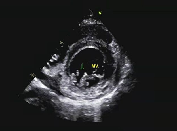
You can also read