NSTEMI in a patient admitted for multiple spinal fractures: a clinical case report
←
→
Page content transcription
If your browser does not render page correctly, please read the page content below
J Pathol Locomot Appar - Riv Patol Appar Locomot - Vol. XIX (1-2) 2020                                      7

NSTEMI in a patient admitted for multiple
spinal fractures: a clinical case report
Fabrizio Nicolò Stefano Perrone, Francesco Geuna, Massimiliano Macrini, Saverio Muscoli, Marco Alfonso Perrone

Division of Cardiology, University of Rome Tor Vergata, Rome, Italy

Correspondence:
Marco Alfonso Perrone, MD, Division of Cardiology University of Rome Tor Vergata
Email: marco.perrone01@gmail.com

DOI: 10.12920/jopola.2020.01

Abstract                                                              Riassunto
Non-ST-elevation myocardial infarction (NSTEMI) is defined by
the presentation of acute chest pain with no persistent ST-segment    L’infarto miocardico senza sopraslivellamento del tratto ST (NSTE-
elevation. It is a common disease in the elder population. There      MI) si presenta con dolore toracico oppressivo in assenza di so-
are some dates that correlate vascular factors to osteoporosis and    praslivellamento del tratto ST. È una patologia frequente nella
the augmented fracture risk in patient with cardiovascular disease    popolazione anziana. Diversi studi hanno dimostrato come, in
but there are few information about a link between a bone fractu-     pazienti con malattia cardiovascolare, esista una correlazione fra
re and NSTEMI. We described a male patient, with no history           fattori vascolari ed osteoporosi ed aumentato rischio di frattura;
of coronary artery disease, hypertension, dyslipidemia. He was        tuttavia vi sono pochi dati sulla correlazione tra frattura ossea ed
admitted to our emergency room after a car accident. A head           NSTEMI. Descriviamo il caso clinico di un uomo, senza storia di
CT showed no active bleeding and a Total Body CT showed no            malattia coronarica, ipertensione o dislipidemia. Il paziente veni-
acute organ damages and vertebral fractures at the D11 and L1-        va trasportato presso il pronto soccorso a seguito di un incidente
L2, confirmed with a MRI of the spine. An orthopedic surgery          automobilistico. La TC cranio non evidenziava emorragie in atto
was scheduled but the following day he started complaining mild       e la TC Total Body evidenziava multiple fratture vertebrali a ca-
chest pain with elevated high sensitive troponin I value. A co-       rico di D11 ed L1-L2 che venivano confermate dalla Risonanza
ronary angiography was performed and percutaneous coronary            Magnetica della colonna vertebrale. Veniva pertanto program-
intervention with placement of two drug eluting stents in LADA        mato l’intervento chirurgico ortopedico ma il giorno successive il
and two in RCA allowed for restoration of the flow. In conside-       paziente sviluppava dolore toracico con aumento delle troponine
ration of the planned surgery, he was initially treated only with     I ad alta sensibilità. Pertanto veniva eseguito uno studio corona-
aspirin and enoxaparin. However, because of two episodes of           rografico e successiva angioplastica percutanea con impianto di
in-stent restenosis, a treatment with aspirin and clopidogrel was     due stent medicati a livello della discendente anteriore e due stent
obligatorily started. A patient with fractures can develop an acute   medicati a livello della coronaria destra, con ripristino del flusso.
coronary syndrome because of several reasons and in this case         In considerazione dell’intervento chirurgico in programma, il pa-
we hypothesize that the stress caused by the trauma have desta-       ziente veniva inizialmente trattato con aspirina ed enoxaparina.
bilized a nonsignificant plaque. DAPT therapy is another element      Tuttavia, a causa di due episodi successivi di restenosi intrastent,
that we have to consider in these patients.                           veniva impostata la terapia antiaggregante con aspirina e clo-
                                                                      pidogrel. Un paziente con fratture può dunque sviluppare una
Keywords: NSTEMI, bone fractures, DAPT, stent restenosis.             sindrome coronarica acuta per diversi motivi ed in questo caso
                                                                      ipotizziamo che lo stress del trauma abbia destabilizzato una
                                                                      placca originariamente non significativa. La doppia terapia an-
                                                                      tiaggregante (DAPT) è un altro elemento da valutare in questa
                                                                      tipologia di pazienti.

                                                                      Parole chiave: NSTEMI, fratture ossee, DAPT, restenosi intra-
                                                                      stent.

Introduction                                                          troponin, and one of the followings: 1) Symptoms of
Acute myocardial infarction (AMI) defines cardio-                     ischemia; 2) new or presumed new significant ST-T
myocyte necrosis with at least one value above the                    wave changes or left bundle branch block on 12-lead
99th percentile of the upper reference limit of the car-              ECG; 3) Development of pathological Q waves on
diac biomarkers, preferably high-sensitivity cardiac                  ECG; 4) Imaging evidence of new or presumed new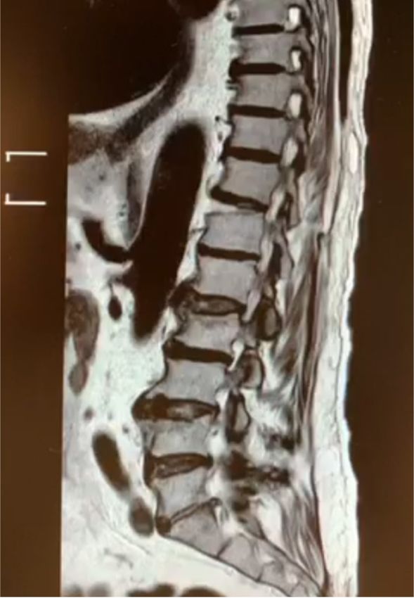
8 Fabrizio Nicolò Stefano Perrone, Francesco Geuna, Massimiliano Macrini, Saverio Muscoli, Marco Alfonso Perrone
loss of viable myocardium or regional wall motion ab- into account the surgical indication. During the recov-
normality; 5) Intracoronary thrombus detected on an- ery in cardiac intensive care unit (CICU) the patient
giography or autopsy(1). Non-ST-elevation myocardial developed a new chest pain episode with increasing in
infarction (NSTEMI) is defined by the presentation of the troponin levels. The emergency coronary angio-
acute chest pain with no persistent ST-segment ele- gram showed sub-occlusion of the DES in RCA and
vation with an electrocardiogram (ECG) that may be of the DES in LADA. Drug-coated balloon (DCB) was
normal or include changes such as transient ST-seg- performed and a DES was implanted in the LADA.
ment elevation, persistent or transient ST-segment The same day, because of a new chest pain episode, an
depression, T-wave inversion, flat T waves or pseu- emergency coronary angiogram was performed and be-
do-normalization of T-waves(2). There are some dates cause of a RCA in-sent restenosis, a DES was implant-
that correlate vascular factors to osteoporosis and ed with a TIMI-3 flow. In consideration of the recurrent
fractures(3), especially about the augmented fracture in-stent restenosis, a treatment with 100 mg aspirin and
risk in patient with cardiovascular disease(4). At the 75 mg clopidogrel was started and the patient was ad-
same time, there are few information about a possible mitted to the Orthopedic Unit to continue the surgical
link between a bone fracture – spontaneous and after a program.
trauma – and an acute coronary syndrome (ACS). We
describe a patient with multiple fractures after a car Discussion
accident who developed NSTEMI. This clinical case describes two apparently unrelated
diseases. Cardiovascular disease (CVD) and especial-
Case Study ly myocardial infarction are the most common cause
We described a case of a 71-year-old man with no car- of hospitalization and this is related with the aging(5).
diological history of coronary artery disease (CAD), At the same time, the same older population can suf-
hypertension, dyslipidemia. He was delivered to the fer more frequently spontaneous or induced fractures
Emergency Room (ER) of our hospital after a car ac- and osteoporotic disease. In literature, few dates are
cident. A head CT scan was immediately performed available about a possible relation between these two
showing a subdural hematoma with no active bleed- classes. They both share the same classic risk factors
ing and no indication to brain surgery. The patient was such as hypertension, smoking and diabetes(6) and the
admitted to the Orthopedic Unit and a Total Body CT physical inactivity after the surgery(7) or the precip-
Scan was performed showing no acute organ damages itation of some cardiovascular risk factors triggered
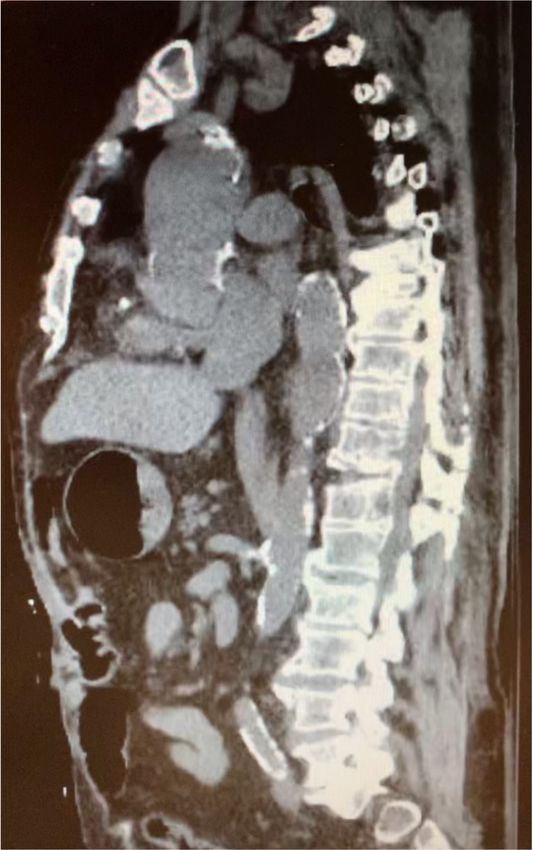
and vertebral fractures at the D11 and L1-L2 (Fig. 1). A by the disease(8) can partially explain the link between
MRI of the spine was subsequentially performed, con- them. On the other hand, it is unclear why, after a car
firming what previously seen at the CT Scan (Fig. 1). accident or a big trauma, there is a bigger prevalence
For this reason, an orthopedic surgery was scheduled. of coronary disease even in a younger population.
The following day, the patient started complaining mild AMI is a possible consequence of a blunt chest trau-
chest pain. The ECG showed inverted T waves in ante- ma, with a direct laceration or injury of the LADA as
rior-lateral leads. The time 0 high-sensitive troponin I the most frequent mechanism(9). Sometimes the ane-
level was elevated at 1086.8 ng/L. A CT Angiography mia secondary to an important bleeding event can de-
was performed and didn’t show acute aortic syndrome terminate a type two myocardial infarction. This case
(AAS). The patient was transferred to the cath-lab and report, instead, describe a myocardial infarction sec-
the coronary angiogram showed a sub-occlusive ste- ondary to an atherosclerosis plaque after a big trauma
nosis of the right coronary artery (RCA) with a TIMI- with fractures. We can only hypothesize that the stress
1 flow (Fig. 3) and a severe stenosis of the proximal caused by the trauma have destabilized a non-signif-
tract of the left anterior descending artery (LADA) icant plaque, leading to a NSTEMI. Other aspect to
with TIMI-1 flow (Fig. 4). Percutaneous coronary in- analyze is the management of the dual antiplatelet
tervention (PCI) with placement of two drug eluting therapy (DAPT) in consideration of the scheduled
stents (DES) in LADA and two DES in RCA allowed surgery. After the first PCI we decided to set a therapy
for restoration of the flow with TIMI 3. Treatment with with an antiplatelet and an anticoagulant. However, in
100 mg aspirin and enoxaparin bid was started, taking consideration of the acute in-stent restenosis episodes,NSTEMI in a patient admitted for multiple spinal fractures: a clinical case report 9
we were forced to set a DAPT and to postpone the tricky diagnosis and administration. The only use of
orthopedic surgery. biomarkers can lead to a wrong or sometimes misun-
derstood diagnosis. The DAPT after a PCI forces to
Conclusion postpone the surgery and the decision of an alternative
In conclusion, AMI after a bone fracture or trauma therapy can cause a in-stent restenosis. For these rea-
is not as rare as it looks like considering the litera- son, the only possibility we have is to analyze every
ture. It’s a relevant argument in consideration of the single case with a multidisciplinary team.
Fig. 1. Spinal CT Scan showing Fig. 2. Sagittal T2 magnetic resonance
fracture at D12. imaging showing fracture at L4.
Fig. 3. Right coronary artery (RCA) showing a
proximal sub-occlusive stenosis.
Fig. 4. Left anterior descending artery (LADA)
showing a severe proximal stenosis.10 Fabrizio Nicolò Stefano Perrone, Francesco Geuna, Massimiliano Macrini, Saverio Muscoli, Marco Alfonso Perrone
References
1. Thygesen K, Alpert JS, Jaffe AS, et al; Executive Group on behalf of Bone Miner Res. 2007 Sep;22(9):1449-54.
the Joint European Society of Cardiology (ESC)/American College 5. Benjamin EJ, Muntner P, Alonso A, et al; American Heart Associa-
of Cardiology (ACC)/American Heart Association (AHA)/World tion Council on Epidemiology and Prevention Statistics Committee
Heart Federation (WHF) Task Force for the Universal Definition of and Stroke Statistics Subcommittee. Heart Disease and Stroke Sta-
Myocardial Infarction. Fourth Universal Definition of Myocardial tistics-2019 Update: A Report From the American Heart Associa-
Infarction (2018). Circulation. 2018 Nov 13;138(20):e618-e651. tion. Circulation. 2019 Mar 5;139(10):e56-e528.
2. Roffi M, Patrono C, Collet JP, et al.; ESC Scientific Document 6. Eastell R, Newman C, Crossman DC. Cardiovascular disease and
Group. 2015 ESC Guidelines for the management of acute coro-
bone. Arch Biochem Biophys. 2010; 503(1):78–83.
nary syndromes in patients presenting without persistent ST-seg-
7. Dharmarajan TS, Banik P. Hip fracture. Risk factors, preoperative
ment elevation: Task Force for the Management of Acute Coronary
assessment, and postoperative management. Postgrad Med. 2006
Syndromes in Patients Presenting without Persistent ST-Segment
Elevation of the European Society of Cardiology (ESC). Eur Heart Jun-Jul;119(1):31-8.
J. 2016 Jan 14;37(3):267-315. 8. Abubakari AR, Bhopal R. Systematic review on the prevalence of
3. Alagiakrishnan K, Juby A, Hanley D, et al. Role of vascular diabetes, overweight/obesity and physical inactivity in Ghanaians
factors in osteoporosis. J Gerontol A Biol Sci Med Sci. 2003 and Nigerians. Public Health. 2008;122:173-182.
Apr;58(4):362-6. 9. Lascault G, Komajda M, Drobinski G, et al. Left coronary artery
4. Samelson EJ, Cupples LA, et al. Vascular calcification in middle aneurysm and anteroseptal acute myocardial infarction following
age and long-term risk of hip fracture: the Framingham Study. J blunt chest trauma. Eur Heart J. 1986;7:538-40.You can also read