Papillon-Lefèvre Syndrome: Report of a case and its management
←
→
Page content transcription
If your browser does not render page correctly, please read the page content below
J Clin Exp Dent. 2012;4(1):e77-81.       		                                       		                Papillon- Lefèvre Syndrome: A case report.

Journal section: Odontostomatology for the disabled or special patients			                                  doi:10.4317/jced.50594

Publication Types: Case Report                                                                              http://dx.doi.org/10.4317/jced.50594

                                        Papillon- Lefèvre Syndrome:
                                     Report of a case and its management

Shabina Sachdeva 1, Namita Kalra 2, Pranav Kapoor 3
1
  MDS. Formerly: Senior Resident, Department of Dentistry, University College of Medical Sciences, New Delhi -110095. Pre-
sently: Assistant Professor, Faculty of Dentistry, Jamia Millia Islamia, New Delhi -110025
2
  MDS. Professor & Head, Department of Dentistry, University College of Medical Sciences, New Delhi-110095
3
  MDS. Formerly: Lecturer, Department of Dentistry, University College of Medical Sciences, New Delhi -110095. Presently:
Assistant Professor, Faculty of Dentistry, Jamia Millia Islamia, New Delhi -110025

Correspondence:
B-4/33, Azad Apartments,
Aurobindo Marg,
New Delhi – 16,
email: drshabinasachdeva@yahoo.co.in

Received: 08/06/2011
Accepted: 17/11/2011

                                                           Sachdeva S, Kalra N, Kapoor P. Papillon- Lefèvre Syndrome: Report of a
                                                           case and its management. J Clin Exp Dent. 2012;4(1):e77-81.
                                                           http://www.medicinaoral.com/odo/volumenes/v4i1/jcedv4i1p77.pdf
                                                            Article Number: 50594          http://www.medicinaoral.com/odo/indice.htm
                                                            © Medicina Oral S. L. C.I.F. B 96689336 - eISSN: 1989-5488
                                                            eMail: jced@jced.es

    Abstract
    Papillon-Lefèvre Syndrome (PLS) is a rare autosomal recessive disorder first described by two French physicians,
    Papillon and Lefèvre in 1924. The disorder is characterized by diffuse palmoplantar keratoderma and precocious
    aggressively progressing periodontitis, leading to the premature loss of deciduous and permanent teeth at a very
    young age. The cutaneous lesions are usually manifested simultaneously with the intra-oral presentations and inclu-
    de keratotic plaques on the palms and soles varying from mild psoriasiform scaly skin to overt hyperkeratosis. The
    etiopathogenesis of the syndrome is relatively obscure and immunologic, genetic or possible bacterial etiologies
    have been proposed. Due to the vast degree of periodontal breakdown involved at such an early age, the dental
    surgeon is often the first to diagnose the syndrome. This paper presents a clinical presentation a 15 year old male
    diagnosed with Papillon- Lefèvre Syndrome

    Key words: Papillon-Lefèvre Syndrome, palmoplantar keratoderma, rapidly progressing periodontitis.

                                                          e77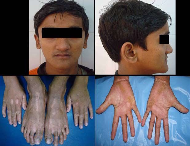
J Clin Exp Dent. 2012;4(1):e77-81.           		                                       		             Papillon- Lefèvre Syndrome: A case report.

Introduction                                                              neously with the intra-oral presentations and present as
Papillon-Lefèvre Syndrome (PLS) or keratosis palmo-                       sharply demarcated erythematous keratotic plaques on
plantaris with periodontopathia is a rare autosomal re-                   the palms and soles, which tend to spread onto the dorsal
cessive disorder characterized by diffuse transgradient                   surfaces.
hyperkeratosis of the palms and soles and severely des-                   This paper presents a brief overview of Papillon-Lefèvre
tructive, rapidly progressive periodontal disease (1).                    syndrome and describes the clinical presentations in a
Associated features may include intra-cranial calcifica-                  case with typical dental and dermatological findings.
tions, susceptibility to bacterial infections and mental
retardation (2, 3).                                                       Case Report
The disorder is first seen in children in the age group of                 A 15 year old male reported to the Department of Den-
1-4 years. No racial or sexual predilection is reported. A                tistry, University College of Medical Sciences, with the
genetic predisposition however exists with greater fre-                   chief complaint of having lost most of his teeth and in-
quency of occurrence in the consanguineous offspring                      ability to chew with the remaining ‘loose’ teeth.
(1, 2).                                                                   Medical history revealed that the patient had been su-
Patients with Papillon-Lefèvre Syndrome often present                     ffering from recurrent skin infections since an early age
with severe gingival inflammation and periodontal des-                    with thickening and subsequent peeling of the skin of
truction soon after the eruption of primary teeth, leading                his hands and feet, for which he had been undergoing
to premature loss of the deciduous dentition. Once the                    intermittent treatment. His grandfather had similar der-
primary dentition exfoliates, the gingiva seems to regain                 matological lesions.
its normal appearance. The eruption of permanent teeth,                   Past dental history revealed that the patient had lost all
however, re-triggers the aggressive periodontitis, which                  his deciduous teeth by about 3 years of age. A number
is unresponsive to conventional periodontal therapy and                   of his permanent teeth also became mobile soon after
results in partial or complete edentulism at a very young                 eruption and were subsequently extracted.
age.                                                                      On clinical examination, the patient presented with a re-
The cutaneous lesions are usually manifested simulta-                     duced facial height and a senile appearance. There was

   Fig. 1. Pre- treatment photographs of the patient showing hyperkeratotic patches on hands and feet.
                                                                        e78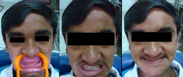
J Clin Exp Dent. 2012;4(1):e77-81. Papillon- Lefèvre Syndrome: A case report.
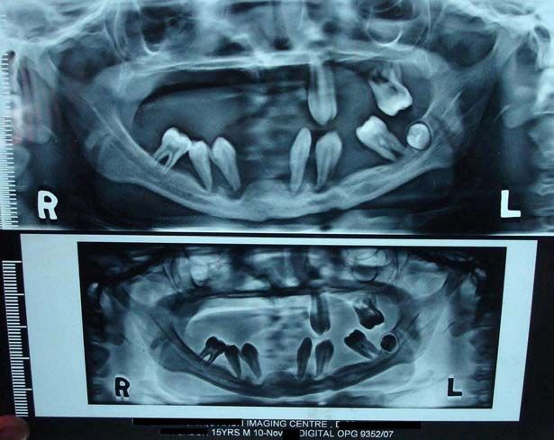
Fig. 2. Panoramic radiograph showing “Floating in air” appearance of teeth.
bilateral palmoplanter keratoderma with symmetric, loss giving the teeth a characteristic “Floating in air”
well-demarcated, yellowish, keratotic plaques on the appearance (Fig. 2).
skin of his palms and soles extending onto the dorsal Routine clinical investigations such as complete blood
surfaces (Fig. 1). hemogram, liver profile, renal profile and ultrasound of
Intra-oral examination revealed that most of his teeth the abdomen and pelvis were all normal. Histopatholo-
were missing and those present were Grade III mobile. gical examination of gingival tissue as well as punch
The Panoramic radiograph demonstrated advanced bone biopsy of skin of palms and soles revealed mild acan-
Fig. 3. Patient rehabilitated with complete dentures.
e79J Clin Exp Dent. 2012;4(1):e77-81. Papillon- Lefèvre Syndrome: A case report.
thosis and dense chronic mononuclear infiltrate in the porary period of healthy gingival tissue is then followed
connective tissue. by another phase of destructive periodontitis once the
An assessment of the patient’s intellect using Bhatia’s permanent teeth erupt. Affected individuals may thus
Battery of Intelligence test revealed that he had an IQ of become partially or completely edentulous in their early
97, which falls in the category of Average Intelligence. teens.
Co-relating the characteristic medical and dental history, The etiopathogenesis of Papillon-Lefèvre syndrome is
intraoral findings and laboratory and histopathological relatively obscure. An immunologic basis has been pro-
investigations, the condition was diagnosed as Papillon- posed and reduced neutrophil, lymphocyte or monocyte
Lefèvre Syndrome. Since all the patient’s remaining function has been reported in some cases (6). Toomes
teeth had very poor prognosis due to extensive bone et al. (7) believe that the syndrome may be genetically
loss, they were planned for extraction. Following due determined and have demonstrated loss-of-function mu-
informed consent, the extractions were carried out in a tations affecting both alleles of the lysosomal protease
phased manner followed by a healing period of 8 weeks, cathepsin-C gene in patients with PLS. The cathepsin-C
which was uneventful. gene, which is located on chromosome 11q14.1-q14.3
Treatment options for prosthodontic rehabilitation were has endopeptidase activity and is expressed in epithe-
discussed and considering the patient’s age and poor lial regions commonly affected by PLS including palms,
socio-economic status, a set of complete dentures was soles, knees, and keratinized oral gingiva. It is also ex-
planned for now, not ruling out the use of dental implants pressed at high levels in various immune cells including
in future. Complete maxillary and mandibular dentures polymorphonuclear leukocytes, macrophages, and their
were delivered (Fig. 3) and the patient was recalled for precursors (8,9). Ryu et al. (10) believe that the severe
follow up and necessary adjustments. periodontal destruction seen in Papillon-Lefèvre syn-
The patient was referred to the Department of Derma- drome may be a result of loss of function mutation in
tology for management of cutaneous lesions where the cathepsin C gene and subsequent dysregulation of
treatment with emollients and Acretenin was initiated. localized polymorphonuclear leucocytes in inflamed pe-
riodontal tissues.
Discussion A possible bacterial etiology has also been proposed
Papillon-Lefèvre Syndrome was originally described by and it is believed that Actinobacillus actinomycetemco-
two French physicians, Papillon and Lefèvre in 1924. mitans, Porphyromonas gingivalis, Fusobacterium nu-
The syndrome is inherited as an autosomal recessive cleatum and Prevotella intermedia may be amongst the
trait and has a reported prevalence of 1-4 cases per mi- organisms involved not only in periodontal breakdown,
llion (1). but also in the cutaneous lesions of Papillon-Lefèvre
The disorder is characterized by diffuse palmoplantar syndrome (11).
keratoderma and rapidly progressing periodontitis lea- The management of cases with PLS requires a multi-
ding to premature loss of both deciduous and permanent disciplinary approach with the active participation of
teeth. A third component of dural calcifications has also the dental surgeon, dermatologist and pediatrician.
been reported by Gorlin et al. (2). Almuneef et al. (3) Treatment of the dental component of the disorder is ai-
recognize pyogenic liver abscess to be a fairly frequent med at eliminating the reservoir of causative organisms
complication of Papillon-Lefèvre syndrome. The case (12). A treatment protocol for the periodontal therapy
presented here, however, did not demonstrate any ab- in patients with Papillon-Lefèvre syndrome has been
normal liver function or ultrasonographic finding. proposed by Ullbro et al. (13). Oral retinoids such as
The dermatological lesions appear first between the acitretin and isotretinoin have proven to be beneficial in
ages of 1 to 4 years and include palmoplantar keratosis, treating both the dental and cutaneous lesions of PLS.
varying from mild psoriasiform scaly skin to overt hy- Retinoid treatment is usually started during the eruption
perkeratosis. The lesions may be aggravated by cold (4). of permanent dentition and is followed till the normal
Our patient gave history of having recurrent pyogenic developmental process is complete (14,15).
skin infections since early childhood, a finding which
has also been reported by Subramanium et al. (5) and Conclusion
may be attributed to the increased susceptibility to bac- Papillon-Lefèvre Syndrome is a devastating disease
terial infections in these patients. process, which, due to the associated cutaneous invol-
The intraoral presentation in Papillon-Lefèvre Syndro- vement and partial or complete edentulism, can severely
me is characterized by severe periodontitis as early as 3 affect the psychological, social and esthetic well being
to 4 years of age. The deciduous teeth develop normally, of the patient at a very young age. The dental surgeon
but their eruption is associated with severe gingival in- is often the first to diagnose the syndrome due to the pe-
flammation and subsequent periodontal destruction lea- riodontopathia involved. A multidisciplinary approach
ding to a premature loss of the primary dentition. A tem- is imperative, with emphasis not only on the dental and
e80J Clin Exp Dent. 2012;4(1):e77-81. Papillon- Lefèvre Syndrome: A case report.
dermatological management, but also on psychological
boost up and counseling of the affected individual.
References
1. Haneke E. The Papillon-Lefèvre syndrome: keratosis palmoplanta-
ris with periodontopathy. Report of a case and review of the cases
in the literature. Hum Genet. 1979; 51: 1-35.
2. Gorlin RJ, Sedano H, Anderson VE. The syndrome of palmar –
plantar hyperkeratosis and premature periodontal destruction of
the teeth. A clinical and genetic analysis of the Papillon-Lefevre
syndrome. J Pediatr. 1964; 65: 895-908.
3. Almuneef M, Al Khenaizan S, Al Ajaji S, Al-Anazi A. Pyogenic li-
ver abscess and Papillon-Lefèvre syndrome: not a rare association.
Pediatrics. 2003; 111: e85-8.
4. Hattab FN, Amin WM. Papillon-Lefèvre syndrome with albinism:
a review of the literature and report of 2 brothers. Oral Surg Oral
Med Oral Pathol Oral Radiol Endod. 2005; 100 : 709-16.
5. Subramaniam P, Mathew S, Gupta KK. Papillon-Lefevre syndro-
me: a case report. J Indian Soc Pedod Prev Dent. 2008; 26:171-4.
6. Firatli E, Gürel N, Efeoglu A, Badur S. Clinical and immunological
findings in 2 siblings with Papillon-Lefèvre syndrome. J Periodon-
tol.1996; 67: 1210-5.
7. Toomes C, James J, Wood AJ, Wu CL, McCormick D, Lench N,
et al. Loss-of-function mutations in the cathepsin C gene result in
periodontal disease and palmoplantar keratosis. Nat Genet. 1999;
23: 421-4.
8. Kurban M, Cheng T, Wajid M, Kiuru M, Shimomura Y, Christia-
no AM. A novel mutation in the cathepsin C gene in a Pakistani
family with Papillon-Lefevre syndrome. J Eur Acad Dermatol Ve-
nereol. 2010; 24: 967–9.
9. Hart TC, Hart PS, Bowden DW, Michalec MD, Callison SA,
Walker SJ, et al. Mutations of the cathepsin C gene are responsible
for Papillon-Lefèvre syndrome. J Med Genet. 1999; 36: 881-770.
10. Ryu OH, Choi SJ, Firatli E, Choi SW, Hart PS, Shen RF, et al.
Proteolysis of macrophage inflammatory protein-1alpha isoforms
LD78beta and LD78alpha by neutrophil-derived serine-proteases.
J Biol Chem.2005; 280 : 17415-21.
11. è.Wara-aswapati N, Lertisirivorakul J, Nagasawa T, Kawashima Y,
Ishikawa I. Papillon-Lefèvre syndrome: Serum immunoglobulin G
(Ig G) subclass antibody response to periodontopathic bacteria: A
case report. J Periodontol. 2001; 72:1747-54.
12. Dhanrajani PJ. Papillon-Lefevre syndrome: clinical presentation
and a brief review. Oral Surg Oral Med Oral Pathol Oral Radiol
Endod. 2009; 108: e1-7.
13. Ullbro C, Brown A, Twetman S. Preventive periodontal regimen in
Papillon-Lefèvre syndrome. Pediatr Dent. 2005; 27: 226-32.
14. Eickholz P, Kugel B, Pohl S, Näher H, Staehle HJ. Combined me-
chanical and antibiotic periodontal therapy in a case of Papillon-
Lefèvre syndrome. J Periodontol. 2001; 72: 542-9.
15. Al-Khenaizan S. Papillon-Lefèvre syndrome: the response to aci-
tretin. Int J Dermatol. 2002; 41: 938-41.
e81You can also read