Hydrochlorothiazide vs Venlafaxine: Drug-induced Bullous Pemphigoid - Cureus
←
→
Page content transcription
If your browser does not render page correctly, please read the page content below
Open Access Case
Report DOI: 10.7759/cureus.4999
Hydrochlorothiazide vs Venlafaxine: Drug-
induced Bullous Pemphigoid
Srikanth Naramala 1 , Hussain Dalal 2 , Sreedhar Adapa 3 , Pallav Patel 4 , Venu Madhav Konala 5
1. Rheumatology, Adventist Medical Center, Hanford, USA 2. Internal Medicine, Internal Medicine
Multi-Specialty Clinic, Houston, USA 3. Nephrology, The Nephrology Group, Visalia, USA 4. Internal
Medicine, Kaweah Delta Medical Center, Visalia, USA 5. Internal Medicine, Ashland Bellefonte Cancer
Center, Ashland, USA
Corresponding author: Srikanth Naramala, dr.srikanth83@gmail.com
Disclosures can be found in Additional Information at the end of the article
Abstract
Pemphigoid group of dermatologic conditions is a group of autoimmune skin disorders
resulting in blistering skin conditions. The two diseases that fall under this category are bullous
pemphigoid and pemphigus vulgaris. While there are many similarities in these two disorders,
there are numerous pathologic and biochemical differences which help us differentiate between
these disorders. In this case report, we report a usual manifestation of bullous pemphigoid in a
72-year-old female secondary to use of a well-known antihypertensive (hydrochlorothiazide)
and/or venlafaxine (anti-depressant).
Categories: Dermatology, Internal Medicine, Rheumatology
Keywords: pemphigus, bullous pemphigoid, hydrochlorothiazide, venlafaxine
Introduction
In pemphigoid diseases of the skin, bullous pemphigoid is an autoimmune disorder most
commonly found in older individuals. The disease predominantly affects patients over the age
of 60. In the United States, approximately six to 13 cases per million patients are diagnosed
every year, with equal incidence in both genders and no racial biases [1]. As described above,
this is an autoimmune phenomenon where we find the presence of autoantibodies at the
dermal-epidermal junction. Although it is an autoimmune disorder, this disorder can also be
caused by the use of systemic drugs, such as certain diuretics, nonsteroidal anti-inflammatory
drugs (NSAIDs), amoxicillin, gliptins and certain biologic agents [2]. As with any drug-induced
reactions, discontinuation of the offending agent is the mainstay of the treatment. After an
extensive review of literature, we report of a drug-induced bullous reaction as a side effect of
one of the most commonly used anti-hypertensive, hydrochlorothiazide and along with
Received 06/19/2019
venlafaxine.
Review began 06/23/2019
Review ended 06/24/2019
Published 06/25/2019
Case Presentation
© Copyright 2019
A 72-year-old Caucasian female with a past medical history of squamous cell cancer of the skin,
Naramala et al. This is an open
access article distributed under the hypothyroidism on Synthroid, and hypertension well-controlled with hydrochlorothiazide for
terms of the Creative Commons the last seven years, was evaluated in rheumatology clinic for oral ulcers. She denies any recent
Attribution License CC-BY 3.0., which travel, insect bites, any exposure to sick contacts, and similar complains in the past. Physical
permits unrestricted use, distribution,
examination was significant for erythematous, ruptured bullae with crusted ulcerated lesion on
and reproduction in any medium,
the dorsal aspect of bilateral foot (Figure 1), scattered maculopapular lesions on bilateral legs, a
provided the original author and
source are credited. single bullous lesion in left axilla, several bullous lesions in the oral cavity, significant hair loss
along with nail changes in fingers and toes (Figures 1, 2).
How to cite this article
Naramala S, Dalal H, Adapa, et al. (June 25, 2019) Hydrochlorothiazide vs Venlafaxine: Drug-induced
Bullous Pemphigoid. Cureus 11(6): e4999. DOI 10.7759/cureus.4999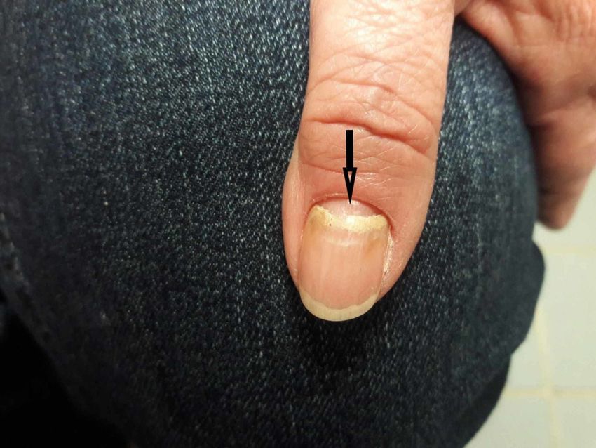
FIGURE 1: Ruptured bullae with open wounds on the bilateral
                                     feet, nail dystrophy with hyperpigmentation.

2019 Naramala et al. Cureus 11(6): e4999. DOI 10.7759/cureus.4999                                  2 of 7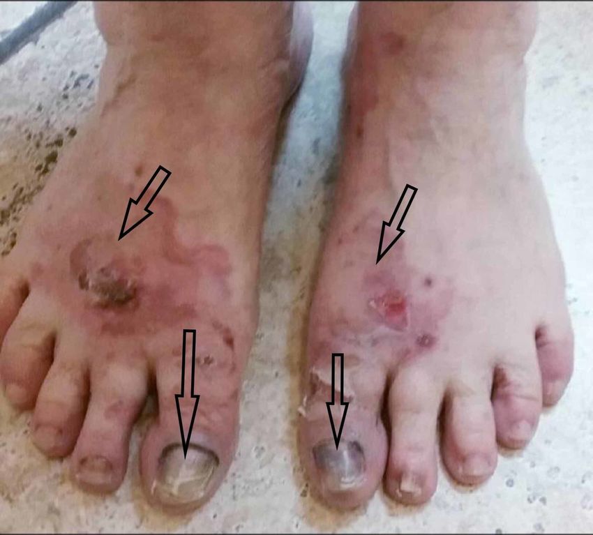
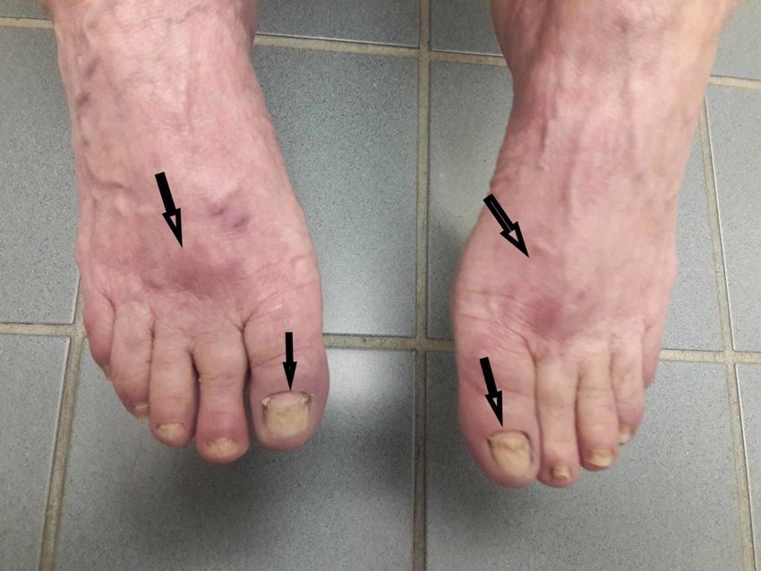
FIGURE 2: Onychomadesis of the nails - shedding of the nails
                                     beginning at the proximal end.

                                   She was initially evaluated by dermatology five months prior to presentation at the
                                   rheumatology clinic for a rash which started on bilateral thighs, later spreading to all over the
                                   body. A skin biopsy of the sub-clavicular region was done which showed nonspecific urticarial
                                   reaction with eosinophilic infiltrates which was thought to be secondary to a drug reaction.
                                   Direct immunofluorescence (DIF) assay was not performed on skin biopsy. Six weeks prior to
                                   the patient noticing the rash, she was started on venlafaxine 75 mg to overcome depression
                                   caused by her husband’s death. The venlafaxine and hydrochlorothiazide were stopped, but the
                                   patient’s rash did not improve. Over the next few months, the patient’s rash progressively
                                   worsened; she went to the emergency multiple times and was discharged on a brief course of
                                   antihistamines and oral steroids with minimal improvement.

                                   She started noticing sores in her oral mucosa and was evaluated by ENT who referred the
                                   patient to rheumatology for evaluation of the autoimmune process. Review of blood work
                                   showed moderate eosinophilia with absolute eosinophil count at 1169/ul (18-500 cells/ul),
                                   which was still persistent but decreased on repeat labs with an absolute eosinophil count of 722
                                   cells/ul (15-500 cells/ul). Extensive autoimmune serologies with antinuclear antibody,
                                   immunofluorescence assay, rheumatoid factor, antineutrophilic cytoplasmic antibody along
                                   with myeloperoxidase and proteinase-3 antibody, immunoglobulins A, G, M, and E,
                                   complements and bullous pemphigoid antibodies (BP 180 and BP 230) were negative. Infectious
                                   workups including HIV, hepatitis B core antibody, hepatitis B surface antigen, hep C antibody,
                                   rapid plasma reagin, and parvovirus IgM were also negative. The patient was then referred to
                                   dermatology for additional biopsy with special instructions to do DIF on the specimen. DIF of
                                   the right foot revealed linear deposits of C3 along the junctional zone and negative
                                   immunoglobulins, consistent with bullous pemphigoid. Blood work was positive for
                                   desmoglein-3 antibody at 161 u/ml (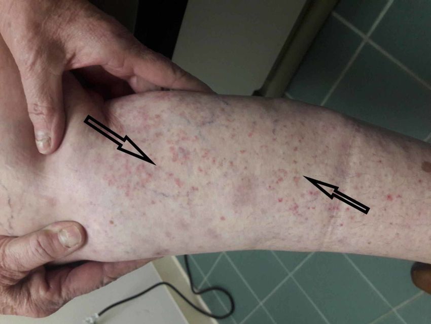
age-matched screening workup including mammogram, pap smear, and colonoscopy which
                                   were negative.

                                   The patient was started on prednisone 40 mg per day with tapper with the resolution of rash
                                   and oral lesions (Figure 3). Her rash and oral lesions recurred on 5 mg per day of prednisone
                                   (Figure 4). Her prednisone dose was increased back to 40 mg per day with a taper plan along
                                   with starting Imuran 100 mg per day. Her prednisone was tapered down and stopped in two
                                   months. Her disease is under control on Imuran 100 mg per day for the last three months.

                                     FIGURE 3: Four weeks post treatment with oral prednisone -
                                     Complete resolution of bullae and open wounds, significant
                                     improvement of nail changes.

2019 Naramala et al. Cureus 11(6): e4999. DOI 10.7759/cureus.4999                                                                  4 of 7
FIGURE 4: Recurrence of rash on 5 mg prednisone.
Discussion
Bullous pemphigoid is an immune-mediated blistering skin disorder characterized by
subepidermal blisters and compromising 80% of cases in pemphigoid group of disorders [1]. It
is characterized by subepithelial blister formation, deposition of immunoglobulins, and
complements at the dermo-epidermal junction and mucosal basement membrane [3]. There is
subsequent damage to the epithelial basement membrane. The binding of these antibodies to
the epithelial basement membrane initiates a series of reactions resulting in separation of the
epidermis and the dermis in the skin (giving rise to the classic bullae) as well as epithelium
from subepithelial tissue in mucous membranes [3]. Typical findings in Bullous pemphigoid
include tense, thick fluid-filled bullae on abdomen, groin, and extremities compared to bullae
of pemphigus vulgaris which are thinner and can be smudged off with pressure. While the
majority of the cases in pemphigoid group of disorders are immune-mediated, the drug-induced
bullous disease should also be given important consideration.
Review of the literature shows that there are about 50 different medications that cause drug-
induced bullous pemphigoid, and there are likely more medications as new therapies become
available [2]. One of the notable medications from the class of diuretics is hydrochlorothiazide.
There have been a handful of case reports in the literature linking hydrochlorothiazide use to
Bullous pemphigoid disorder, noting that onset of clinical features in most cases occurring in
as little as six weeks after initiation of therapy. In only one case report published by Garcia
Sanchez et al., the onset of symptoms was “several years” after the initiation of
hydrochlorothiazide, but it was unclear about the exact duration of HCTZ therapy in the
patient [4].
Based on the Naranjo scoring system for adverse drug reaction, we propose a high probability of
this patient’s Bullous pemphigoid reaction to hydrochlorothiazide and venlafaxine [5].
2019 Naramala et al. Cureus 11(6): e4999. DOI 10.7759/cureus.4999 5 of 7Hydrochlorothiazide takes higher probability as several cases in the literature have linked HCTZ
therapy to bullous pemphigoid reaction, but venlafaxine is also a likelihood given that the
patient was just started on this anti-depressant six weeks before the reaction. While there is no
documentation in literature with venlafaxine-induced bullous pemphigoid, given the timeline
of events and the scoring probability of this adverse drug reaction, venlafaxine could also be
considered a causative agent.
Skin biopsy is most definitive in diagnosing bullous pemphigoid. Serological testing to detect
bullous pemphigoid antibodies can be useful in supporting the diagnosis. Bullous pemphigoid
is usually diagnosed with increased levels of bullous pemphigoid antigen 180 (BP180) and
bullous pemphigoid antigen 230 (BP230) [3]. In a retrospective study by Roussel et al. the
sensitivity and specificity of detecting combined BP180 and BP230 antibodies by ELISA was
87% and 88%, respectively [6]. However, in our patient, both the levels of BP 180 and BP 230
were normal. On the other hand, the level of desmoglein-3 antibody was elevated, with the
level of desmoglein-1 antibody being in normal limits. Both these antibodies are usually
elevated in patients with pemphigus vulgaris. Although this is rare, there have been case
reports, for example, Sami et al. reporting of a presence of elevated desmoglein-3 antibody in
patients who have been diagnosed histopathologically as Bullous pemphigoid [7].
Based on the clinical presentation and the findings of skin biopsy, we made a diagnosis of
Bullous pemphigoid in our patient, possibly induced by hydrochlorothiazide and/or venlafaxine
use.
Conclusions
Bullous pemphigoid is the most common form of pemphigoid disorders, usually diagnosed in
patients over the age of 60. It is an autoimmune phenomenon, which in some cases can be
triggered by medication use. There are over 50 different types of medications which are known
to cause Bullous pemphigoid. The use of hydrochlorothiazide and/or venlafaxine can also be a
contributing factor in inducing a similar reaction. As with most drug-induced reactions,
cessation of the offending agent tends to be the mainstay of treatment. Additional
immunosuppressive treatment should be considered in refractory cases similar to our patient.
Additional Information
Disclosures
Human subjects: Consent was obtained by all participants in this study. Conflicts of interest:
In compliance with the ICMJE uniform disclosure form, all authors declare the following:
Payment/services info: All authors have declared that no financial support was received from
any organization for the submitted work. Financial relationships: All authors have declared
that they have no financial relationships at present or within the previous three years with any
organizations that might have an interest in the submitted work. Other relationships: All
authors have declared that there are no other relationships or activities that could appear to
have influenced the submitted work.
References
1. Bullous pemphigoid. (2018). Accessed: June 10, 2019:
https://www.ncbi.nlm.nih.gov/books/NBK535374/.
2. Stavropoulos PG, Soura E, Antoniou C: Drug‐induced pemphigoid: a review of the literature . J
Eur Acad Dermatol Venereol. 2014, 28:1133-1140. 10.1111/jdv.12366
3. Clinical features and diagnosis of bullous pemphigoid and mucous membrane pemphigoid .
(2019). Accessed: June 13, 2019: http://www.uptodate.com/contents/clinical-features-and-
diagnosis-of-bullous-pemphigoid-and-mucous-membrane-pemphigoid....
2019 Naramala et al. Cureus 11(6): e4999. DOI 10.7759/cureus.4999 6 of 74. García Sanchez VC, Calle Romero Y, de la Peña Parra E, Lorenzo Borda S: Hydrochlorothiazide
induced bullous pemphigoid. (Article in Spanish). Semergen. 2013, 39:214-217.
10.1016/j.semerg.2012.02.002
5. Naranjo CA, Busto U, Sellers EM, et al.: A method for estimating the probability of adverse
drug reactions. Clin Pharmacol Ther. 1981, 30:239-245. 10.1038/clpt.1981.154
6. Roussel A, Benichou J, Randriamanantany ZA, et al.: Enzyme-linked immunosorbent assay for
the combination of bullous pemphigoid antigens 1 and 2 in the diagnosis of bullous
pemphigoid. Arch Dermatol. 2011, 147:293-298. 10.1001/archdermatol.2011.21
7. Sami N, Bhol KC, Beutner EH, Plunkett RW, Leiferman KM, Ahmed AR: Diagnostic features of
pemphigus vulgaris in patients with bullous pemphigoid. Dermatology. 2002, 204:108-117.
10.1159/000051827
2019 Naramala et al. Cureus 11(6): e4999. DOI 10.7759/cureus.4999 7 of 7You can also read