Looking after Your Eyesight - 14 April 2018 - Open Meeting - Goring and ...
←
→
Page content transcription
If your browser does not render page correctly, please read the page content below
Slide 1

GORING AND WOODCOTE MEDICAL PRACTICE
PATIENT PARTICIPATION GROUP (PPG)

Open Meeting

 Looking after Your Eyesight

 14 April 2018
Slide 2

Agenda
• The New Practice Website
  • Julia Beasley

• Ophthalmology from the GP perspective
 • Dr Jessica Reed
• The Consultant view
 • Mr Martin Leyland
Slide 3

The New Practice Website
The new website is at the same URL as before:

https://www.goringwoodcotemedicalpractice.nhs.uk/
OPEN PPG MEETING

       OPHTHALMOLOGY
      SATURDAY 14TH APRIL 2018

Mr Martin Leyland BSc MB ChB MD FRCOphth
Dr Jessica Reed MB BS BSc DRCOG MRCGP
OPHTHALMOLOGY IN PRIMARY CARE
• Blepharitis Minor Eye Conditions Service
(MECS) Oxfordshire
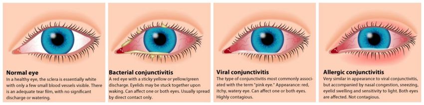
• Conjunctivitis ✓
✓
Foreign bodies
Red/gritty/watery eyes
✓ Flashes/floaters
✓ Ingrowing eyelashes
• Orbital cellulitis
❌Painful red eyes
❌Significant ocular trauma
• Ophthalmic Shingles ❌Transient loss of vision
❌Problems following recent ocular
surgery
• Red flags Robert Stanley in Wallingford
Hayselden and Partners in Wallingford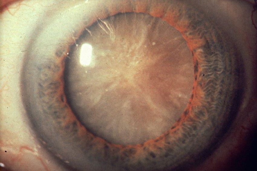
ASSESSMENT IN PRIMARY CARE

• Take a history and identify symptoms

• Observation – asymmetry, redness, pupils

• Check visual acuity

• Check ocular movements

• Stain the surface of the eye

• Direct ophthalmoscopy
ANATOMY

BLEPHARITIS
•   Inflammation of the eyelids

•   Causes crusting, itchy and redness/swelling of lid margins

•   Anterior (base of eyelashes) or posterior (meibomian
    glands)

•   Not an infection/contagoius, possibly a reaction to normal
    bacteria growing on the skin

•   Associated with seborrhoeic dermatitis and rosacea

•   Lid hygiene

•   Topical antibiotics, oral antibiotics

•   May cause infections (keratitis)/ulcers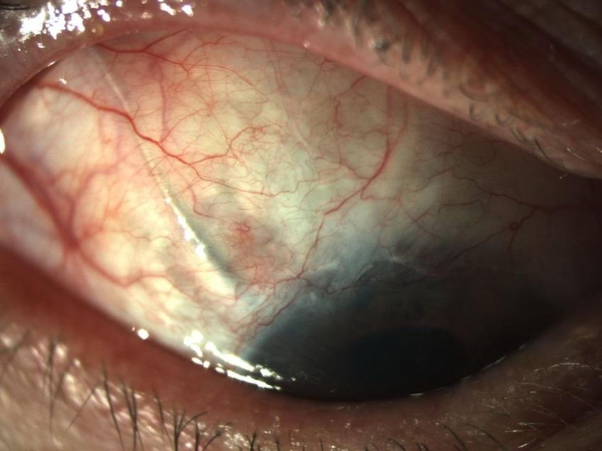
CONJUNCTIVITIS

• Very common!
• Seek advice from the pharmacist
• Usually viral… and contagious
• Should not be painful and should not affect your vision
• If bacterial – chlormaphenicol/levofloxacin
• If allergic – sodium cromoglicate

PRESEPTAL (PERIORBITAL) CELLULITIS
                                  • Quite common, less serious than orbital cellulitis

                                  • Infection anterior to the orbital septum

                                  • Eye lids are red and swollen

                                  • More common in young children

• Can be caused by upper respiratory tract or sinus infection

• Commonly a streptococcus infection

• Treatment with co-amoxiclav

• Not to be confused with orbital cellulitis….
ORBITAL CELLULITIS
• Much more serious
• Again, predominantly affects children
• Infection has spread beyond the septum into the orbit
❗ Reduced vision
❗ Chemosis
❗ Painful eye movements
❗ Restricted eye movements
❗ Proptosis
• Requires urgent assessment by eye casualty or ENT for IV antibioticsOPHTHALMIC SHINGLES • Shingles is caused by reactivation of Varicella Zoster (Chicken pox virus) • Ophthalmic branch of the trigeminal nerve (15% of all cases of shingles) • Blistering rash with numbness, pain and tingling, does not cross the midline • Hutchinson’s sign – nasociliary branch of the trigeminal nerve is affected, making eye involvement more likely (50%) • Complications – iritis, scelritis, keratitis and glaucoma • Treatment is with antivirals eg. Aciclovir • If the eye is involved, eye casualty assessment is needed
RED FLAGS IN PRIMARY CARE

❗ Painful, red eye

❗ Sudden loss of vision

❗ Significantly reduced visual acuity

❗ Painful eye movements

❗ Loss of colour vision

❗ Photophobia
USEFUL RESOURCES

• Patient UK

• Moorfields Eye Hospital

• NHS Choices
Ophthalmology

          Martin Leyland
Consultant Ophthalmologist Royal
Berkshire and Oxford Eye Hospitals
 www.berkshireeyesurgery.co.uk
Content • Ophthalmology referral • The big 4: – Glaucoma – Diabetes – Age-related macular degeneration – Cataract • Looking after your eyes
Referral: who’s who?
• Ophthalmologists
  – medical doctors specialising in eyes; usually surgeons
• Ophthalmic opticians = Optometrists
  – prescribe, fit and sell glasses; also have training in eye
    disease
• Orthoptists
  – specialise in assessment of eye movement
    abnormalities (e.g. squint) and children’s vision
    measurement
Referral: how?
Hospital Eye
Ophthalmic
? Service
Routine referral
for complex
A&E conditions &
surgery
Urgent referral
‘Choose & Book’
[Main A&E ‘after
hours’]
RBH by referral
OEH ‘walk-in’ Intermediate care
‘Soon’ appointments for
minor conditions
Berkshire Harmonie by
referral
Oxford MECS referral or
self-arrangedThe normal eye
www
RetinaGlaucoma
Glaucoma
• Damage to the optic nerve due to high pressure
  of fluid within the eye
• Diagnosis:
  – Appearance of optic nerve
     • But wide range of normal appearances
  – Measurement of eye pressure
     • Some people have high pressure but never get glaucoma,
       others have the condition despite normal pressure
  – Assessment of visual field
     • Not an easy test to do and misses early damage
Treatment of glaucoma • Identify the condition before it causes symptoms (damage cannot be reversed) – Visit optometrist every 1-2 years after age 50 – Earlier if history of early onset in close family • Lower the eye pressure to prevent further damage – Eye-drops – Surgery
Glaucoma eye-drops
• Increase fluid outflow
– Latanoprost ‘Xalatan’, Bimatoprost
‘Lumigan’
• Reduce fluid production
– -blockers: timolol
– CA inhibitors: dorzolamide
– -agonists: brimonidine
• Combination drops
– Timolol plus latanoprost or dorzolamidePutting eye drops in • Main problem with efficacy of eyedrops is poor compliance (not putting the drops in) • One drop is enough! • Pull down lid and drop into conj sac • Occlude nasolacrimal duct if taste unpleasant • Bottle-holders available in pharmacy • Preservative free if more than 4 a day or allergic/toxic
Diabetic Retinopathy
Diabetic Retinopathy
• Damage to micro-blood vessels within the
  retina caused by high blood sugar
• Early detection allows better treatment
• High blood sugar causes
  – Blood vessel leakage (DMO)
  – Blood vessel closure (ischaemia)
  – Reactive production of new blood vessels which
    bleed, leak and scar
Diabetic eye screening
• Berkshire Diabetic Eye • Oxfordshire Diabetic
Screening Programme Eye Screening
• In GP practices Programme
• In optometry practices
Diabetics >= 12 years old, screening service notified by GP
Drops to dilate pupils
Digital photography
Images assessed by computer software and by non-medical graders
Quality control/training by RBH and OEH
Standards set by NHS Diabetic Eye Screening ProgrammeLooking for sight threatening retinopathy • 31% of all images graded have ‘retinopathy’, 1:10 require referral to hospital. • Mild case with one micro aneurysm - no referral • Severe case with new retinal vessels and haemorrhage - urgent referral and seen within 1 week
Retinal artery
                                    Retina
   Retinal vein

Optic nerve ‘disc’
                           Macula            Fovea
M1 : Sight threatening maculopathy
M1 : sight 1.64%
threatening
cases =Maculopathy
R1M1R3: new vessels on optic disc
0.43% of cases = R3Treatment of retinopathy • Secondary prevention by weight loss, blood sugar and blood pressure control • Argon laser pan-retinal photocoagulation for proliferative disease • Focal argon laser or intravitreal injections for DMO (macular oedema)
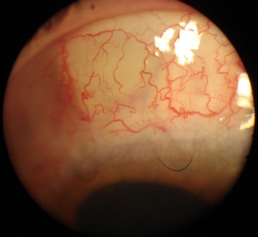
Age-related Macular Degeneration
Age related Macular Degeneration
                (AMD)
• An eye disease that progressively destroys the macula,
  the central portion of the retina, impairing central vision
• Age is the main risk factor
   – Presents after the age of 50, more common after 60
   – 1 in 500 between age of 55-65 have some form of AMD
   – 1 in 8 people above the age of 85
• The commonest cause of central visual loss in the
  developed world
• AMD accounts for almost 50% of blind registration in
  England and Wales
Two main forms of AMD: Dry and wet
Severe
visual loss3
Dry AMD2 Geographic
(85-90%) Atrophy2
Drusen
Wet
Formation1
AMD2
(90%)
Wet AMD2 Disciform
(10-15%) Scar2
39Symptoms of dry AMD • Blurred vision: especially reading, close-work • Minor distortion • Dark patch in central vision • Gradually progressive over years • Never lose peripheral vision
Dry atrophic AMD

Progression slow
and variable

No treatment
available
Secondary prevention of AMD
• Age Related Eye Disease Study (AREDS)
• Vitamins A,C,E and zinc (anti-oxidants) in high
  doses
  • ~20% reduction in progression in cases with at high risk of it
    (moderate disease in both eyes or severe disease in one eye)
• Ocuvite, Preservision, Macushield etc.
• Buy over the counter (not prescription)
• Smoking (oxidants ++) doubles risk of AMD sight-
  loss
Symptoms of wet AMD • Painless visual loss • Distortion • Missing patch/blur in central vision • May progress over days or weeks
Retinal artery
Retina
Retinal vein
Optic nerve ‘disc’
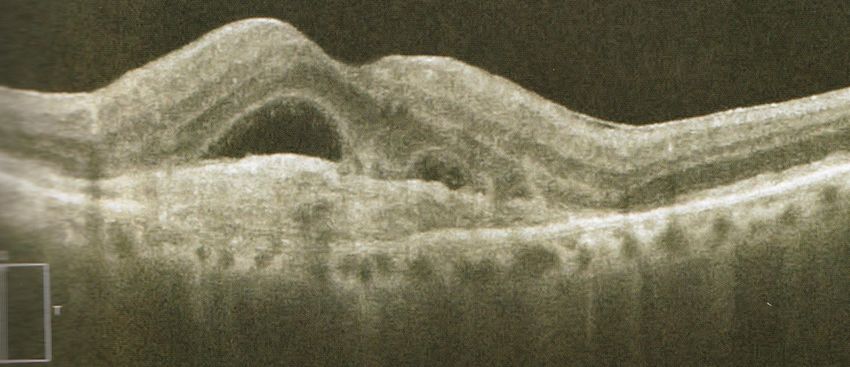
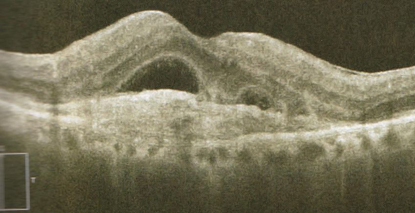
Macula FoveaWet AMD
Fluid/blood   Mass of   Distorted
under         new       retina
retina        blood
              vessels
Wet AMD
Treatment of wet AMD
• Antibodies (synthetic
  biological molecules that bind
  specifically to one protein)
  block vascular endothelial
  growth factor (VEGF)
• Large molecule that cannot
  get into the eye except by
  direct injection
• Ranibizumab (Lucentis),
  Bevacizumab (Avastin),
  Aflibercept (Eyelea)
Treatment Schedule
                               Month
           1   2   3   4   5   6   7   8   9   10   11 12
Lucentis

 Eylea

           1   2   3       5       7       9        11
X Number of Injections
neededEfficacy • Poor efficacy if acuity
End-stage    AMD
    Treatments of end-stage AMD
•   still largely
    Implantable   ineffective
                Miniature        and
                          Telescope
    experimental
    IMT
• Argus II Retinal Prosthesis System
• RPE cell transplant
    Intraocular lens-based
    approaches are very expensive
    and do not work
LVAs, Blind registration, information and self-help
Eye Clinic
Liaison Officer
(ECLO)
Macular disease
society
RNIBCataract
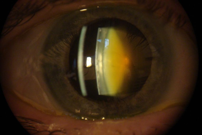
Symptoms
•   Gradual onset over months/years
•   Frequent changes of glasses prescription
•   Blur
•   Glare
•   Loss of contrast sensitivity
•   Loss of colour
•   Ghosting/double vision
• Worldwide most common cause of blindness
• 350,000 cataract operations/year in NHS
• Most common surgical procedure in UK
• In Oxfordshire & Berkshire “Cataract surgery is
  only routinely commissioned for patients who,
  after correction (e.g. with glasses), have a visual
  acuity of 6/12 or worse in their cataract-affected
  eye”
• Recent NICE guideline nice.org.uk/guidance/ng77
  states (1.2.2) “Do not restrict access to cataract
  surgery on the basis of visual acuity”
Looking after your eyes
• Diabetes
   – Diet/weight control to prevent onset
   – Close control of blood sugar once diabetic
   – Annual diabetic eye screening
• Glaucoma
   – Annual or biannual checks at optometrists from
     age 50, earlier (and free) if 1st degree relative
   – Take glaucoma treatment regularly
• AMD
   – Stop smoking
   – Good diet with anti-oxidants (vitamin
     supplements only proven in people with existing
     high-risk AMD)
Trauma Nailgun
• Goggles with DIY, gardening,
squash, badminton
• Caution +++ with alkali
• Irrigate +++ if any splashes
PaintballThat’s all, thank you for listening!
You can also read