Scoliosis: Symptoms, Evaluation and Treatment - Department of Orthopaedics - Nationwide ...
←
→
Page content transcription
If your browser does not render page correctly, please read the page content below
Department of Orthopaedics

  Scoliosis: Symptoms,
Evaluation and Treatment
What is Scoliosis?
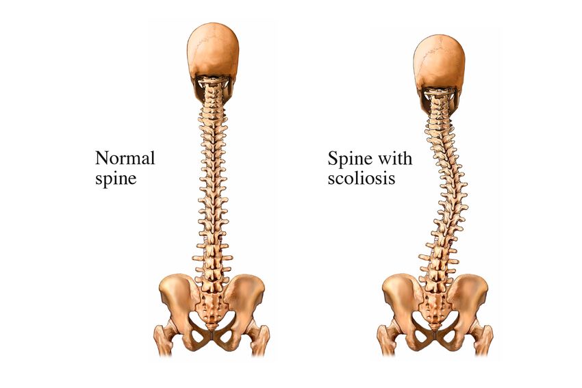
Scoliosis is a lateral deviation and rotation away from the midline. A normal spine is straight in the frontal
plane but is curved in the sagittal plane, meaning that there is a thoracic kyphosis (outward curve of the upper
back) and a lumbar lordisis (inward curve of the lower back). The scoliotic spine deviates from the midline
and rotates maximally at the apex of the curve. Increased curvature with rotation can enhance the appearance
of kyphosis when, in fact, it can be due to rib and chest deformity. When severe, this effect can compromise
cardiac and/or pulmonary function.
The apex (point) of the curve defines its center and is the most laterally deviated disc or vertebra. Curves may
be described as primary or secondary. The primary curve is the first to develop; however, two or three curves of
equal severity may exist making the determination of the primary curve difficult. Secondary (compensatory)
curves develop after the formation of the primary curve as a means of balancing the head and trunk over the
pelvis. Similar compensation also occurs in the sagittal plane, which can exaggerate the kyphosis or lordosis.
2Classifications of Scoliosis
There are three main types of scoliosis; idiopathic, neuromuscular/syndrome-related and congenital. Scoliosis
can also be categorized by age of onset. Curve progression risks include gender, remaining skeletal growth,
curve location and magnitude. Curve progression is most rapid during peak skeletal growth, like during
early infancy and adolescence. There is an increased incidence in family members of affected individuals.
There is a seven times greater chance of a sibling having scoliosis and a three times greater chance if a parent
has scoliosis. Therefore, if one family member has scoliosis, particular attention should be paid to evaluate
subsequent children in that family.
Normal Spine Spine with Scoliosis
Figure 1 and 2
Idiopathic Scoliosis
Approximately 80 percent of patients with scoliosis have idiopathic scoliosis. The prevalence with a curve
greater than 10 degrees in childhood and adolescent population has been reported to be from 0.5 to 0.3 per
100 and larger curves greater than 30 degrees ranging from 1.5 to 3 per 1,000. Idiopathic scoliosis can be
delineated as infantile (0-3 years of age), juvenile (4 to 10 years of age), adolescent (11 to 17 years of age) and
adult (over 18 years of age).
3Infants present with a left thoracic curve in almost 90 percent of cases. The male-to-female ratio is 3-to-2.
This can be associated with abnormal skull findings, hip dysplasia, congenital heart disease and developmental
delay. Scoliosis in infants can be resolving or progressive. Most curves are self-limited and 90 percent resolve
spontaneously. The few that are progressive can be difficult to treat. Spinal fusion to stop curve progression
is not an option in this group of children because the thoracic height would cease to develop and lung
development would be restricted. Bracing and/or Risser casting are two of the current medical treatments for
these children. The brace or cast is changed as the child grows, in an attempt to stop progression of the curve,
but does not improve the curve.
The age when juvenile idiopathic scoliosis develops is one of the most important factors in determining the
natural history of the disorder, with early onset cases more likely to be progressive. Scoliosis before the
adolescent growth spurt is more likely to have an underlying spinal cord abnormality as the cause of the
deformity with the incidence as high as 20 percent in this group. Juvenile scoliosis encompasses about 8 to 16
percent of cases of idiopathic scoliosis. Boys seem to be affected earlier than girls. If curves reach 30 degrees,
they are almost always progressive if left untreated. The rate of progression is 1 to 3 degrees per year before
age 10 and sharply increases to 4.5 to 11 degrees per year after that age. Literature suggests that 95 percent of
these children will eventually need a spinal fusion.
Adolescents are the most common group of idiopathic scoliosis. They usually develop a curve after reaching 10
years old, associated with the rapid growth of adolescence. Roughly 2 percent of adolescents have a scoliosis
greater than or equal to 10 degrees. The ratio of boys to girls is equal for minor curves, but it is dominated by
girls as the curve magnitude increases, reaching 1-to-8 for those requiring treatment.
Neuromuscular/Syndrome-Related Scoliosis
Disorders of either neuropathic or myopathic etiology make up a large proportion of the non-idiopathic
causes of pediatric scoliosis. Tumors of intra- or extra-spinal origin should also be considered. In some patients,
scoliosis is a result of poor muscle control, muscular weakness, or paralysis related to a specific disease, illness
or injury. Included in this category are syringomyelia, marfan syndrome, myelomeningocele, tethered cord,
neurofibromatosis, muscular dystrophy, cerebral palsy, poliomyelitis, arthrogryposis and traumatic cord injuries.
Congenital Scoliosis
Congenital scoliosis is defined as curvature of the spine resulting from vertebral element malformation,
segmentation of the vertebrae, or a combination of the two. Most of this development occurs during the
third to sixth week in utero. Despite the opportunities for error, congenital malformations are relatively rare.
The elements of the spinal column develop at the same time as several other major organ systems such as the
bladder,kidneys and heart. As a result, there is an association between congenital vertebra anomalies and other
malformations. If a child has been found to have abnormal vertebrae, a thorough cardiac and urologic work-
up is indicated.
4Warning Signs
Idiopathic scoliosis can go unnoticed in a child because it is rarely painful in the formative years. Therefore,
parents should watch for the following warning signs of scoliosis when their child is about 8 years of age:
• Uneven shoulders
• Prominent shoulder blade or shoulder blades
• Uneven waist
• Elevated hips
• Head is not centered directly above the pelvis
• Leaning to one side
• Backache or lower-back pain
• Fatigue
• Spine curves abnormally to the side (laterally)
Some schools sponsor scoliosis screenings. Although only a physician can accurately diagnose scoliosis, school
screenings can help alert parents to the presence of the warning signs in their child.
Treatment for Scoliosis
Scoliosis cannot be prevented, but the treatment options are varied for children with this musculoskeletal
disorder. Each child is thoroughly evaluated and a treatment plan is determined based on their individual
diagnosis and prognosis.
Watchful Waiting
There is no form of therapy, stretching or exercise that has been proven to benefit scoliosis. Therefore, many
patients are simply monitored by “watchful waiting.” Patients with curves less than 25 degrees should be
monitored every four to 12 months, depending on age and growth rate, with a physical exam and repeat X-rays.
Bracing/Casting
As curves progress above 25 to 40 degrees in a child who is still growing, bracing is usually recommended to
help slow or stop the progression of the curve. Individualized patient treatment will dictate the type of brace
and wearing schedule.
In very immature children with progressive curves, a Risser body cast can be applied in an attempt to improve
flexibility until they can convert to brace wear. Again, the objective is to limit the curve progression and once
the child has matured, a spinal fusion may be indicated.
5Growth Modulation
A growth modulation procedure may be indicated if a skeletally immature patient has a curve that has progressed
despite bracing and/or casting. There are several types of growth modulation procedures. The ones most
commonly performed include insertion of growing rods, VEPTRs, and MAGEC rods.
Growing Rods - The term “growing rod” loosely means instrumentation without fusion. This procedure
is used in young patients with rapidly progressive curves despite aggressive brace therapy. A limited fusion
is performed proximally and distally with hooks to hold a more subcutaneous rod in place. Subsequent
distraction (minor surgical procedure to lengthen the rod) is performed every six to 12 months during the
child’s growth phase. Eventually, these children will have a spinal fusion, and in addition, may be braced
during growth.
VEPTR - A Vertical Expandable Prosthetic Titanium Rib (VEPTR) is a Synthes device indicated for
treatment of thoracic insufficiency syndrome in skeletally immature patients. This is defined as the
inability of the thorax to support normal organ growth. Young patients with scoliosis with or without
rib anomalies or congenitally malformed vertebrae fall into this category. Again, this is only a temporary
measure that enables the thoracic cavity and spine to grow until a definitive spinal fusion.
MAGEC Rods - The MAGEC (MAGnetic Expansion Control) system is a new growth modulation
procedure that utilizes a magnetic rod that is implanted in the patient. This rod is fixated proximally and
distally in the spine very similar to traditional growing rods. However, the magnetic actuator within the
rod can be controlled by an external remote control. This allows the patient’s spine to be lengthened in
the clinic instead of in the operating room, therefore reducing some of the complications that can occur.
The MAGEC rod cannot be used on every patient, but does offer a new innovative option for patients
that qualify.
The Tether™ - Vertebral Body Tethering System is different than typical scoliosis surgery, called a “spinal
fusion,” which involves implanting stiff metal rods along either side of the spine to straighten the
undesirable curves. Rather than stiff metal rods, The Tether™ - Vertebral Body Tethering System uses a
strong, flexible cord to pull on the outside of a scoliosis curve to straighten out the spine. The bones, or
“vertebra” in a scoliotic spine are wedge shaped, tall on one side and short on the other. When the vertebra
are pulled by the cord, it puts pressure on the tall side of the vertebra on the outside of the curve. This
pressure slows the growth on the tall side of the vertebra, so that the short side can grow and catch up.
After surgery, the spine may continue to straighten even more over time as the patient grows.
6Spinal Fusion Surgery
The main goal of a spinal fusion is to improve spinal alignment and to prevent curve progression. A spinal
fusion includes placement of instrumentation in addition to arthrodesis (fusion). At Nationwide Children’s,
posterior spinal fusions are most commonly performed. Some patients require anterior arthrodesis with
concurrent posterior instrumentation and fusion. Posterior spinal instrumentation includes placing pedicle
screws or laminar hooks attached to rods, which hold the spine in a corrected position.
Before Fusion After Fusion
Figure 3 and 4
7When to Refer to Nationwide Children’s Hospital Orthopedic Department
• Examination by a family physician or pediatrician has taken place and a curvature greater than 10 degrees
has been discovered.
• Most cases of adolescent idiopathic scoliosis (less than 20 degrees) require no treatment, but should be
checked often, about every four to six months in a skeletally immature child.
• Depending on the clinical exam and radiographs, further tests may be warranted, such as an MRI.
• As curves get worse (above 25 to 30 degrees in a child who is still growing), bracing is usually recommended
to help slow the progression of the curve. The selection of the brace and the manner in which it is used
depends on many factors, including the specific characteristics of the curve. The exact brace will be decided
on by the patient and orthopedist.
• Curves of 45-50 degrees or greater usually require surgery because curves this pronounced have a high risk
of progression even after bone growth stops. Surgery involves correcting the curve through instrumentation
and fusing the bones in the curve together. A brace may be required to stabilize the spine after surgery.
• When referring a patient, please have the patient bring all imaging studies done at other facilities. If
possible, include PA and Lateral Standing Spine films.
Referrals and Consultations
The Orthopedic Center at Nationwide Children’s accepts
referrals from across the U.S. and internationally.
Online: NationwideChildrens.org/Orthopedics
Phone: (614) 722-5175 | Fax: (614) 355-1395
Physician Direct Connect Line for 24-hour urgent physician consultations:
(614) 355-0221 or (877) 355-0221
W337702
Published 06/14/2021You can also read