THERAPEUTIC MANAGEMENT OF DOGS AFFECTED WITH CANINE PARVO VIRUS (CPV) INFECTION
←
→
Page content transcription
If your browser does not render page correctly, please read the page content below
International Journal of Science, Environment                               ISSN 2278-3687 (O)
and Technology, Vol. 6, No 5, 2017, 2797 – 2803                                  2277-663X (P)

      THERAPEUTIC MANAGEMENT OF DOGS AFFECTED WITH
            CANINE PARVO VIRUS (CPV) INFECTION
            M. Bhargavi1, B. Shobhamani2, K. Nalini Kumari3 and Ch. Srilatha4
    1
      PhD scholar, Department of Veterinary Medicine, NTR College of Veterinary Science,
                                         Gannavaram
 2            3
   Professor, Professor and Head, Department of Veterinary Medicine, College of Veterinary
                                       Science, Tirupati
    4
      Professor & Head, Department of Veterinary Pathology, College of Veterinary Science,
                                           Tirupati
 Sri Venkateswara Veterinary University (SVVU), Tirupati – 517502, Andhra Pradesh, India
                E-mail: bhargavireddy71@gmail.com (*Corresponding Author)

Abstract: Among the viruses, canine parvovirus was considered as highly contagious and
causes acute haemorrhagic gastroenteritis and myocarditis in dogs. In the present study,
faecal samples from twenty four dogs suspected for canine parvovirus (CPV) with symptoms
of vomiting, bloody diarrhoea and fever were tested with sandwich lateral flow
immunochromatography kit. Among them, seven patients positive for CPV infection showed
fruitful recovery after treatment with ceftriaxone with tazobactam, ondansetron, ethamsylate
and fluid therapy.
Keywords: Canine parvovirus, Dogs, treatment.

Introduction

In dogs, infection caused by canine parvovirus 2 is one of the most significant viral causes of
acute hemorrhagic enteritis and myocarditis, and is one of the most important pathogenic
viruses. This highly contagious, often fatal disease is caused by strains of CPV-2 (2, 2a, 2b
and 2c) (Greene and Decaro., 2012). Clinical signs range from nonspecific signs such as
anorexia, depression, lethargy, and fever to typical signs like vomition and mucoid to
haemorrhagic bloody diarrhoea. When left untreated, CPV infection progresses rapidly and
causes severe dehydration, disseminated intravascular coagulation, bacterial translocation,
and sepsis, with a mortality rate that exceeds 90%. With prompt recognition and aggressive
supportive therapy, survival rates approach 80 to 95 percent (Crawford and Sellon.,2010).
Because specific treatment against parvoviral enteritis remains elusive, supportive care and
basic therapeutic principles are still applicable for the management. Best management
Received Aug 9, 2017 * Published Oct 2, 2017 * www.ijset.net
2798 M. Bhargavi, B. Shobhamani, K. Nalini Kumari and Ch. Srilatha strategy requires admission and aggressive treatment with crystalloid fluids, synthetic and natural colloids, correction of hypoglycemia and any electrolyte disturbances (Goddard and Leisewitz.,2010). Hence, the present study was undertaken to study the therapeutic management of canine paroviral enteritis in dogs. Materials and Methods A total of twenty four dogs presented with signs suggestive of canine parvoviral infection such as vomitions, bloody diarrhoea, fever, weakness, inappetence, lethargy etc., were selected for the present study. Faecal samples were collected with sterile swabs and tested with sandwich lateral flow immunochromatography kit (Scan VetTM PARVO from M/S INTAS Pharmaceuticals Ltd., Matoda- 382210, Ahmedabad, India). Among them, seven patients positive for the canine parvoviral infection were subjected to detailed clinical examination from the day of presentation upto clinical recovery (0th day to 5th day). Whole blood and serum were collected from the patients on the day of presentation (0th day) and 5th day to study the alterations in haematobiochemical parameters. The patients were treated with the following drug regimen: ceftriaxone with tazobactam @ 20 mg/kg b.wt., intravenously twice a day, ondansetron @ 0.2 mg/kg b.wt., intravenously twice a day upto the remission of emesis, ethamsylate @ 10 mg/kg b.wt., was given intravenously in cases with severe haemorrhagic enteritis, fluid therapy was initiated with isotonic Ringer’s lactate (RL) as the initial choice for replacement along with which 5 % dextrose normal saline (DNS), potassium chloride and hetastarch were supplemented in required cases. Apart from them, dietary recommendations were advised for the patients. The patients were monitored regularly for 5 days. The results obtained were subjected to statistical analysis as per the procedures described by Snedecor and Cochran (1994) and by using SPSS (15.0) software and Graph pad Prism software (6.0”version). Results and Discussion In the present study, out of twenty four pups, seven pups positive for CPV infection with sandwich lateral flow immunochromatography assay (ScanvetTM PARVO) as shown in Figure No.1 were randomly selected. Parenteral administration of broadspectrum antibiotics is warranted in parvoviral enteritis because of the combination of severe disruption of epithelial
Therapeutic Management of Dogs Affected with ….                   2799

barrier (potentially allowing entry of bacteria into the blood stream) and peripheral
neutropenia increasing the risk of sepsis (Rewerts and Cohn., 2000). In the present study,
patients were treated with ceftriaxone in combination with tazobactam. In dogs with
parvoviral enteritis, parenteral administration of third generation cephalosporins can be used
as sole treatment alternative to achieve the desired spectrum (Greene and Decaro., 2012).
Tazobactam is a β- lactamase inhibitor (Sandhu.,2013). Earlier parvoviral infected dogs were
treated with ceftriaxone in combination with tazobactam with variable results (Roy et al.,
2010). Ondansetron (antiemetic) used in the present study is a serotonin (5HT3) receptor
antagonist that acts peripherally and centrally to inhibit vomiting (Mantione and Otto,, 2005).
Ondansetron has been used in small animals suffering from refractory vomiting that has not
responded to other antiemetics, vomiting induced by chemotherapy, parvovirus and hepatic
lipidosis. Earlier ondansetron was administered for the control of emesis in parvoviral
affected dogs (Bhat et al., 2013 and Yatoo et al., 2013). Ethamsylate, a systemic haemostatic
agent that reduces capillary bleeding was used in the present study. Following systemic
administration, the mean bleeding time is significantly reduced; mainly used for the treatment
of capillary haemorrhage and haematemesis (Sandhu.,2013). Fluid therapy was indicated to
correct dehydration, reestablish effective circulating blood volume, as well as to correct
electrolyte and acid- base disturbances and is the main stay of managing puppies with
parvoviral enteritis (Prittie., 2004). In the present study, lactated Ringer solution was
administered initially as it is a balanced electrolyte solution and is isotonic to blood (Goddard
and Leiseweitz., 2010), earlier reported by Dongre et al (2013) and Yatoo et al (2013). In the
present study, hypoglycaemic puppies (2) were also supplemented with 5 % dextrose normal
saline solution, which may be necessary to prevent hypoglycemia once the initial critical
hypoglycemia has been addressed (Prittie.,2004). In the present study, hypokalemic pups (2),
fluids were supplemented with potassium chloride based on their serum potassium levels
(
2800                M. Bhargavi, B. Shobhamani, K. Nalini Kumari and Ch. Srilatha

obtained better results. In the present study, two severely dehydrated pups were supplemented
with hetastarch, a nonprotein synthetic colloid at the rate of 10 ml/kg/day. Savigny and
Macintire (2010) used hetastarch in CPV affected dogs. Besides above, dietary
recommendations like food should be withheld in CPV infected puppies until the diarrhoea
and vomiting subside (Nandi and Kumar., 2010), because feeding can exacerbate vomiting
(Rewerts and Cohn., 2000). Affected pups with vomiting and diarrhoea were typically
maintained NPO (nothing by mouth) to rest the gastrointestinal tract or “Nil per os” for 24-
72 hrs has been recommended (Goddard and Leisewitz 2010). Diets in the initial feeding
period should be easily digestible and low in fat because villus structure and function may
require a number of days to return to normal (McCandlish., 1998). Pattern of clinical
recovery of the patients in this study is presented in Table 1. and Figure 2. One pup which
was febrile (1050 F), severely dehydrated (STT- 10 secs) and with bloody diarrhoea collapsed
on 2nd day of therapy. The mean haematological and serum biochemical values of pups before
and after therapy in comparison with healthy control dogs are presented in Table 2.
Conclusion
Based on the findings of present study, it was concluded that the prominent clinical signs
noticed among the affected pups were dullness, anorexia, bloody, foul smelling diarrhoea,
varying degrees of dehydration, pyrexia and tachycardia. After therapy, normalcy was
attained in majority of haematobiochemical parameters. Clinical recovery was evident in all
the patients and was influenced by the age, time of presentation and severity of
haematobiochemical alterations apart from the clinical management with broad spectrum
antibiotics, symptomatic and supportive therapy.
References
[1] Bastan I, Kurtede A and Ozen D 2013 Prognostic usefulness of some parameters in dogs
with canine parvovirus. Ankara Univ Vet Fak Derg 60: 53-58.
[2] Bhat AA, Wadhwa DR and Khan MA 2013 Therapeutic management of canine parvo
viral (CPV) gastroenteritis. Veterinary Practitioner 14(1): 96-97.
[3] Brown AJ and Otto CM 2008 Fluid therapy in vomiting and diarrhea. Vet Clin
North Am Small Anim Pract 38(3):653–75.
Therapeutic Management of Dogs Affected with ….                 2801

[4] Crawford PC and Sellon RK 2010 Canine viral diseases. In: Ettinger J S and Feldman E C,
Text Book of Veterinary Internal Medicine, W.B. Saunders Company, Philadelphia, pp.
958-962.
[5] Dongre J, Mehta HK and Maheswari P 2013 Rapid diagnosis and clinical management of
canine parvovirus infection Intas polivet 14 (1): 155-156.
[6] Goddard A and Leisewitz AL 2010 Canine Parvovirus. Vet Clin Small Anim 40:
1041–1053.
[7] Greene C E and Decaro N 2012 Canine viral enteritis. In: Greene C E, Infectious diseases
of the dog and cat, Fourth edition, W.B. Saunders, Elsevier, pp. 67-74.
[8] Mantione N L and Otto C M 2005 Characterization of the use of antiemetic agents in dogs
with parvoviral enteritis treated at a veterinary teaching hospital: 77 cases (1997-2000).
Journal of the American Veterinary Medical Association 227(11):1787-93
[9] McCandlish I 1998 Canine parvovirus infection. In:Gorman E, Canine Medicine and
Therapeutics, fourth edition, Blackwell science limited, pp. 127-130.
[10] Nandi S and Kumar M 2010 Canine parvovirus: current perspective. Indian Journal of
Virology 21(1): 31-44.
[11] Prittie J 2004 Canine parvoviral enteritis: a review of diagnosis, management, and
prevention. J Vet Emerg Crit Care 14(3):167–176.
[12] Rewerts JM and Cohn LA 2000 CVT update: Diagnosis and treatment of canine
parvovirus. In: Kirk RW Current Veterinary Therapy XIII, Small Animal Practice, W.B.
Saunders, Philadelphia, London, pp 629- 632.
[13] Roy S, Roy M and Sagar KA 2010 Haemato-biochemical and therapeutic studies of
canine parvoviral infection. Intas Polivet 11(2): 344-347.
[14] Sandhu H S 2013 Essentials of Veterinary Pharmacology and theraputics, second edition,
Kalyani publishers.
[15] Savigny MR and Macintire DK 2010 Use of oseltamivir in the treatment of canine
parvoviral enteritis. Journal of Veterinary Emergency and Critical Care 20(1) :132–142.
[16] Snedecor G and Cochran W G 1994 Statistical methods. Eighth edition, IOWA State
University Press, Ames, IOWA, USA.
2802 M. Bhargavi, B. Shobhamani, K. Nalini Kumari and Ch. Srilatha
[17] Yatoo M I, Jhambh R, Melepad D P and Dimri U 2013 Parvovirus infection in spitz pup:
A case report. Research Journal for Veterinary Practitioners 1(4): 41-42.
Figure 1.A. Positive result B. Negative result
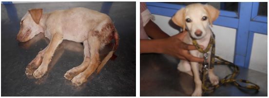
Figure 2. 0th day 5th day of therapy
Table 1. Pattern of clinical recovery
Parameter 0th day 1st day 2nd day 3rd day 4th day 5th day
General Dullness 7 7 7 3 1 -
activity Normal - - - 3 5 6
Anorexia 5 5 4 - - -
Appetite Inappetence 2 2 3 3 1 -
Satisfactory - - - 3 5 6
Whitish frothy 2 2 2 - - -
Yellowish 1 2 1 - - -
Vomiting Plain watery 1 2 1 - -
Hematemesis 3 1 - - - -
Absent - - 3 6 6 6
Greenishyellow, foul
2 - - - - -
smell
Diarrhoea Bloody, foul smell 5 7 7 2 - -
Semisolid,foul smell - - - 1 - -
Absent - - - 3 6 6
Dehydration Mild 3 4 4 2 1 1Therapeutic Management of Dogs Affected with …. 2803
Moderate 2 2 2 1 - -
Severe 2 1 1 - - -
Normal - - - 3 5 5
Fever 3 3 3 1 - -
Temperature
Normal 4 4 4 5 6 6
Tachycardia 4 2 2 - - -
Heart rate
Normal 3 5 5 6 6 6
Table 2. Mean haematological and serum biochemical values of pups before and after
therapy (Mean+S.E)
Parameter Control group (n=6) CPV positive pups
Before therapy After therapy
(0th day) (5th day)
Hb (gm%) 12.92+0.25a 11.37+1.46a 9.25+0.68b
PCV (%) 38.25+0.37a 29.3+2.26b 25.22+1.48b
TEC (m/cumm) 6.28+0.89a 5.2+0.76a 4.01+0.31a
TLC/cumm 8533.33+379.18a 12314+2970.04a 8966.67+675.61a
DLC
N (%) 70.0+0.73a 78.5+2.94b 73.83+1.11ab
L (%) 22.67+0.67a 13.83+2.31b 20.33+0.84a
M (%) 4.17+0.31a 3.67+1.2a 3.5+0.34a
E (%) 3.8+0.49a 3.83+0.72a 3.4+0.51a
Platelets (/cumm) 456166.66+22918.2a 275285.7+61853.75b 419500+46484.98a
Blood urea nitrogen 10.47+1.13a 38.7+10.53b 13.5+1.62a
(BUN) (mg/dl)
Total serum protein 7.17+0.09a 6.52+0.52a 6.61+0.45a
(gm/dl)
Blood glucose (mg/dl) 100.67+6.69a 115.86+14.31a 113.17+4.79a
Serum potassium (mmol/l) 4.82+0.11a 3.7+0.22b 4.45+0.34ab
Serum sodium (mmol/l) 144.03+0.81a 140.71+4.13a 143.25+0.75a
Serum chloride (mmol/l) 111.33+1.06a 103.71+1.87b 110.61+1.46a
Means followed by same superscript(s) don’t differ significantlyYou can also read