Effect Of Tfl(Tensor Fascia Lata) Muscle Stretching In Medial Compartment Osteoarthritis Knee Patients: A Randomized Control Trial
←
→
Page content transcription
If your browser does not render page correctly, please read the page content below
IOSR Journal of Dental and Medical Sciences (IOSR-JDMS)
e-ISSN: 2279-0853, p-ISSN: 2279-0861. Volume 10, Issue 2 (Sep.- Oct. 2013), PP 17-22
www.iosrjournals.org

  Effect Of Tfl(Tensor Fascia Lata) Muscle Stretching In Medial
   Compartment Osteoarthritis Knee Patients: A Randomized
                          Control Trial
                                            Dr Nishant H Nar,
             MPT(ortho ), HOD and clinical physiotherapist Civil hospital, Rajkot, Gujarat, India

Abstarct:
Background:-
Osteoarthritis is a chronic, localized joint disease affecting approximately one-third of adults, with the disease
prevalence increasing with advancing age. OA affects many joints including the large, weight bearing joints of
the hips and knees and also the spine, hands, feet and shoulders. The knee is the most common weight bearing
joint affected by OA, with the disease predominantly affecting the medial compartment of the tibio-femoral joint.
Patients with knee OA frequently report symptoms of knee pain and stiffness as well as difficulty with activities
of daily living such as walking, stair-climbing and house keeping.
Objectives:-
To compare the effectiveness of TFL muscle stretching exercises and conventional physiotherapy treatment with
conventional physiotherapy treatment alone in people with unilateral medial compartment knee osteoarthritis.
Materials and Methodology:-
Study included 30 (Thirty) subjects with unilateral medial compartment knee OA, aged 45 years or above. The
subjects were randomly divided into 2 groups: Group -A and Group -B. The subjects were treated for a period
of 6 weeks, 6 days a week, once daily. Pain was assessed by VAS score and physical function was assessed by
WOMAC Index of Osteoarthritis.
Results:-
The results were analyzed by Wilcoxon Signed Rank Test. Group A showed significant improvement in pain
(T=120, p
Effect Of Tfl(Tensor Fascia Lata) Muscle Stretching In Medial Compartment Osteoarthritis Knee
strategies in the management of the condition [9, 10]. However, there is an absence of high quality evidence to
support the use of such therapies [9].
There are several reasons for the development of OA including age, being overweight, heredity factors,
and joint damage from a previous injury or during early development of a joint. Increased loading across the
joint has been implicated in the progression of knee OA severity [11]. In knee OA, the medial tibiofemoral
compartment is the most common site of disease. The susceptibility of the medial compartment to OA
development may relate to greater load distribution (i.e., 60–80%) to the medial than the lateral compartment,
even in healthy knees, during gait. Excessive medial compartment loading is widely believed to contribute to
medial OA progression. Because direct measurement of knee load is invasive, external knee adduction moment
during gait, a correlate of medial load, has been used in knee OA studies [12]. The role of gait analysis in the
quantification of dynamic joint load has received much attention in the literature in light of the difficulty in
performing in vivo measurement of joint loading during movement [13, 14, 15]. From the research, the external knee
adduction moment, an indirect measure of load in the medial compartment of the tibio-femoral joint [16], has
emerged as an important and widely accepted biomechanical marker of knee load.
Cross-sectional studies demonstrate that patients with knee OA have a higher peak knee adduction moment
during walking when compared to healthy age-matched controls [17, 18]. It is also likely that the higher prevalence
of medial compared with lateral tibiofemoral joint OA is the result of differences in the relative loading within
the tibiofemoral joint. The external knee adduction moment determines load distribution across the medial and
lateral tibial plateaus [12, 19, 20], with force across the medial compartment almost 2.5 times that of the lateral [16].
It has also been reported that for patients with knee OA, the magnitude of the adduction moment is predictive of
clinical outcomes such as severity of knee pain and radiographic disease [21, 22].
A variety of exercise programs for knee OA have been described in the literature. These have included general
aerobic exercise programs such as walking or cycling as well as more specific programs involving strengthening
of particular muscle groups and/or flexibility exercises.
The primary aim of this study is to determine whether stretching of TFL muscle in people with medial
compartment knee OA can reduce knee pain and improve physical function. It is hypothesized that a 6-week
programme of stretching of TFL muscle(tensor fascia lata muscle) will improve pain and physical function in
people with medial compartment knee OA.
Aims And Objective:
1) To determine the effectiveness of TFL muscle stretching in people with medial compartment knee
osteoarthritis.
2) To compare the effectiveness of TFL muscle stretching and conventional treatment with conventional
treatment in people with medial compartment knee osteoarthritis.
II. Study Design And Materials:
Study Design
An Experimental study was conducted to study the effects of TFL muscle stretching exercises in patients with
osteoarthritic knee joints.
Study Setting
All patients were referred from Orthopaedic Out-patient Department, Civil Hospital, rajkot to Physiotherapy
Department, Civil Hospital, Rajkot where they all were treated during study period.
Sample Selection
The sample size consisted of 30 (thirty) patients, who were diagnosed with unilateral medial compartment
tibiofemoral OA, as per the Inclusion Criteria and the Exclusion Criteria.
Study Duration
The total duration of the study was 6 months. The subjects were treated for a period of 6 weeks, 6 days a week,
one session daily.
Sample Size
The sample size of 30 (thirty) patients was divided in to two groups.
Group A: 15 patients.
Group B: 15 patients.
Age Group
45 years or older.
Gender: Both sexes
Male: 14
Female: 16
www.iosrjournals.org 18 | PageEffect Of Tfl(Tensor Fascia Lata) Muscle Stretching In Medial Compartment Osteoarthritis Knee
Selection Criteria:
Inclusion Criteria:-
1. Age greater than or equal to 45 years.
2. Unilateral medial compartment tibiofemoral OA without involvement of any other compartment of knee
joint.
3. Duration of symptoms: Chronic according to IASP classification.
4. At least some difficulty in daily function due to knee OA.
5. Both genders are included.
6. Kellgren-Lawrence radiographic grade I, II and III.
7. Patients who are able to comprehend commands.
8. Willingness to participate in the study.
Exclusion Criteria:-
1. History of trauma within one year to affected knee joint.
2. Associated with any other pathological condition such as neoplasm, osteomyelitis, vascular problem etc.
3. Low back pain radiating to knee joint.
4. Knee surgery or intra articular corticosteroid injection within 6 months to affected knee joint.
Materials Used In The Study:-
Consent form, universal goniometer, vas scale, weight cuffs, WOMAC Index of Osteoarthritis, Examination
Table, Short-wave Diathermy Machine, Kodak C875 Zoom Digital Camera, Paper, Pencil, Scale, Pins.
Description Of The Tools:-
Visual Analog Scale (VAS):
WOMAC Index of Osteoarthritis:
III. Methodology:
Ethical clearance was obtained from the Ethical Clearance Committee of civil hospital ,rajkot prior to
the study. Those who fulfilled the inclusion criteria were taken up for the study. The whole procedure of the
study was explained to all the subjects. A written informed consent of all the subjects was taken prior to the
study. All the subjects were assessed as per the assessment form.
30 (thirty) subjects were taken for the study with diagnosis of unilateral medial compartment knee OA;
14 male and 16 female. They were randomly divided in to two groups for the study. Each subject of the study
was treated for a period of 6 weeks, 6 days a week, one session daily. An assessment was done prior to starting
of treatment and weekly assessment was taken for these subjects.
Exercise Protocol
All the subjects were informed in detail about the type and nature of the study. The subjects were
divided in to two groups; Group A and Group B, 15 patients in each group. All the subjects were randomly
selected and assigned in to each group.
Group A:
The subjects in Group A were given TFL muscle stretching exercises and conventional physiotherapy treatment.
Group B:
The subjects in Group B were given conventional physiotherapy treatment.
CONVENTIONAL PHYSIOTERAPY TREATMENT FOR BOTH GROUP -A & GROUP -B: - (ref)
A. SHORT WAVE DIATHERMY:
B. STRETCHING EXERCISES:
1) Standing calf stretch
2) Supine hamstring stretch
3) Prone quadriceps stretch
C. RANGE OF MOTION EXERCISES:
1) In long sitting position, knee mid-flexion to end range extension
2) In long sitting position, knee mid-flexion to end range flexion
3) Stationary bicycle
www.iosrjournals.org 19 | PageEffect Of Tfl(Tensor Fascia Lata) Muscle Stretching In Medial Compartment Osteoarthritis Knee
D. STRENGTHENING EXERCISE:
1) Static quadriceps sets in knee extension
2) In high sitting position knee mid-flexion to end range extension with weight cuff
3) In prone position knee end range extension to mid-flexion with weight cuff
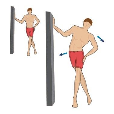
TFL MUSCLE STRETCHING EXERCISES FOR GROUP -A:-
Subjects in Group A were given a series of three exercises designed to stretch TFL muscles, 6 days a week
for 6 weeks.
Type of exercise
1) In side lying position therapist will do flexion at knee joint, adduction and external rotation at hip joint
And other two methods of self stretching exercise:
2) In supine lying cross leg stretching
3) In standing cross leg strtching
The whole study was extended for a period of 6 months. The duration of treatment programme for each
subject was 6 weeks. All the thirty (30) subjects completed the whole treatment programme of 6 weeks with out
any discomfort.
IV. Results:
Table 1 show the gender distribution of the 30 subjects who participated in the study. In the Group A
where the subjects underwent hip abductor muscle strengthening exercises and conventional physiotherapy
treatment had 8 males and 7 females and in the Group B where the subjects underwent conventional
physiotherapy treatment alone had 6 males and 9 females. There was no significant predominance of sex.
Table 1 Gender Distribution of the Subjects:
Gender Group A Group B
Male count 8 6
% 53.33% 40%
Female count 7 9
% 46.66% 60%
Total 15 15
Table 2 displays the statistics of age distribution of the 30 subjects. Among the 30 subjects, the mean age of 15
subjects in Group A was 51.33 with a standard deviation (SD) of 5.2326, and the mean age of 15 subjects in
Group B was 52 with a standard deviation of 5.0142. No significant age difference was seen across the two
groups.
Table 2 Age Distribution of the Subjects:
Group N Mean SD
Group A 15 51.33 5.2326
Group B 15 52 5.0142
www.iosrjournals.org 20 | PageEffect Of Tfl(Tensor Fascia Lata) Muscle Stretching In Medial Compartment Osteoarthritis Knee
Wilcoxon Signed Rank Test (1,2) was applied in Group A and in Group B for with-in group analysis and it is
as follows:
In Group A, results showed significant improvement on VAS score (T = 120 > 95, p < 0.05).
In Group A, results showed significant improvement on WOMAC score (T = 120 > 95, p < 0.05).
In Group B, results showed significant improvement on VAS score (T = 91 > 74, p < 0.05).
In Group B, results showed significant improvement on WOMAC score (T = 120 > 95, p < 0.05).
Wilcoxon Sum Rank Test (Mann Whitney ‘U’ Test) (1,2) was applied for between-group comparison of
Group A and Group B, and it is as follows:
On comparing Group A and Group B for post-treatment VAS score, results showed significant difference in
improvement (z = -2.82, p = 0.0052).
On comparing Group A and Group B for post-treatment WOMAC score, results showed significant difference
in improvement (z = -3.56, p = 0.0004).
For Group A:
Score Pre Post T p<
Mean + SD Mean + SD
VAS 7 + 1.690 2 + 1.463 120 0.05
WOMAC 66.66 + 6.986 27.66 + 4.237 120 0.05
For Group B:
Score Pre Post T p<
Mean + SD Mean + SD
VAS 6.93 + 1.387 4.066 + 1.907 91 0.05
WOMAC 67.13 + 6.577 37.46 + 6.356 120 0.05
On comparing Group A and Group B:
Score z Value p Value
VAS -2.82 0.0052
WOMAC -3.56 0.0004
The ‘z’ values (corresponding to ‘p’) are highly significant which suggest that TFL muscle stretching exercises
are effective in reduction of pain and improvement of physical function along with conventional physiotherapy
treatment.
V. Conclusion:
There was an experimental study comparing effectiveness of TFL muscle stretching and conventional
physiotherapy treatment in osteoarthritis knee patients.
There was a statically significant difference in both experimental and conventional groups.
Hence, Null Hypothesis of no significant effect of TFL muscle stretching exercises can be rejected and
Alternative Hypothesis of , there is an additive effect of TFL muscle stretching exercises on reduction of pain
and improvement of physical function can be accepted.
Acknowledgements:
I would like to thank my teachers and guide and all staff of physiotherapy department, civil hospital and I am
grateful to all my patients for their kind cooperation and willingness to participate in this study, without whom
this study would not have materialized.
Interest Of Conflict:
The authors perceive no conflict of interest in this study.
References:
[1]. Felson DT, Naimark A, Anderson J, Kazis L, Castelli W, Meenan RF: The prevalence of knee osteoarthritis in the elderly. The
Framingham Osteoarthritis Study. Arthritis and Rheumatism 1987, 30:914-918.
[2]. Australia A: Painful Realities: The economic impact of arthritis in Australia in 2007. 2007.
[3]. Hamerman D: Clinical implications of osteoarthritis and aging. Annals of the Rheumatic Diseases 1995, 54:82-85.
[4]. Badley E, Wang P: Arthritis and the aging population: projections of arthritis prevalence in Canada 1991 to 2031. Journal of
Rheumatology 1998, 25(1):138-144.
[5]. Ledingham J, Regan M, Jones A, Doherty M: Radiographic patterns and associations of osteoarthritis of the knee in patients referred
to hospital. Ann Rheum Dis 1993, 52(7):520-526.
[6]. Iorio R, Healy WL: Unicompartmental arthritis of the knee. J Bone Joint Surg Am 2003, 85-A (7):1351-1364.
www.iosrjournals.org 21 | PageEffect Of Tfl(Tensor Fascia Lata) Muscle Stretching In Medial Compartment Osteoarthritis Knee
[7]. Guccione AA, Felson DT, Anderson JJ, Anthony JM, Zhang Y, Wilson PWF, Kelly-Hayes M, Wolf PA, Kreger BE, Kannel WB:
The effects of specific medical conditions on the functional limitations of elders in the Framingham study. American Journal of
Public Health 1994, 84:351-358.
[8]. Dieppe PA, Ebrahim S, Martin RM, Juni P: Lessons from the withdrawal of rofecoxib. Bmj 2004, 329(7471):867-868.
[9]. Jordan K, Arden N, Doherty M, Bannwarth B, Bijlsma J, Dieppe P, Gunther K, Hauselmann H, Herrero-Beaumont G, Kaklamanis P,
et al.: EULAR recommendations 2003: an evidence based approach to the management of knee osteoarthritis: report of a task force
of the Standing Committee for International clinical Studies Including Therapeutic Trials (ESCISIT). Annals of the Rheumatic
Diseases 2003, 62:1145-1155.
[10]. OA ASo: Recommendations for the medical management of osteoarthritis of the hip and knee. 2000 update. Arthritis and
Rheumatism 2000, 43(9):1905-1915.
[11]. Miyazaki T, Wada M, Kawahara H, Sato M, Baba H, Shimada S: Dynamic load at baseline can predict radiographic disease
progression in medial compartment knee osteoarthritis. Ann Rheum Dis 2002, 61:617-622.
[12]. Hurwitz D, Sumner D, Andraicchi T, Sugar D: Dynamic knee loads during gait predict proximal tibial bone distribution. Journa l of
Biomechanics 1998, 31:423-430.
[13]. Andriacchi T, Lang P, Alexander E, Hurwitz D: Methods for evaluating the progression of osteoarthritis. J Rehabil Res Dev 2000 ,
37(2):163-170.
[14]. Andriacchi T, Mundermann A: The role of ambulatory mechanics in the initiation and progression of knee osteoarthritis. Current
Opinion in Rheumatology 2006, 18:514-518.
[15]. Sharma L, Kapoor D, Issa S: Epidemiology of osteoarthritis: an update. Current Opinion in Rheumatology 2006, 18:147-156.
[16]. Schipplein OD, Andriacchi TP: Interaction between active and passive knee stabilizers during level walking. Journal of Orthopaedic
Research 1991, 9:113-119.
[17]. Bailunas A, Hurwitz D, Ryals A, Karrar A, Case J, Block J, Andriacchi T: Increased knee joint loads during walking are present in
subjects with knee osteoarthritis. Osteoarthritis & Cartilage 2002, 10:573-579.
[18]. Hurwitz D, Ryals A, Case J, Block J. Andriacchi T: The knee adduction moment during gait in subjects with knee osteoarthritis is
more closely correlated with static alignment than radiographic disease severity, toe out angle and pain. J Orthop Res 2002, 20:101-
108.
[19]. Jackson B, Teichtahl A, Morris M, Wluka A, Davis S, FM C: The effect of the knee adduction moment on tibial cartilage volume and
bone size in healthy women. Rheumatology 2004, 43:311-314.
[20]. Wada M, Maezawa Y, Baba H, Shimada S, Sasaki S, Nose Y: Relationships among bone mineral densities, static alignment and
dynamic load in patients with medial compartment knee osteoarthritis. Rheumatology 2001, 40:499-505.
[21]. Shrader M, Draganich L, Pottenger L, Piotrowski G: Effects of knee pain relief in osteoarthritis on gait and stair-stepping. Clinical
Orthopaedics and Related Research 2004, 421:188-193.
[22]. Sharma L, Hurwitz DE, Thonar E, Sum JA, Lenz ME, Dunlop DD, Schnitzer TJ, Kirwanmellis G, Andriacchi TP: Knee Adduction
Moment, Serum Hyaluronan Level, and Disease Severity in Medial Tibiofemoral Osteoarthritis. Arthritis & Rheumatism 1998,
41(7):1233-1240.
www.iosrjournals.org 22 | PageYou can also read