HEAT: A SOFTWARE ASSISTANT FOR THE ANALYSIS OF LV REMODELING AFTER MYOCARDIAL INFARCTION IN 4D MR FOLLOW-UP STUDIES
←
→
Page content transcription
If your browser does not render page correctly, please read the page content below
HeAT: A Software Assistant for the Analysis
          of LV Remodeling after Myocardial Infarction
                  in 4D MR Follow-Up Studies

        D. Säring1 , A. Stork2 , S. Juchheim2 , G. Lund3 , G. Adam2 , H. Handels1

                            1
                          Department of Medical Informatics,
                 2
                Department of Diagnostic and Interventional Radiology,
      University Medical Center Hamburg-Eppendorf, 20246 Hamburg, Germany
              3
                Röntgeninstitut Düsseldorf, 40476 Düsseldorf, Germany

                            Email: d.saering@uke.uni-hamburg.de

      Abstract: Spatio-temporal Cine-MR image sequences of the heart contain informa-
      tion about shape and motion changes, spatial delayed-enhanced (DE)-MR enables
      identification of pathological structures after myocardial infarction. In this paper the
      Heart Analysis Tool (HeAT) for the quantitative analysis of 4D MR image sequences
      of infarct patients is used to analyze the characteristics of left ventricular (LV) remod-
      eling extracting regional and global parameters in the acute (base-line) and approxi-
      mately 15 months past acute (follow-up) phase. The software assistant HeAT provides
      the combined evaluation of Cine- and DE-MR image data. Partitioning of the my-
      ocardium in segments enables the analysis with high local resolution. Corresponding
      segments are generated and used for intra patient comparison. First results of 10 infarct
      patients indicate an exact analysis of morphological and functional characteristics of
      the infarcted myocardium. Based on the visualization and statistical interpretation of
      these analysis LV remodeling can be quantified. Quantitative analysis can help to find
      predictive parameters describing LV remodeling having an impact on further therapy.

1   Introduction

Myocardial infarction causes the development of myocardial necrosis. The necrotic area
loses its ability to contract and the remaining heart muscle needs to compensate for that
weakened area. Loss of contractile function is attributed to left ventricular remodeling.
LV remodeling is an important element in the progression of cardiac insufficiency. Re-
modeling is characterized by wall thinning, chamber dilation and increased end-diastolic
and end-systolic volumes [J+ 03]. It is an important clinical problem to get quantitative
parameters characterizing LV remodeling.
New magnetic resonance (MR) imaging techniques provide the means for a non-invasive
evaluation of anatomic and functional parameters of the beating heart. In this paper, 4D
Cine-MR and 3D delayed enhancement (DE)-MR image sequences are used for this pur-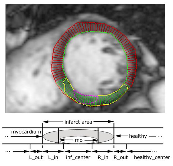
pose. Cine-MR with high spatial and temporal resolution enable accurate measurements of
the myocardial shapes. DE-MR is effective in identifying the presence and extent of my-
ocardial infarction. DE-MR also has the potential to allow identification of microvascular
obstruction (MO). Presence of MO is associated with greater myocardial damage, with LV
remodeling and increased risk of mortality and morbidity [W+ 98]. Analyzing myocardial
variation, e.g. infarct shrinkage, revitalization of shocked tissue, requires detailed eval-
uation of the transition from healthy myocardium to diseased myocardium (borderzone).
Thus, anatomical information from Cine-MR and pathological information from DE-MR
images have to be combined.
Due to the quantity of MR image data, analyzing 4D MR data by hand is time-consuming
and in most cases not reproducible. The computer-assisted extraction of these measure-
ments is reproducible and fast. Several approaches for assessing LV wall motion are pub-
lished e.g. [SRD94]. In contrast to these approaches the Heart Analysis Tool (HeAT) in-
corporates myocardial shape and motion information derived from 4D Cine-MR, as well as
acute infarction information (e.g. MO) from DE-MR. Local analysis in high user-defined
resolution is enabled by dividing the myocardium into small myocardial segments. Fur-
thermore a unique labeling of these segments enables inter/intra patient comparison. De-
rived quantitative parameters can be stored and analyzed by medical experts.

2   Methods

In the Department of Diagnostic and Interventional Radiology, University Medical Cen-
ter Hamburg-Eppendorf, two MR techniques are used to acquire spatio-temporal image
data of patients after myocardial infarction and approximately 15 months later. Generally,
these images are generated in 4D MR short axis studies (Cine-MR) and 3D DE-MR. All
images are acquired with a 1.5T system by using electrocardiographic triggering and dur-
ing breath-hold sequences. The Cine-MR images have a resolution of 224 x 256 x Z (Z
differs between 7 and 12) voxels. These sequences usually contain 16 up to 20 time frames
per R-R interval. The DE-MR was performed in all short-axis LV sections by using T1-
weighted turbo fast low-angle shot sequence. The inversion time was 220-300 msec and
the resolution is 224 x 256 for each slice. About 200 Cine-MR images and 7-12 DE-MR
images for each patient were acquired.
The software assistant HeAT enables the import of 4D Cine-MR, 3D DE-MR and XML
contour files. First, a segmentation of anatomical structures (e.g. endocard, epicard, pap-
illaries, right ventricle) at endiastolic and endsystolic phase in 4D Cine-MR is provided.
Selection of corresponding slices in Cine- and DE-MR using Mutual Information provides
automatic comparative visualization of both modalities. Registration of corresponding
slices enables direct myocardial contour overlay on DE-MR to support the data-driven
segmentation of pathological structures (infarct area, MO).
In addition to the extraction of global parameters (e.g. LV ejection fraction (LVEF), stroke
volume (SV)) a segment model dividing the myocardium in a variable number of myocar-
dial segments enables extraction of local parameters with high resolution.
HeAT was executed on a standard PC with Intel Pentium 4 CPU 3.00 GHz, 2 GB memory
and NVIDIA Quadro4 graphic card. To avoid re-implementing of state-of-the-art methods
we implemented HeAT in C++ utilizing ITK and VTK with a problem-oriented adaptation.

2.1     Pre-processing of the MR image sequences

Pre-processing is necessary before the automatic algorithms for parameter extraction can
be applied to evaluate the effect of LV remodeling. The analysis is based on the segmen-
tation of myocardial and infarct structures. Furthermore the registration of corresponding
Cine- and DE-MR slices enables the combined analysis of the outlined structures.

2.1.1    Segmentation of anatomical and pathological structures

The automatic segmentation of anatomical structures in cardiac MR images is a difficult
task due to artifacts caused by turbulent blood flow, which leads to a loss of signal and
temporary occlusion of object boundaries. In recent years, several methods with different
approaches for segmentation of the left ventricle were developed, e.g. model-based seg-
mentation [Par03]. At the moment the focus of the development of HeAT is the quantita-
tive analysis. Therefore, a basic tool for manual contour drawing was used for identifying
endocard and epicard in different phases of the heart cycle in HeAT. Additionally, 3D visu-
alization of the segmented structures is generated in real-time. Unfortunately, in Cine-MR
image sequences the localization and quantification of pathological areas like the infarct
area and MO is not possible. But in DE-MR data these structures can be distinguished
using a data-driven segmentation approach described in [G+ 02]. Outlined contours are
stored in XML-files.

2.1.2    Registration of Cine- and DE-MR data

One objective of HeAT is to combine Cine- and DE-MR contour information. Direct
transfer of myocardial contours to the DE-MR image sequences causes two problems.
Correspondence: DE-MR sequences contain only one frame per cardiac loop. In or-
der to minimize heart-moving artifacts this frame is acquired in a neutral cardiac phase
(end-diastolic phase). DE-MR imaging requires approximately 220 ms. Cine-MR im-
age sequences usually contain 16 to 20 time frames, each frame is acquired in about 10
- 50 ms. Thus, for each DE-MR image several temporal Cine-MR images of the same
spatial position exist. For combination of anatomical and pathological information the
definition of corresponding Cine- and DE-MR data is required. In this paper we address
the correspondence problem using Mutual Information (MI) as similarity measure for the
entire images. The corresponding image pair is selected automatically by the maximum
MI value. Alignment: The imaging of Cine- and DE-MR data requires different settings,
e.g. inversion time. Generally, these settings leads to different field of views and/or dif-
ferent patient positions. So, an alignment of corresponding Cine- and DE-MR slices is
necessary. To address this problem a semi-automatic rigid registration using MI is ap-
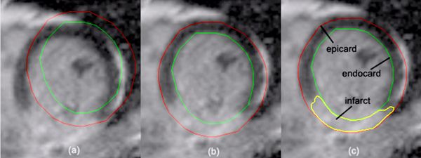
plied. After pre-registration, the user can manually adjust shifting and scaling of DE-MR.
Thus, information of both datasets is combined by transferring the Cine-MR contours to
the corresponding registered DE data.

Figure 1: Visualization of the results of direct contour overlay (Cine-MR myocardial contours on
DE-MR image data) before (a), and after (b) registration. Segmentation of the infarct area is pre-
sented in (c).

2.2    Automatic computation of quantitative parameters

Detailed cardiac analysis of patient image data after myocardial infarction requires a high
local resolution. Therefore, a standardised arrangement in 17 segments was proposed by
the American Heart Association [C+ 02]. For analyzing the characteristics of LV remodel-
ing, local resolution of the 17 segment model is insufficient. So, HeAT supports a segment
model with a variable number of segments in dependence on the centerline method [S+ 86].
Based on the endocardial and epicardial contours a centerline was computed and there-
after the myocardium was divided in even spaced segments. Unified segment numbering
according to significant anatomical structures (wall position of the right ventricle in an-
teriorseptal section) enables identification of corresponding segments (Fig.2(a)). Manual
selection of the anatomical structure is time-consuming and user-dependent. Therefore,
the starting point is automatically defined at 45◦ to a horizontal line from the geometri-
cal midpoint of the ventricle in anterolateral direction. Provided that all images are equal
oriented, segments of different phases and different patients but with the same segment
number can be identified as corresponding. Thus, these segments are usable for inter- and
intra-patient comparison. The local quantitative parameters are computed automatically
for each myocardial segment. Additionally, global parameters (e.g. LVEF, myocardial
mass LVDM) were calculated. For evaluation of LV remodeling 13 regions of interest
were automatically selected, e.g.

      • infarct-center (infcenter ) (central one-third of the infarct circonverence)
      • infarct-periphery (Lin ,Rin ) (border one-third of the infarct circonverence)
• peri-infarct zone (Lout ,Rout ) (10% of the myocardial circonverence adjacent to the
infarct border).
Each region is defined as a set of segments (see Fig.2(b)). Union of infarct-periphery and
peri-infarct zone represents the border zone.
(a) (b)
Figure 2: Schematically representation of anatomical and pathological structures as well as segment
lines and segment numbering (a). Definition of ROIs in the infarct surrounding area (b).
3 Results
In HeAT Cine- and DE-MR datasets of 10 patients after myocardial infarction (base-line
(BL), follow-up (FU)) were analyzed by two medical experts in all slices for each pa-
tient. Based on the segmentation with HeAT one dataset can be analyzed in about 10
minutes. For the acquirement of the local parameters, e.g. endocardial movement or in-
farct transmurality a myocardial partitioning in 100 segments was used. The automatically
computed parameters were displayed in tables (Fig.4) and as curves in a datasheet (Fig.3).
This enables a clinical interpretation of characteristics describing LV remodeling at the
first glance. Main results of the first 10 patients are discussed in the following.
The analysis of the global parameters shows at BL an inverse correlation between the
infarct size and left ventricular ejection fraction (LVEF) (r = 0.65, P < 0.05), reflecting
that patients with large infarction had worse left ventricular function. Mean infarct size
decreased from 25 ± 11 gr at BL to 17 ± 7 gr at FU (P < 0.01). Mean LV mass also
decreased from 118 ± 25 gr at BL to 103 ± 19 gr at FU (P < 0.05). There was aFigure 3: Regression plot (infarct size change to base-line infarct size) of 10 patients(a).
strong correlation between the initial infarct size at BL and reduction of infarct size at FU
(r = 0.80, P < 0.01), indicating that infarct shrinkage is determined by the BL infarct
size.
Figure 4: Datasheets of global (b) and local (c) parameters of pat #6
The analysis of the regional parameters shows the mean diastolic wall thickness of the
infarcted tissue decreased from 7.7 ± 1.4 mm at BL to 6.7 ± 1.5 mm at FU (P < 0.05), in-
dicating wall thinning of the infarcted myocardium. Mean infarct transmurality decreased
from 71 ± 13 % at BL to 65 ± 11 % at FU (P < 0.05). The number of the infarcted
segments showed no significant change from BL (39 ± 17 segments) to FU (43 ± 22
segments, P = not significant), indicating that thinning and not stretching occurs in the
infarcted myocardium.
4 Discussion
For cardiac analysis and visualization a huge range of tools, e.g. IDL exist. In contrast to
these tools HeAT enables a combined analysis of anatomical and functional information ininfarct regions and healthy tissue based on registered Cine- and DE-MR image sequences.
Furthermore, computation of parameters in user-defined regions of interest with high local
resolution enables a detailed evaluation of LV remodeling. The manual segmentation is
time-consuming and its results are user-dependent. Therefore automatic or semi-automatic
methods will be integrated to improve the segmentation process.
Data analysis using the software assistant HeAT quantifies significant shrinkage of the
infarcted area from BL to FU. The initial infarct size determines the later reduction of
infarct size. The shrinkage of the infarction is mainly characterized by thinning of infarct
transmurality. In this study additional 20 patient datasets will be evaluated concerning LV
remodeling in the next step.
Intra-patient comparison of parameters in corresponding local areas (BL to FU) can be
useful to predict the course of disease (e.g. regeneration of shocked tissue can be identi-
fied by an increased endocardial movement and myocardial thickening). These results of
inter-patient comparisons can be useful for predicting the course of disease for an acute
myocardial infarct patient.
References
[C+ 02] M D Cerqueira et al. Standardized Myocardial Segmentation and Nomenclature for Tomo-
graphic Imaging of the Heart. Circulation - American Heart Association (AHA) Scientific
Statement, pages 105–539, 2002.
[G+ 02] B L Gerber et al. Accuracy of Contrast-Enhanced Magnetic Resonance Imaging in Pre-
dicting Improvement of Regional Myocardial Function in Patients After Acute Myocar-
dial Infarction. Circulation, pages 1083–1089, Aug 2002.
[J+ 03] B M Jackson et al. Border zone geometry increases wall stress after myocardial infarction:
contrast echocardiographic assessment. Heart Circ Physiol, (284):475 – 479, 2003.
[Par03] N Paragios. Shape-based Segmentation and Tracking in Cardiac Image Analysis. IEEE
Trans. on Medical Image Analysis, pages 402–407, 2003.
[S+ 86] F H Sheehan et al. Advantages and applications of the centerline method for characteriz-
ing regional ventricular function. Circulation, (74):293–305, Aug 1986.
[SRD94] P Shi, G Robinson, and J Duncan. Myocardial motion and function assessment using 4D
images. pages 148–159. IEEE Visualization in Biomedical Computing, 1994.
[W+ 98] K C Wu et al. Prognostic significance of microvascular obstruction by magnetic resonance
imaging in patients with acute myocardial infarction. Circulation, (97):765–772, 1998.You can also read