Mucocele of the Appendix: Case Report & Review of Literature - ClinMed International Library
←
→
Page content transcription
If your browser does not render page correctly, please read the page content below
ISSN: 2378-3397
                                                                                               Abiyere et al. Int J Surg Res Pract 2021, 8:125
                                                                                                          DOI: 10.23937/2378-3397/1410125
                                                                                                                            Volume 8 | Issue 1
                    International Journal of                                                                                     Open Access

                    Surgery Research and Practice
Case Report

Mucocele of the Appendix: Case Report & Review of Literature
Abiyere OH1,3*, Adewara O2, Akute OO1 and Babatunde O1
1
 Department of Surgery, Federal Teaching Hospital, Nigeria
2
 Department of Obstetrics & Gynaecology, Federal Teaching Hospital, Nigeria                                                     Check for
                                                                                                                                updates
3
 Department of Surgery, Afe Babalola University, Nigeria

*Corresponding author: Dr. Abiyere OH, Department of Surgery, Federal Teaching Hospital, PMB 201, Ido-Ekiti, Ekiti
State, Nigeria

    Abstract                                                             Introduction
    Appendiceal Mucocele is a rare disease. Sometimes, it is                 Appendiceal mucocele is an obstructive dilatation of
    discovered accidentally and sometimes mimic acute appen-             the appendiceal lumen caused by intraluminal accumu-
    dicitis. Correct diagnosis before surgery is very crucial for        lation of mucoid material. It is a rare disease. The inci-
    the selection of adequate surgical treatment to avoid Intra-
    operative and Postoperative complication. Ultrasonography            dence is 0.2% to 0.7% of all appendectomy specimens
    and particularly, computed Tomography should be used                 [1-3]. This transformation is caused by one of four pat-
    extensively for this purpose. If mucocele of the appendix is         terns of epithelial proliferation: Retention cyst, mucosal
    treated incorrectly Pseudomyxoma peritonei which is char-            hyperplasia, mucinous cystadenoma and mucinous cys-
    acterized by malignant process may develop.
                                                                         tadenocarcinoma [4,5].
    We present a case, which was discovered accidentally
    while evaluating the patient for another condition. She is a             This disease does not have a typical clinical picture.
    47-year-old woman, who presented at the surgical out-pa-             Sometimes the patient has pain in the lower right quad-
    tient department with clinical features suggestive of chronic        rant of the abdomen; therefore a surgeon may mistake
    cholecystitis, had abdominal ultrasonographic scan which             it for acute appendicitis. Sometimes it is discovered in-
    showed Cholelithiasis and a cystic tubular swelling in the
    right lower abdomen, which was reported as a right adnexal           cidentally while evaluating a patient for another con-
    cyst. Open surgery was performed. At the time of surgery,            dition as in this case. Acute appendicitis is one of the
    multiple stones (3) in the gall bladder and a cystic mass of         most common surgical diseases [3,6,7].
    the appendix with dimensions 13.0 × 3.5 cm, with thinned
    walls without perforation were discovered. No discharge                  It is important to differentiate between these two
    was found in the peritoneal cavity. Diagnosis of mucocele            pathologies before surgery and select adequate surgical
    of the appendix was suspected. Only appendectomy was                 tactics. If treated improperly, the mucocele may prog-
    performed because no pathologic process was found in the
                                                                         ress, epithelial cells may escape into the peritoneal cav-
    base of the appendix and lymph nodes were not increased
    in size.                                                             ity and Pseudomyxoma peritonei may develop which
                                                                         has a high mortality [7]. We present the case which was
    Also had cholecystectomy. Histopathologic diagnosis was
    retention cyst (Simple appendiceal mucocele). After 4                discovered intraoperative.
    months, the patient is doing well.
                                                                         Case Report
    Keywords
                                                                             A 47-year-old woman was referred to the Surgical
    Mucocele, Appendix, Gallbladder, Cholelithiasis, Retention           Outpatient Department. Her complain was pain in the
    cyst (simple appendiceal mucocele)
                                                                         right hypochondrial region, worsen by fatty meal. The
                                                                         pain has been on and off for the past 9 months. No pain
                                                                         in the right lower abdomen. No anorexia. Examination
                                                                         of the abdomen showed mild tenderness in the right hy-

                                       Citation: Abiyere OH, Adewara O, Akute OO, Babatunde O (2021) Mucocele of the Appendix: Case
                                       Report & Review of Literature. Int J Surg Res Pract 8:125. doi.org/10.23937/2378-3397/1410125
                                       Accepted: February 15, 2021; Published: February 17, 2021
                                       Copyright: © 2021 Abiyere OH, et al. This is an open-access article distributed under the terms of the
                                       Creative Commons Attribution License, which permits unrestricted use, distribution, and reproduction
                                       in any medium, provided the original author and source are credited.

Abiyere et al. Int J Surg Res Pract 2021, 8:125                                                                                • Page 1 of 4 •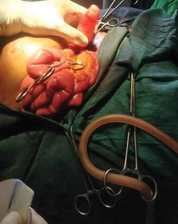
DOI: 10.23937/2378-3397/1410125 ISSN: 2378-3397
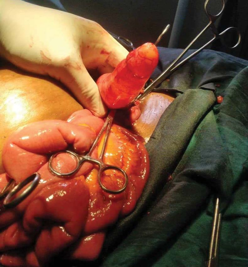
Figure 1 and Figure 2: Intraoperative finding of appendiceal mucocele with normal appendix base.
was no complication in the postoperative period. Four
months after Surgery the patient is feeling well.
Discussion
Mucocele of the appendix was first described by
Rokitansky [8]. It is a descriptive and unspecific term to
define the cystic dilation of the appendix caused by the
accumulation of mucus secretion. This process is slow
and gradual, with no signs of infection inside the organ.
It results from lumen obstruction in the appendix, which
is secondary to the inflammatory or neoplastic prolifer-
ation of the appendix mucosa or lesion in the caecum,
adjacent to the appendiceal ostium. While some arti-
cle, confirm its prevalence among women [9,10] other
demonstrate a higher incidence among men [11].
Mucocele of the appendix is divided into four patho-
logical subgroups based on the epithelial characteristic
[12,13].
Figure 3: Intraoperative distended appendix with mucin The first group consist of a simple retention cyst sec-
without perforation. ondary to occlusion of the appendix by faceolith, scar
tissue from previous inflammation or in rare cases due
pochondrium. No palpable mass or swelling in the right to endometriosis [14]. It has a normal or flattened ep-
iliac fossa. Ultrasonography showed multiple stones (3) ithelium, moderate luminal dilation up to 2 cm and it
in the gallbladder and right adnexal cystic swelling. The constitutes about 20% of all appendiceal mucocele [14].
working diagnosis was symptomatic Cholelithiasis and The case presented falls under this group. However, it
right ovarian cyst. Open surgery (Laparotomy) was per- was much larger than expected for this pathologic type.
formed. At the time of surgery, thickened gallbladder The second group with hyperplastic epithelium and
with multiple stones (3) within it and a cystic mass of moderate luminal dilatation: This constitutes about 20%
the appendix, 13.5 × 3.5 cm, thinned wall, without per- of all mucocele of the appendix [14].
foration were discovered Figure 1, Figure 2 and Figure 3. The third group is benign mucinous cystadenoma:
The ovaries were normal. No discharge was found This is characterized by tubular adenomatous epitheli-
in the peritoneal cavity. A mucocele of the appendix um with varying degree of epithelial atypia. It produces
was suspected only appendectomy was performed be- large amount of mucin with prominent luminal dilata-
cause no pathologic process was found in the base of tion of up to 6 cm. it is the most common form, consti-
the appendix. The patient also had cholecystectomy. tuting about 50% cases and with associated 20% risk of
Histopathologic diagnosis was retention cyst (simple perforation [13,14]. The fourth group encompasses the
appendiceal mucocele) and chronic cholecystitis. There malignant mucinous cystadenocarcinoma; character-
Abiyere et al. Int J Surg Res Pract 2021, 8:125 • Page 2 of 4 •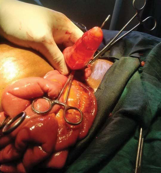
DOI: 10.23937/2378-3397/1410125                                                                            ISSN: 2378-3397

ized by glandular stromal invasion and/or tumour cells       rate exploration of the abdomen is advised due to the
in peritoneal implants i.e. Pseudomyxoma peritonei. It       well-known association between the appendiceal muco-
sometimes resembles mucinous carcinoma of the colon.         cele and other mucin-secreting cells such as colon and
It constitutes about 11-20% of all cases with 6% risk of     ovarian cancers [21].
spontaneous rupture [13,14]. Cystadenoma and cysta-
denocarcinoma are neoplastic appendiceal mucocele,
                                                             Conclusion
constituting about 35% of all primary neoplasm of the            Mucocele of the appendix is a rare disease with vague
appendix [11,14].                                            symptoms. Abdominal ultrasonographic scan important
                                                             diagnostic tool, but histopathology is needed for defini-
    The clinical presentation of the disease does not have
                                                             tive diagnosis. Surgery for benign appendiceal mucocele
a specific picture. It often flows asymptomatically as in
                                                             has an excellent long-term prognosis.
this case. In about 50% of cases it is discovered acciden-
tally during radiologic and endoscopic examination or        Consent
at surgery. However, a patient’s clinical symptoms may
                                                                The authors certify that they have obtained all ap-
include pain, palpable abdominal mass, nausea, vomit-
                                                             propriate patient consent forms. In the form the patient
ing, weight loss, gastrointestinal bleeding, and signs of
                                                             has given her consent for images and other clinical in-
intussusceptions of the intestine [9,13,14].
                                                             formation to be reported in the journal. The patient un-
    Pre-operative diagnosis of appendiceal mucocele is       derstand that her name will not be published and due
very important for the selection of an adequate surgical     efforts will be mode to conceal her identity.
method to prevent peritoneal dissemination to prevent
intraoperative and postoperative complication and re-        Funding
peated surgery [13].                                            Nil.
   Sonographic examination is considered the first line      Conflict of Interest
diagnostic modality that can probably differentiate be-
nign and malignant mucocele [15]. An appendicular               There are no conflicts of interest.
diameter of 15 mm or more has been determined the            References
threshold for diagnosis of mucocele with a sensitivity of
                                                             1. Rangarajan M, Palanivelu C, Kavalakat AJ, Parthasarathi R
83% and a specificity of 92%. Computerized tomography           (2006) Laparascopic appendectomy for mucocele of the ap-
(CT) scan is important to confirm the diagnosis and to          pendix. Report 8 cases. Indian J Gastroenterol 25: 256-257.
evaluate the extent of the disease.                          2. Marudanayagam R, Wilhams GT, Rees BI (2006) Review of
    Fine needle aspiration cytology (FNAC) is not usually       the pathological results of 2600 appendectomy specimens.
                                                                J Gastroenterol 41: 745-749.
recommended as it increases the risk of perforation and
dissemination in to peritoneal cavity [16]. Colonoscopy      3. Ruiz-Tovar J, Teruel DG, Gastineires VM, Dehesa AS,
usually reveals an elevation of the appendicular orifice.       Quindos PL, et al. (2007) Mucocele of the appendix. World
                                                                J Surg 31: 542-548.
In addition, a yellow mucous discharge would be visible
as well. Colonoscopy is also important for the diagnosis     4. Aho A, Heinonen R, Lauren P (1973) Benign and malignant
                                                                mucocele of the appendix. Histological types and progno-
of synchronous or metachronous cancers when present.            sis. Acta Chir Scand 139: 392-400.
   Conventional surgery is generally preferred to lapa-      5. Higa E, Rosai J, Pizzimbono CA, Wise L (1973) Mucosal
roscopic approach as the latter increases the risk of rup-      hyperplasia, mucinous cystadenoma, and mucinous cysta-
ture, but laparoscopic appendectomy is performed for            odenocarcinoma of the appendix. A re-evaluation of appen-
                                                                diceal “mucocele”. Cancer 32: 1525-1541.
selected patients which confer the advantages of min-
imal-access surgery, including the avoidance of a large      6. Pickhardt PJ, Levy AD, Rohrmann CA Jr, Kende AL (2002)
incision, a better cosmetic outcome and a short con-            Primary neoplasms of the appendix manifesting as acute
                                                                appendicitis: CT findings with pathologic comparison. Ra-
valescent period [17-20]. Simple appendectomy is the            diology 224: 775-781.
choice for patients with benign mucocele as suggested
                                                             7. Sugarbaker PH (2009) Appendiceal epithelial neoplasms
by the presence of a normal caecum and appendicular             and pseudomyxoma peritonei, a District clinical entity with
base and no evidence of perforation as in our patient.          District treatments in Bland ICJ. General surgery principle
As in our case presented, the patient had open appen-           and international practice. London-Limited, Springer, 885-
dectomy as the pathology was incidentally found intra-          893.
operative while performing open cholecystectomy. In          8. Rokitansky CF (1842) A manual of pathological anatomy.
our centre, there is little or no laparoscopic setting and      Philadelphia, Blancardand.
minimal laparoscopic skill, being a semi-urban to rural      9. Misdraji J, Yantiss RK, Graeme Cook FM, Balis UY, Young
setting in a low income region. Right hemicolectomy is          RH (2003) Appendiceal mucinous neoplasms. A clinic-
recommended when malignant mucocele is suspected                pathologic analysis of 107cases. Am J Surg Pathol 27:
                                                                1089-1103.
by the presence of a perforated mucocele, enlarged
mesenteric lymph node or a positive cytology. An accu-       10. Stocchi L, Wolff BG, Larson DR, Harrington JR (2003) Sur-

Abiyere et al. Int J Surg Res Pract 2021, 8:125                                                               • Page 3 of 4 •
DOI: 10.23937/2378-3397/1410125                                                                                      ISSN: 2378-3397

    gical treatment of appendiceal mucocele. Arch Surg 138:          17. Hung Lau, Wai KY, Loong F, Lee F (2020) Laparoscopic
    585-590.                                                             resection of an appendiceal mucocele. Surg Laparosc En-
                                                                         dosc Percutan Tech 12: 367-370.
11. Kim SH, Lim HK, Lee WJ, Lim JH, Byun JY (1998) Mu-
    cocele of the appendix: Ultrasonographic and CT findings.        18. Saveri S, Mandrioli M, Birindelli A, Biscardi A, Donato L, et
    Abdom Imaging 23: 292-296.                                           al. (2015) Single-incision laparoscopic appendectomy with
                                                                         a low-cost technique and surgical-glove port: “How to do it”
12. Mpapho J, Pako M, Gezahen A, Sheikh O, Johamel R
                                                                         with comparison of the outcomes and costs in a consecu-
    (2017) A case report of a giant appendiceal mucocele and
                                                                         tive single-operator series of 45 cases. J Am Coll Surg 222:
    literature review. Pan Afr Med J 28: 1-6.
                                                                         e15-e30.
13. Akbulut S, Tas M, Sogutcn N, Arikanoglu Z, Basbug M, et
                                                                     19. Mandrioli M, Inaba K, Piccinini A, Biscardi A, Sartelli M, et
    al. (2011) Unusual histopathological findings in a appen-
                                                                         al. (2016) Advances in laparoscopy for acute care surgery
    dectomy specimen. World J Gastroenterol 17: 1961-1970.
                                                                         and trauma. World J Gastroenterol 22: 668-680.
14. Idris LO, Olaofe OO, Adejumbe OM, Kolawole AO, Jimoh AK
                                                                     20. Saverio S, Mandrioli M, Sibilio A, Smerieri N, Lombardi R,
    (2015) Giant mucocele of the appendix in pregnancy: A case
                                                                         et al. (2014) A cost-effective technique for laparoscopic
    report and review of literature. Int J Surg Case Rep 9: 95-97.
                                                                         appendectomy: Outcomes and costs of a case-control pro-
15. Lien WC, Huang SP, Chi CL, Liu KL, Lin MT, et al. (2006)             spective single-operator study of 112 unselected consec-
    Appendiceal outer diameter as an indicator for differentiat-         utive cases of complicated acute appendicitis. J Am Coll
    ing appendiceal mucocele from appendicitis. Am J Emerg               Surg 218: e51-e65.
    Med 24: 801-805.
                                                                     21. Karakaya K, Barut F, Emre AU, Ucan HB, Cakmak GK, et
16. Pickhardt PJ, Levy AD, Rohrmann CA Jr, Kende AL (2003)               al. (2008) Appendiceal mucocele: Case reports and review
    Primary neoplasms of the appendix: Radiologic spectrum               of current literature. World J Gastroentenol 14: 2280-2283.
    of diseases with pathologic correlation. Radiographics 23:
    645-662.

Abiyere et al. Int J Surg Res Pract 2021, 8:125                                                                         • Page 4 of 4 •
You can also read