An Effective Use of Methylene Blue Dye for Guiding Resection of Nasal Dermoid Sinus Cyst
←
→
Page content transcription
If your browser does not render page correctly, please read the page content below
ISSN: 2378-3397
                                                                                              Karmakar et al. Int J Surg Res Pract 2021, 8:126
                                                                                                          DOI: 10.23937/2378-3397/1410126
                                                                                                                            Volume 8 | Issue 1
                     International Journal of                                                                                    Open Access

                     Surgery Research and Practice
Research Article

An Effective Use of Methylene Blue Dye for Guiding Resection
of Nasal Dermoid Sinus Cyst
Shilpi Karmakar1*, Arun Kumar Singh2,3 and Saurabh Karmakar4
1
 M.Ch in Plastic Surgery, Post Graduate Department of Plastic Surgery, King George’s Medical University,
India
                                                                                                                                Check for
2
 M.Ch in Plastic Surgery, Professor and Vice Chancellor, Atal Bihari Vajpayee Medical University, India                         updates
3
 Former Head, Department of Plastic Surgery, King George’s Medical University, India
4
 Associate Professor, Department of Pulmonary Medicine, All India Institute of Medical Sciences, India

*Corresponding author: Dr. Shilpi Karmakar, M.Ch in Plastic Surgery, Post Graduate Department of Plastic Surgery, King
George’s Medical University, Lucknow, 226003, India, Tel: 919628000023

    Abstract                                                              Introduction
    Background: Nasal dermoid sinus cyst (NDSC) is a rare                    Nasal dermoid sinus cysts (NDSC) are uncommon,
    congenital lesion, typically presenting at birth or in early in-      occurring in 1 in 20000 to 40000 live births [1]. Here
    fancy. Treatment of NDSC includes complete resection of               we demonstrate an effective surgical approach involv-
    the mass and the sinus tract, which may extend intracra-
    nially. Incomplete resection of the track is associated with          ing intraoperative visualization of the sinus track with
    high recurrence. We were looking for methods to delineate             methylene blue (MB) dye to facilitate complete resec-
    the NDSC track, without increasing the financial burden to            tion and reduce the incidence of recurrence.
    the patient.
    Method: The patient was a 12-year-old female who came
                                                                          Method and Result
    with a recent onset nasal swelling, which developed near a                A 12-year-old female presented with a swelling over
    punctum present since birth. Intraoperatively, we cannulat-           nasal dorsum of few days duration (Figure 1 and Figure
    ed the sinus and injected methylene blue (MB) dye through
    it. The dye served as a visual guide for the dissection.              2). She had a pit over dorsum of nose, since birth, from
                                                                          which occasionally greyish, cheesy, sebaceous material
    Result: Complete excision could be achieved. No complica-
    tions or recurrence has been noted after more than a year.
                                                                          was expressed, for which medical attention was never
                                                                          sought. There was no history of nasal infection or ab-
    Conclusion: We advocate the use of MB for precise and
                                                                          scess.
    complete excision of NDSC. The method needs to be ex-
    plored in a larger series.                                                The swelling was non tender, smooth, diffuse, glob-
    Keywords                                                              ular, not adherent to skin, the deeper margins were
                                                                          insinuated between the nasal bones. There were no
    Nasal dermoid sinus cyst, Dermoid, Methylene blue, Intra-
    cranial, Recurrence, Complete resection                               signs of inflammation. A midline punctum was noted.
                                                                          A provisional diagnosis of NDSC was made. Contrast en-
    Abbreviations                                                         hanced computed tomography (CECT) of head and neck
    NDSC: Nasal Dermoid Sinus Cyst; MB: Methylene Blue;                   was done. CECT scan detected a small volume of fluid
    CECT: Contrast Enhanced Computed Tomography; CT:                      along the nasal dorsum (Figure 3). There was no evi-
    Computed Tomography; MRI: Magnetic Resonance Imaging
                                                                          dence of bony erosion along anterior cranial fossa and
                                                                          crista galli appeared to be normal for the age (Figure 4).

                                        Citation: Karmakar S, Singh AK, Karmakar S (2021) An Effective Use of Methylene Blue Dye for
                                        Guiding Resection of Nasal Dermoid Sinus Cyst. Int J Surg Res Pract 8:126. doi.org/10.23937/2378-
                                        3397/1410126
                                        Accepted: March 08, 2021; Published: March 10, 2021
                                        Copyright: © 2021 Karmakar S, et al. This is an open-access article distributed under the terms of the
                                        Creative Commons Attribution License, which permits unrestricted use, distribution, and reproduction
                                        in any medium, provided the original author and source are credited.

Karmakar et al. Int J Surg Res Pract 2021, 8:126                                                                                • Page 1 of 5 •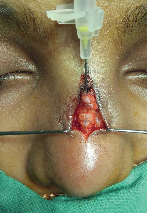
DOI: 10.23937/2378-3397/1410126 ISSN: 2378-3397
Figure 3: Axial cut of CT scan, showing fluid collection on
dorsum of nose.
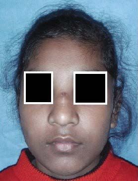
Figure 1: Frontal view of patient showing punctum on dor-
sum of nose.
Figure 2: Right lateral view of patient, showing swelling
on dorsum of nose. Figure 4: Axial cut of CT scan, showing normal crista galli.
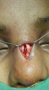
Patient was planned for excision of the NDSC. Written deformity in the nasal bones (Figure 8). A percutaneous
consent for photography, for publication of photographs osteotomy of nasal bones was performed. Layered clo-
and for surgery was taken. Neurosurgeon was kept on sure of the incisions was performed.
standby in case the track extended intracranially. The
Wound healed well without any complications. The
patient was taken under general anaesthesia. Sinus track
excised tissue was sent for histopathological examina-
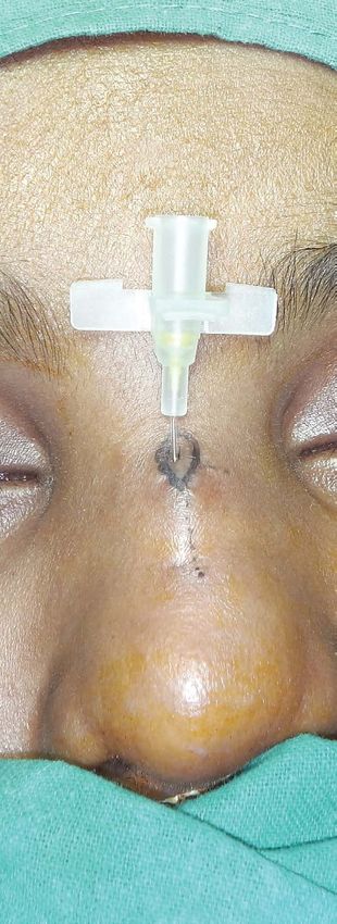
was cannulated using 24 G cannula (Figure 5). 0.2 mL
tion, which confirmed the diagnosis of NDSC. The pa-
methylene blue dye was injected through the cannula. A
tient is in our follow up for more than five years now
vertical elliptical incision was given around the punctum
and extended towards the sinus track (Figure 6). Careful and there has been no recurrence.
sharp dissection was done around the track. 1.2 centi- Discussion
metre sinus track was excised in toto (Figure 7). After ex-
cision, no dye was seen in the field. There was open roof Cruvelier first described a nasal dermoid cyst [2].
Karmakar et al. Int J Surg Res Pract 2021, 8:126 • Page 2 of 5 •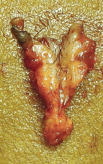
DOI: 10.23937/2378-3397/1410126                                                                             ISSN: 2378-3397

                                                                            Figure 7: The excised sinus track.

 Figure 5: Intraoperative picture showing cannulation of the
 sinus track and marking of incision.

                                                                 Figure 8: Intraoperative picture showing total excision
                                                                 of sinus track and open roof deformity of nasal dorsum.
                                                                 There is no dye in the field.

                                                                    The prenasal theory, by Grunwald attempts to ex-
                                                               plain the development of NDSC [1]. Between 4 to 8
                                                               weeks of gestation, fonticulus frontalis separates nasal
                                                               and frontal bones, and a prenasal space separates the
                                                               nasal bones and cartilage [1,3]. A diverticulum of dural
                                                               mater extends from the anterior cranial fossa through
  Figure 6: Intraoperative picture showing dissection of the   the foramen cecum (which lies anterior to the crista gal-
  sinus track.                                                 li) into the prenasal space. It touches the skin of the nose
                                                               briefly and quickly involutes [1,3]. The nasal and frontal
Various terms used to describe the lesion-dermoid,             bones fuse, obliterating the fonticulus frontalis. Finally,
dermoid sinus, dermal cyst-led to confusion. Sessions          the foramen caecum is filled with fibrous tissue [1,3].
coined the term NDSC to include all lesions containing
ectoderm and mesoderm located in the nose [2].                    NDSC occurs when skin is pulled along with the re-
                                                               gressing dural diverticulum [1,3]. A sinus tract may form

Karmakar et al. Int J Surg Res Pract 2021, 8:126                                                                 • Page 3 of 5 •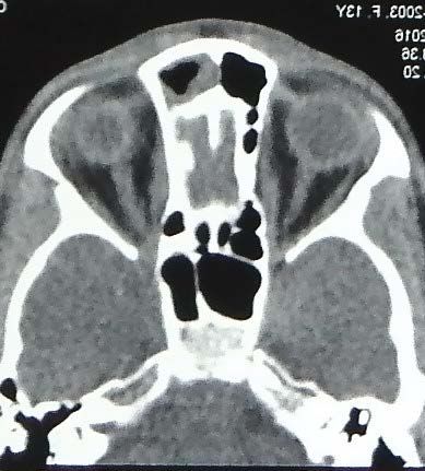
DOI: 10.23937/2378-3397/1410126                                                                               ISSN: 2378-3397

anywhere along the course of the diverticulum [1,3].           [12]. The time and labour added to the surgery is far
Frequency of intracranial connection have been variably        outweighed by the benefits of complete resection.
reported by Wardinsky (45%), Sessions (31%), Rahbar
(19%) and Bradley (4%) [1,2].
                                                               Conclusion
                                                                  NDSC is a rare congenital lesion, which often poses
    The possibility of a transcranial extension neces-
                                                               diagnostic and surgical dilemma. Extent of surgery is
sitates a thorough radiographic evaluation by CT and
                                                               guided by the sinus track and an intracranial approach
magnetic resonance imaging (MRI) [1]. It was believed
                                                               may be needed. Intraoperatively we were guided by
that CT findings of wide foramen cecum and bifid cris-
                                                               MB in our dissection. MB has a long history of use, for a
ta galli, are suggestive of intracranial extension [4]. A
                                                               myriad list of conditions and has a safe record. The dye
number of false positive findings led Penslar to review
                                                               has the potential to reduce surgical trauma and improve
the radiographic images and conclude that an enlarged
                                                               success rate. The technique needs to be explored in a
foramen caecum and a bifid crista galli did not always
                                                               larger series.
correlate with intracranial extension [4]. However, a
normal foramen caecum and crista galli certainly rules         Acknowledgement
out intracranial disease [3].
                                                                  The author acknowledges the faculty and staff of the
   On T1 weighted images of MRI, NDSC appear hyper-            parent institute.
intense [5]. It must be distinguished from the intraosse-
ous fat deposition of crista galli that occurs with normal     Conflicts of Interest
maturation in most children, by age of 5 years [3].               None declared.
    When there is a dorsal ostium, a vertical elliptical in-   Funding
cision has been preferred by Rahbar, which we too, have
used [1].                                                         None.

   Intracranial extension must be assumed until prov-          References
en otherwise [1,3]. Incomplete resection is associated         1. Rahbar R, Shah P, Mulliken JB, Robson CD, Perez-Atayde
with meningitis and recurrence in up to 15% of cases [1].         AR, et al. (2003) The presentation and management of na-
Sessions and Pensler advocated that craniotomy can be             sal dermoid: A 30-year experience. Arch Otolaryngol Head
                                                                  Neck Surg 129: 464-471.
avoided if there is only fibrous track at the cranial base
[2,4]. An epithelium lined track would be stained by MB        2. Sessions RB (1982) Nasal dermal sinuses: New concepts
                                                                  and explanations. Laryngoscope 92: 1-28.
dye and revealed, whereas a fibrous track would not.
Thus we used the dye as a tool to guide dissection.            3. Lowe LH, Booth TN, Joglar JM, Rollins NK (2000) Midface
                                                                  anomalies in children. Radiographics 20: 907-922.
   A search of literature revealed other surgeons too
                                                               4. Penslar JM, Bauer BS, Naidich TP (1988) Craniofacial der-
used the method for similar indications. Phalen, et al.           moids. Plast Reconstr Surg 82: 953-958.
found that in about 20% of their cases, preoperative im-
                                                               5. Rubayi S (2015) General operative management and post-
aging was not consistent with intraoperative findings,            operative care. Reconstructive plastic surgery of pressure
and they used MB to delineate the extent of dermoid               ulcers. Springer, Berlin, Heidelberg, 67-79.
cyst [6]. Verma and Saha used MB to facilitate dissection      6. Phelan AL, Christine MJ, Ceschini AS, Henry CR, Mackay
of the dermoid sinus [7,8]. Histological examination of           DR, et al. (2017) Sparing a craniotomy: The role of intraop-
frozen sections of the tissue from base of track to detect        erative methylene blue in management of midline dermoid
dermal elements, is another method to determine com-              cysts. Plast Reconstr Surg 139: 1445-1451.
pleteness of resection [9].                                    7. Verma M, Singh AK (2018) Approach to dorsal nasal sinus:
                                                                  A rare case report. J Rare Disord Diagn Ther 4: 11.
    Use of MB to stain tissues for dissection is not new.
                                                               8. Saha VP, Ghosh D, Dutta S, Saha S, Basu AK (2017) Mid-
MB has been used to stain cysts, sinuses and ulcers of            line nasal dermoid-A series of thirteen cases and review of
pressure sore, pilonidal sinus and ganglion [5,10,11].            literature. Bengal J Otolaryngol Head and Neck Surg 25:
Dorafshar used the dye to detect minute sinuses in burn           154-159.
wounds [12]. MB is used to stain suspected cancerous           9. Henry C, Mackay D, Dias M (2017) Nasal glioma and crani-
tissues of uterus, cervix, oral cavity, liver and colon           al facial dermoid. Dermatology.
[13,14]. It was the first synthesized drug used for treat-     10. Su Y, Xie Y, Qin J, Nan G (2015) Visualisation of the wrist
ing malaria [15]. MB has been used to facilitate remov-            ganglion capsule by methylene blue staining as an aid for
al of accidentally embedded metallic foreign bodies in             complete resection in children. J Hand Surg Am 40: 685-687.
children [15].                                                 11. Ardelt M, Kocijan R, Dittmar Y, Fahrner R, Rauchfuss F, et
                                                                   al. (2016) Effects of methylene-blue staining on the extent
    Extensive systemic use have shown it to be a safe              of pilonidal sinus excision. J Wound Care 25: 342-347.
drug [12,13]. It can be sterilized by autoclaving and is
                                                               12. Dorafshar AH, Gitman M, Henry G, Agarwal S, Gottlieb LJ
stable after high temperature and high pressure steril-            (2010) Guided Surgical debridement: Staining tissues with
ization [13]. The dye is inexpensive and widely available          methylene blue. J Burn care Res 31: 791-794.

Karmakar et al. Int J Surg Res Pract 2021, 8:126                                                                 • Page 4 of 5 •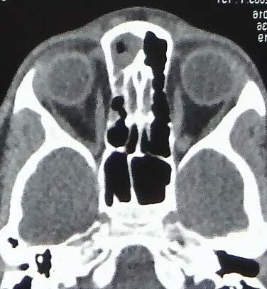
DOI: 10.23937/2378-3397/1410126                                                                                        ISSN: 2378-3397

13. Li A, Zhang X, Zhang Y, Jin W, Zhang S (2016) Application       15. Su Y, Nan G (2016) Using methylene blue as a marker to
    value of in vivo nerve staining in nerve sparing radical hys-       find and remove tiny metallic foreign bodies embedded in
    terectomy. Womens Health Gynecol 2.                                 the soft tissues of children: A randomized controlled trial. Int
                                                                        J Surg 29: 43-48.
14. Shou-Wang C, Shizhoug Y, Wenping L, Geng C, Wanqing
    G, et al. (2015) Sustained methylene blue staining to guide
    anatomic hepatectomy for hepatocellular carcinoma: Initial
    experience and technical details. Surgery 158: 121-127.

Karmakar et al. Int J Surg Res Pract 2021, 8:126                                                                          • Page 5 of 5 •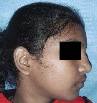

You can also read