Normal anatomic variants of paranasal sinus region studied by computed tomography
←
→
Page content transcription
If your browser does not render page correctly, please read the page content below
Normal anatomic variants of paranasal sinus region ……                  Zanco J. Med. Sci., Vol. 24, No. (2), August, 2020
                                             https://doi.org/10.15218/zjms.2020.022

            Normal anatomic variants of paranasal sinus region studied by
                              computed tomography

                       Received: 21/1/2019                                             Accepted: 22/4/2019

                                                  Shawnam Nasih Dawood1*

                                                          Abstract
Background and objective: Variation in paranasal sinus anatomy, as shown on
computed tomographic scans, is of potential significance as it may pose risks during
surgery or predispose to certain pathologic conditions. This study aimed to investigate the
frequency of anatomic variants of the nasal cavity and paranasal sinuses in our population
and determine their relationship with gender and find out the association of these variants
with mucosal abnormalities.
Methods: This was a cross-sectional study conducted at the College of Medicine, Hawler
Medical University from October 2017 to January 2019. A review of computed tomography
scans of the paranasal sinuses of 300 patients was done; special attention was directed
toward identifying bony anatomic variants and mucosal abnormalities.
Results: Frequent variants were: Agger nasi cells (72%), nasal septal deviation (71.7%),
Haller cells (70.7%), concha bullosa (61%), elongation of uncinate process (69.7%) and
different variants of the sphenoidal sinus (78.8%). The frequency of variants did not differ
significantly with respect to gender, except for sphenoidal variants along with Keros types
and asymmetry of the ethmoidal roof. A significant association was found between middle
concha variants and inferior turbinate enlargement (P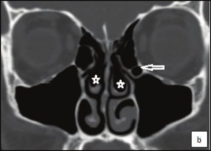
Normal anatomic variants of paranasal sinus region ……      Zanco J. Med. Sci., Vol. 24, No. (2), August, 2020
                                  https://doi.org/10.15218/zjms.2020.022

and orbits.17-20 The frequency of these                  (CB), paradoxical (false) middle concha,
variants may differ among the different                  hypoplasia; (3) ethmoid air cells: Agger
ethnic groups.21There is no data on                      nasi cells, Haller cells, and Onodi cells;
anatomic variants of the PNS region in our               (4)ethmoid uncinate process: elongation
population in the literature review. This                of the upper edge, pneumatization;
study aimed to investigate the frequency of              (5) sphenoid sinus variants and (6) other
a number of anatomic variants of the PNS                 variants: maxillary or frontal sinus septa or
region in our population, find out their                 hypoplasia. The depth of olfactory fossa
gender distribution and the association of               was classified according to the grading
these variants with mucosal abnormalities,               system proposed by Keros into three
and compare the results with previous                    types.22 The asymmetry of ethmoidal roof
investigations conducted in different                    and incidence of aerated crista galli was
populations. The end goal was to gather                  also investigated. The summation of the
this knowledge in an effort to reduce the                unilateral and bilateral abnormalities has
rate of complications of endoscopic sinus                been reported as the prevalence of
surgery.                                                 variants without considering the "half-head"
                                                         as a separate entity. The images were
Methods                                                  also analyzed for the presence of
This was a cross-sectional study conducted               mucosal abnormalities, including PNS
at the College of Medicine, Hawler Medical               mucosal thickening and inferior turbinate
University from October 2017 to January                  enlargement indicative of rhinosinusitis.
2019. The study protocol was approved by                 Statistical Analysis
the Research Ethical Committee of the                    The statistical package for the social
college. It comprised a review of 350                    sciences software, version 21 (Chicago; IL,
cases with unenhanced PNS CT scans at                    USA) was used for data entry and analysis.
Rizgary teaching hospital using Siemens                  A descriptive approach was used to
SOMATOM Emotion16-slice multidetector                    determine frequency and percentages
CT scanner. All scans were performed for                 while in analytic approach (Chi-square test)
evaluation of a symptom related to the PNS               was used for categorical variables.
region. After excluding those below 18                   A P value of
Normal anatomic variants of paranasal sinus region ……      Zanco J. Med. Sci., Vol. 24, No. (2), August, 2020
                                  https://doi.org/10.15218/zjms.2020.022

a number of the anatomic variants are                    Haller cells (70.7%), CB (61%) and
shown in Table 1. Frequent variants                      different forms of sphenoid variants
were Agger nasi cells (72%), NSD (71.7%),                (78.7%) as shown in Figures 1 and 2.

Table 1: Frequency of certain anatomic variants in the studied sample.
Anatomic Variants                                         Number                         % (N=300)

Nasal septal variants

NSD                                                          215                            71.7
Septal spur                                                  102                            34.0

Septal pneumatization                                        50                             16.7

Middle turbinate variants

CB                                                           183                            61.0
Paradoxical turbinate                                        57                             19.0

Hypoplastic turbinate                                         3                              1.0

Aggernasi cells                                              216                            72.0

Haller cells                                                 212                            70.7

Onodi cells                                                  134                            44.7

Ethmoid Uncinate process
Elongation                                                   209                            69.7

Pneumatization                                                9                              3.0

Frontal sinus agenesis or hypoplasia                         35                             11.7

Keros classification

Type I                                                       93                             31.0

Type II                                                      200                            66.7

Type III                                                       7                             2.3

Ethmoidal roof asymmetry                                     130                            43.3

Sphenoidal variants                                          236                            78.7

Crista galli pneumatization                                  17                              5.7

Maxillary sinus
Hypoplasia                                                    4                              1.3
Septum                                                        75                            25.0

                                                    3
                                                   189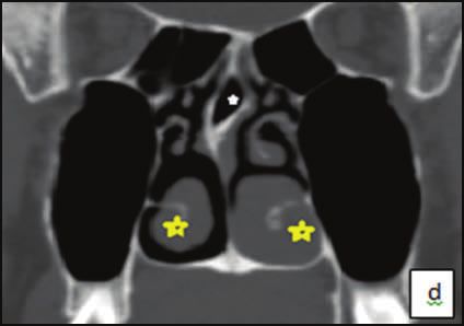
Normal anatomic variants of paranasal sinus region …… Zanco J. Med. Sci., Vol. 24, No. (2), August, 2020
https://doi.org/10.15218/zjms.2020.022
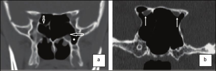
Figure 1: Coronal non-contrast computed tomography images in the bone window.
(a) Bilateral Agger nasi cells (white arrows) and nasal septal deviation to the right (yellow
arrow); (b) Bilateral concha bullosa (stars) and Haller cell (white arrow); (c) Nasal septal
deviation to the right with spur; (d) Septal pneumatization (white star) and bilateral inferior
turbinate enlargement more severe at the left (yellow stars).
Figure 2: Coronal CT images of sphenoid sinus variants: (a) pneumatization of the left
pterygoid processes (star) with protrusion and partial dehiscence of the left vidian nerve
(horizontal arrow) and septum deviated to the right (vertical arrow); it is seen to insert over
the bone covering the right internal carotid artery; (b) protrusion and partial dehiscence of
the optic nerves (arrows).
4
190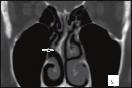
Normal anatomic variants of paranasal sinus region ……       Zanco J. Med. Sci., Vol. 24, No. (2), August, 2020
                                  https://doi.org/10.15218/zjms.2020.022

The frequency of anatomic variants did not                Keros types and ethmoidal roof asymmetry
differ significantly with respect to gender,              (P
Normal anatomic variants of paranasal sinus region …… Zanco J. Med. Sci., Vol. 24, No. (2), August, 2020
https://doi.org/10.15218/zjms.2020.022
Mucosal abnormalities representing than females. The relationship between
rhinosinusitis were detected in 268 (89.3%) certain anatomic variants and mucosal
of 300 patients, including inferior turbinate abnormalities is shown in Tables 3 and 4.
hypertrophy and mucosal thickening of A significant association was found
the sinuses. The prevalence of mucosal between the middle turbinate variants and
thickening was significantly higher in males the presence of mucosal abnormalities.
Table 3: Association of certain anatomic variants with mucosal thickening of the sinuses.
Variables Mucosal thickening of the sinuses Total (N=300) P value
Absent Present
No. (%) No. (%) No. (%)
Gender
Male 54(38.8) 85(61.2) 139(100) 0.001
Female 94(58.4) 67(41.6) 161(100)
Nasal septal deviation
Absent 37(43.5) 48 (56.5) 85(100) 0.206
Present 111(51.6) 104 (48.4) 215(100)
Concha bullosa
Absent 52(44.4) 65(55.6) 117(100) 0.176
Present 96(52.5) 87(47.5) 183(100)
Agger nasi cells
Absent 39(46.4) 45(53.6) 84(100) 0.53
Present 109(50.5) 107(49.5) 216(100)
Haller cells
Absent 39(44.3) 49(55.7) 88(100) 0.263
Present 109(51.4) 103(48.6) 212(100)
Onodi cells
Absent 83(50) 83(50) 166(100) 0.797
Present 65(48.5) 69(51.5) 134(100)
Table 4: Association of certain anatomic variants with inferior turbinate enlargement.
Variables Inferior Turbinate Enlargement Total P value
Absent Present No. (%)
No. (%) No. (%)
Gender
Male 20(14.4) 119(85.6) 139(100) 0.024
Female 40(24.8) 121(75.2) 161(100)
Nasal septal deviation
Absent 17(20.5) 66 (79.5) 83(100) 0.897
Present 43(19.8) 174(80.2) 217(100)
Concha bullosa
Absent 34(29.1) 83(70.9) 117(100) 0.002
Present 26(14.2) 157(85.8) 183(100)
Agger nasi cells
Absent 20(23.8) 64(76.2) 84(100) 0.304
Present 40(18.5) 176(81.5) 216(100)
Haller cells
Absent 11(12.5) 77(87.5) 88(100) 0.036
Present 49(23.1) 163(76.9) 212(100)
Onodi cells
Absent 36(21.7) 130(78.3) 166(100) 0.41
Present 24(17.9) 110(82.1) 134(100)
6
192Normal anatomic variants of paranasal sinus region …… Zanco J. Med. Sci., Vol. 24, No. (2), August, 2020
https://doi.org/10.15218/zjms.2020.022
Discussion the frequency of pneumatization of the
The paranasal sinus region is subject to nasal septum is (0-18) %.6,29,33 Haller cells
a large variety of lesions. Normal anatomic (Figure 1 b) are ethmoid air cells that
variants in this region are important as they extend laterally over the medial aspect of
may have pathological consequences or the roof of the maxillary sinus; they may
maybe the source of difficulty/ complication cause narrowing of the infundibulum.27
during surgery. Certain anatomic variants In the present study, Haller cells were
are thought to be predisposing factors frequent variants and found in 70.5% of
for the development of sinus diseases, the scans (Figure), but other authors have
and thus it becomes necessary for the reported a wide variation in the prevalence
radiologist to be aware of these variants, rates such as 25%,1 9.1%,33 8%,34 and
especially if the patient is a candidate 33%.31,35 Onodi cell was found in 44.7%
for functional endoscopic sinus surgery of the scans in the current study. Other
(FESS).23 The most common anatomic studies reported Onodi cell in 8%1 and
variant in this study was Agger nasi cell 14%6 of their samples. Onodi cell is the
(72%), similar to a study done in Turkey.6 most posterior ethmoid air cell that extends
Other authors have reported prevalence laterally. This extension is near the carotid
rates of 88%,24 96.5%,25 and 94.1%.26 canal and close to the optic nerve, which
The Aggernasi cell (Figure 1 a) is the most emphasizes the clinical importance of
anterior of the anterior ethmoid cells, considering this anatomic variation prior to
found anterior and superior to the middle any attempt for invasive intervention.27
turbinate attachment to the lateral wall.27 Middle turbinate or middle concha variants
Enlargement of Agger nasi cells has been were common in the present study
found to correlate with a decrease in CT in (81.7%). Concha bullosa (CB) is one of the
the anterioposterior size of the nasofrontal most frequently found anatomic variants.
recess, involved in the frontal sinus The incidence of CB in the population
drainage pathway. Failure to address ranges from 13 to 73% in literature;36 the
Agger nasi disease can contribute to the differences reported may have been
failure of the primary surgery.28 The second influenced by the aeration degree, and
most common anatomic variant was the lower rates suggest that only large
deviation of the nasal septum. The NSD turbinates may have been taken into
(Figure 1 a) is an asymmetric bowing of the consideration. The present study has
nasal cartilaginous septum. Such bowing adopted the definition by Zinreich et al.,37
may compress the middle turbinate in who have considered any pneumatization
a lateral fashion, which may lead to the degree as CB and was found in 61.4% of
narrowing of the middle meatus. 27 In this the scans (Figure 1 b). Other studies have
study, any deviation from the midline was found a prevalence of 30.6% by Alsowey
regarded as NSD without considering et al.,32 31.7% by Kalaiarasi et al.,36 30%
the degree of deviation determined by and 51% by two different studied in
connecting the lines being drawn Turkey.6,29 The middle turbinate usually
downward from the crista galli and upward curves medially toward the nasal septum.
from the nasal eminence, and it was found However, the resultant anatomic variant is
in 71.7% of the scans. Earlier studies known as a paradoxical middle turbinate
reported different rates, for example, 89.7% when the turbinate curves laterally. Such
by Kaya et al.,6 81.8% by Kaplanoglu a variant can narrow or obstruct the nasal
et al.,29 68.2% by Sharma et al.,30 60% cavity, middle meatus, or infundibulum.27
by Reddy et al.,31 and 48.8% by Alsowey Again, the prevalence reported by
et al.32 Other variants of the nasal different authors may diverge because
septum included septal spur (34%) and some consider any involved portion of
pneumatized septum (17.3%). In literature, the turbinate as paradoxical curvature.
7
193Normal anatomic variants of paranasal sinus region …… Zanco J. Med. Sci., Vol. 24, No. (2), August, 2020
https://doi.org/10.15218/zjms.2020.022
In contrast, others may consider this There were other uncommon variants
variation only in cases where the whole with crista galli pneumatization in 5.7%,
turbinate is unusually curved towards maxillary sinus hypoplasia in 1.3%, and
the opposite side. In the present study, uncinate process pneumatization in 3%
57 patients were found with paradoxical of cases. The dissimilarities observed
middle turbinates (19%) near to a study between our findings and the results of
done in India 18%.31 Roman et al.1 have previous investigations may be attributed
found this variant in 8% of their cases, to racial, geographic, and hereditary
Adeel et al.2 in 14.3% and Kaya et al.6 disparities, differing sensitivity of data
in 4.3%. The uncinate process (UP) is acquisition, and discrepant definitions for
a structure that has multiple variations a few diagnostic variants. In this study,
between individuals. The superior the frequency of variants did not differ
attachment of the UP has three major significantly with respect to gender
variations that help determine the anatomic (P >0.05) in agreement with Reddy et al.31
configuration of the frontal recess and its and Yadav et al.38 except for sphenoidal
drainage.27 The present study investigated variants along with Keros types and
the shape and pneumatization of the UP. ethmoidal roof asymmetry (PNormal anatomic variants of paranasal sinus region …… Zanco J. Med. Sci., Vol. 24, No. (2), August, 2020
https://doi.org/10.15218/zjms.2020.022
Conclusion associated with recurrent acute rhinosinusitis.
Laryngoscope 2010; 120:631–4.
Anatomic variants of the paranasal sinus 11. İla K, Yilmaz N, Öner S, Başaran E, Öner Z.
region were common in our population; the Evaluation of superior concha bullosa by
most frequent ones were those involving computed tomography. Surg Radiol Anat 2018;
40:841–6.
the nasal septum, ethmoid and sphenoid
12. Kanowitz SJ, Nusbaum AO, Jacobs JB,
sinuses. The study of these variants is Lebowitz RA. Superior turbinate pneumatization
important for the management of paranasal in patients with chronic rhinosinusitis: prevalence
sinus region disease. on paranasal sinus CT. Ear Nose Throat J 2008;
87:578–9.
Competing interests 13. Som PM, Park EE, Naidch TP, Lawson W.
Crista galli pneumatization is an extension of the
The author declares no competing
adjacent frontal sinus. Am Soc Neuroradiol 2009;
interests. 30:31–3.
14. Comer BT, Kincaid NW, Smith NJ,Wallace JH,
References Kountakis SK.Frontal sinus septations predict
1. Roman RA, Hedeşiu M, Gersak M, Fidan F, the presence of supraorbital ethmoid cells.
Băciuţ G, Băciuţ M. Assessing the prevalence of Laryngoscope 2013; 123:2090–3.
paranasal sinuses anatomical variants in patients 15. Smith KD, Edwards PC, Saini TS, Norton NS.
with sinusitis using Cone Beam Computer The prevalence of concha bullosa and nasal
Tomography. Clujul Med 2016; 89(3):419–21. septal deviation and their relationship to maxillary
2. Khojastepour L, Mirhadi S, Mesbahi SA. sinusitis by volumetric tomography. Int J Dent
Anatomical Variations of Ostiomeatal Complex in 2010; 2010:9–13.
CBCT of Patients Seeking Rhinoplasty. J Dent 16. Kim HJ, Jung CM, Lee JW, Tae KY, Kahng H,
(Shiraz) 2015; 16(1):42–8. Sung KH, et al. The relationship between
3. Azila A, Irfan M, Rohaizan Y, Shamim AK. anatomic variations of paranasal sinuses and
The prevalence of anatomical variations in chronic sinusitis in children. Acta Otolaryngol
osteomeatal unit in patients with chronic 2006; 126:1067–72.
rhinosinusitis. Med J Malaysia 2011; 66:191–4. 17. Mikami T, Minamida Y, Koyanagi I, Baba T,
4. Stallman JS, Lobo JN, Som PM. The incidence of Houkin K. Anatomical variations in
concha bullosa and its relationship to nasal septal pneumatization of the anterior clinoid process.
deviation and paranasal sinus disease. AJNR J Neurosurg 2007; 106:170–4.
2004; 25:1613–8. 18. Hamid O, El Fiky L, Hassan O, Kotb A, El Fiky S.
5. Fadda GL, Rosso S, Aversa S, Petrelli A, Anatomic variations of the sphenoid sinus and
Ondolo C, Succo G. Multiparametric statistical their impact on trans-sphenoid pituitary surgery.
correlations between paranasal sinus anatomic Skull Base 2008; 18:9–15.
variations and chronic rhinosinusitis. Acta 19. Hewaidi G, Omami G. Anatomic variation of
OtorhinolaryngolItal 2012; 32:244–51. sphenoid sinus and related structures in Libyan
6. Kaya M, Çankal F, Gumusok M, Apaydin N, population: CT scan study. Libyan J Med 2008;
Tekdemir I. Role of anatomic variations of 3(3):128–33.
paranasal sinuses on the prevalence of sinusitis: 20. Kitaguchi Y, Takahashi Y, Mupas-Uy J, Kakizaki
Computed tomography findings of 350 patients. H. Characteristics of Dehiscence of Lamina
Niger J Clin Pract 2017; 20:1481–8. Papyracea Found on Computed Tomography
7. Mathew R, Omami G, Hand A, Fellows D, Before Orbital and Endoscopic Endonasal
Lurie A. Cone beam CT analysis of Haller Surgeries. J Craniofac Surg 2016; 27(7):662–5.
cells: prevalence and clinical significance. 21. Badia L, Lund VJ, Wei W, Ho WK. Ethnic
Dentomaxillofac Radiol 2013; 42:20130055 variation in sinonasal anatomy on CT-scanning.
8. Nouraei SA, Elisay AR, Dimarco A, Abdi R, Rhinology 2005; 43:210–4.
Majidi H, Madani SA, et al. Variations in 22. Gupta P, Ramesh P. Radiological observation of
paranasal sinus anatomy: implications for the ethmoid roof on basis of keros classification and
pathophysiology of chronic rhinosinusitis and its application in endonasal surgery. Int J Anat
safety of endoscopic sinus surgery. J Otolaryngol Res 2017; 5:4204–7.
Head Neck Surg 2009; 38(1):32–7. 23. Reddy UD, Dev B. Pictorial essay: Anatomical
9. Tomovic S, Esmaeili A, Chan NJ, Choudhry OJ, variations of paranasal sinuses on multidetector
Shukla PA, Liu JK, et al. High-resolution computed tomography-How does it help FESS
computed tomography analysis of the surgeons? Indian J Radiol Imaging 2012;
prevalence of Onodi cells. Laryngoscope 2012; 22(4):317–24.
122(7):1470–3. 24. Kubota K, Takeno S, Hirakawa K. Frontal recess
10. Alkire BC, Bhattacharyya N. An assessment anatomy in Japanese subjects and its effect on
of sinonasal anatomic variants potentially the development of frontal sinusitis: computed
9
195Normal anatomic variants of paranasal sinus region …… Zanco J. Med. Sci., Vol. 24, No. (2), August, 2020
https://doi.org/10.15218/zjms.2020.022
tomography analysis. J Otolaryngol Head Neck Nose And Paranasal Sinuses In Multidetector
Surg 2015; 44(1):21. Computed Tomography. JIOM 2017; 39(1):49–
25. Choby G, Thamboo A, Won TB, Kim J, Shih LC, 54.
Hwang PH. CT analysis of frontal cell prevalence 39. Gupta AK, Gupta B, Gupta N, Tripathi N.
according to the International Frontal Sinus Computed Tomography of Paranasal Sinuses:
Anatomy Classification. Int Forum Allergy Rhinol A Roadmap to Endoscopic Surgery. Clin
2018; 8:825–30. RhinolInt J 2012; 5(1):1–10.
26. Han D, Zhang L, Ge W, Tao J, Xian J, Zhou B. 40. Gibelli D, Cellina M, Gibelli S, Oliva AG,
Multiplanar computed tomographic analysis of Termine G, Sforza C. Anatomical variants of
the frontal recess region in Chinese subjects sphenoid sinuses pneumatisation: A CT scan
without frontal sinus disease symptoms. ORL J study on a Northern Italian population. Radiol
Otorhinolryngol Relat Spec 2008; 70:104–12 Med 2017; 122:575–80.
27. Vaid S, Vaid N. Normal Anatomy and Anatomic 41. Farmer SEJ, Eccles R. Chronic inferior turbinate
Variants of the Paranasal Sinuses on Computed enlargement and the implications for surgical
Tomography. Neuroimaging Clin N Am 2015; intervention. Rhinology 2006; 44:234–8.
25(4):527–48.
28. Bradley DT, Kountakis SE. The role of agger nasi
air cells in patients requiring revision endoscopic
frontal sinus surgery. Otolaryngol Head Neck
Surg 2004; 131:525–7.
29. Kayalioglu G, Oyar O, Govsa F. Nasal cavity
and paranasal sinus bony variations: a computed
tomographic study. Rhinology 2000; 38:108–13.
30. Sharma BN, Pant OB, Lohani B. Computed
tomography in the evaluation of pathological
lesions of paranasal sinuses. J Nepal Health Res
Counc 2015; 13(30):116–20.
31. Reddy A, Kakumanu PK, Kondragunta C,
Gandra NR. Role of computed tomography in
identifying anatomical variations in chronic
sinusitis: An observational study. West Afr J
Radiol 2018; 25:65–71.
32. Alsowey AM, Abdulmonaem G, Elsammak A,
Fouad Y. Diagnostic Performance of
Multidetector Computed Tomography (MDCT)
in Diagnosis of Sinus Variations. Pol J Radiol
2017; 82:713–25.
33. Adeel M, Shaheryar M, Rajput M, Akhter S,
Ikram M, Arain A, et al. Anatomical variations of
nose and para-nasal sinuses; CT scan review.
JPMA 2013; 63:47–52.
34. Riello APFL, Boasquevisque EM. Anatomical
variants of the ostiomeatal complex: tomographic
findings in 200 patients. Radiol Bras 2008;
41(3):149–54.
35. Dafalla S Seyed M, Elfadil N Elmustafa O,
Hussain Z. A Computed Tomography-Aided
Clinical Report on Anatomical Variations of the
Paranasal Sinuses. Int J Med Res Health Sci
2017; 6(2):24–33.
36. Kalaiarasi R, Ramakrishnan V, Poyyamoli S.
Anatomical Variations of the Middle Turbinate
Concha Bullosa and its Relationship with Chronic
Sinusitis: A Prospective Radiologic Study. Int
Arch Otorhinolaryngol 2018; 22(3):297–302.
37. Stallman JS, Lobo JN, Som PM. The Incidence
of Concha Bullosa and Its Relationship to Nasal
Septal Deviation and Paranasal Sinus Disease.
Am J Neuroradiol 2004; 25(9):1613–8.
38. Yadav R, Ansari M, Humagain M, Mishra D.
Assessment Of Anatomical Variations Of
10
196You can also read