Three-dimensional simulation of human teeth and its application in dental education and research
←
→
Page content transcription
If your browser does not render page correctly, please read the page content below
Original Article
                                                                                    http://mjiri.iums.ac.ir                                    Medical Journal of the Islamic Republic of Iran (MJIRI)
                                                                                                                                                                     Iran University of Medical Sciences

                                                                                   Three-dimensional simulation of human teeth and its application
                                                                                                 in dental education and research

                                                                                                                      Maryam Koopaie*1, Sajad Kolahdouz2
Downloaded from mjiri.iums.ac.ir at 7:29 IRST on Tuesday November 12th 2019

                                                                                    Received: 2 April 2016                 Accepted: 12 November 2016                        Published: 24 December 2016

                                                                                 Abstract
                                                                                   Background: A comprehensive database, comprising geometry and properties of human teeth, is needed for
                                                                                 dentistry education and dental research. The aim of this study was to create a three-dimensional model of human
                                                                                 teeth to improve the dental E-learning and dental research.
                                                                                   Methods: In this study, a cross-section picture of the three-dimensional model of the teeth was used. CT-Scan
                                                                                 images were used in the first method. The space between the cross- sectional images was about 200 to 500 mi-
                                                                                 crometers. Hard tissue margin was detected in each image by Matlab (R2009b), as image processing software.
                                                                                 The images were transferred to Solidworks 2015 software. Tooth border curve was fitted on B-spline curves,
                                                                                 using the least square-curve fitting algorithm. After transferring all curves for each tooth to Solidworks, the sur-
                                                                                 face was created based on the surface fitting technique. This surface was meshed in Meshlab-v132 software, and
                                                                                 the optimization of the surface was done based on the remeshing technique. The mechanical properties of the
                                                                                 teeth were applied to the dental model.
                                                                                   Results: This study presented a methodology for communication between CT-Scan images and the finite ele-
                                                                                 ment and training software through which modeling and simulation of the teeth were performed. In this study,
                                                                                 cross-sectional images were used for modeling. According to the findings, the cost and time were reduced com-
                                                                                 pared to other studies.
                                                                                   Conclusion: The three-dimensional model method presented in this study facilitated the learning of the dental
                                                                                 students and dentists. Based on the three-dimensional model proposed in this study, designing and manufactur-
                                                                                 ing the implants and dental prosthesis are possible.

                                                                                 Keywords: Three-Dimensional Modeling, Dental Education, Finite Element Analysis.

                                                                                 Cite this article as: Koopaie M, Kolahdouz S. Three-dimensional simulation of human teeth and its application in dental education and
                                                                                 research. Med J Islam Repub Iran 2016 (24 December). Vol. 30:461.

                                                                                   Introduction                                                         ment of those patients who are referred to
                                                                                   Nowadays, the status of the oral health is                           dental schools or based on practicing on the
                                                                                 one of the great challenges in the field of                            standard plastic teeth. Difficulty of regular
                                                                                 health, treatment and medical education. In                            access to human samples and deficiency of
                                                                                 this context, specialized training of the staff                        expert observers are the limitations of tradi-
                                                                                 and students is of prime importance. One of                            tional methods. This study found that using
                                                                                 the most important educational needs of the                            the virtual three-dimensional tooth model
                                                                                 dental students is evaluation of patients'                             appeared to be more efficient than plastic
                                                                                 teeth and oral cavity. In this study, the                              models (1).
                                                                                 teaching principles of prevention, diagnosis                             Using the virtual three-dimensional mod-
                                                                                 and treatment of the oral cavity disorders                             el of the teeth and gums, as well as enhanc-
                                                                                 were the learning priorities. During dentis-                           ing the quality of dental education will re-
                                                                                 try education, students practice many dental                           duce learning time. Dentists need models
                                                                                 procedures. The traditional approach in                                for frequent modifications to reach the best
                                                                                 dentistry education is based on the treat-                             prosthesis and to have a more accurate ex-
                                                                              ____________________________________________________________________________________________________________________
                                                                               1
                                                                                . (Corresponding author) Assistant Professor, Department of Oral and Maxillofacial Medicine, School of Dentistry, Tehran University of
                                                                               Medical Sciences, Tehran, Iran. m-koopaie@tums.ac.ir & maria_koopaie @yahoo.com
                                                                               2
                                                                                . Master of Science in Mechanical Engineering, Department of Mechanical Engineering, Amirkabir University of Technology, Tehran, Iran.
                                                                               sajjadkolahdouz@gmail.com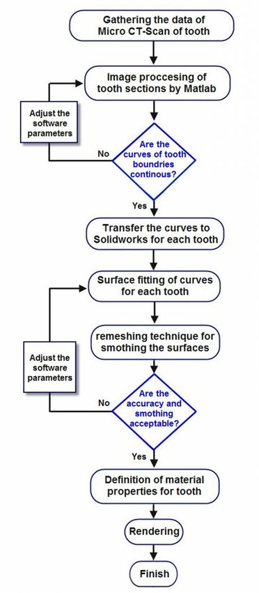
3-D simulation of dental teeth in education

                                                                              amination, and appropriate design. Dental          this method is cost-effective, it appears to
                                                                              modeling and simulation to verify the accu-        have major weaknesses when it comes to
                                                                              racy of the prostheses and to meet dental          different structures of the teeth (enamel and
                                                                              students’ training needs seems irrefutable         dentine), and material properties definition
                                                                              (2,3). Three-dimensional models in various         (2).
                                                                              aspects of dental treatment, including oral          Another approach is based on the Voxel
                                                                              surgery, implants and orthodontics can be          method in which material properties and
Downloaded from mjiri.iums.ac.ir at 7:29 IRST on Tuesday November 12th 2019

                                                                              very useful (4). Most common problems in           different teeth structures are defined. Alt-
                                                                              the field of dentistry are dental decays and       hough the apparent looks of the mesh mod-
                                                                              periodontal diseases (5).Providing a high-         el are better, the Voxel model is more suit-
                                                                              resolution three-dimensional model that            able for the dental finite element analysis
                                                                              consists of all the aspect of oral cavity          due to the definition of the properties of the
                                                                              could be very effective (6).                       teeth (17). Wang et al. found a method
                                                                                Using a three-dimensional model can be           based on the triangular mesh for the dental
                                                                              an alternative method for the conventional         modeling and simulation. They used laser
                                                                              non-imaging method, which reduces costs,           scanner to measure the teeth and the dia-
                                                                              time and increases the compliance of a final       metric shortest distance for triangular mesh
                                                                              prosthesis. The purpose of this study was to       generation (1). In this study, a new method
                                                                              present a three-dimensional model of the           was used based on measuring the teeth
                                                                              teeth to be used in the field of education,        curves by image processing. This method is
                                                                              research and treatment. Furthermore, this          based on the surface automatic mesh gener-
                                                                              method could be effective in reducing the          ation, and is a combination of the two
                                                                              educational cost and time, and decreasing          methods of Voxel and mesh generation.
                                                                              the experimental test for the researchers            A comprehensive knowledge of the teeth
                                                                              (3). Reverse engineering is an important           leads to the more accurate virtual three-
                                                                              technique to manufacture the industrial            dimensional modeling. A database that con-
                                                                              products, and may be used in dental model-         tains geometry and properties of human
                                                                              ing (7-11). Accuracy and precision are the         teeth are needed for dentistry education and
                                                                              challenges of reverse engineering of dental        dental researchers. A three-dimensional
                                                                              modeling. One of the applications of mod-          model of dental teeth can improve dental
                                                                              eling is using the model in the finite ele-        research and specialized training (18,19) as
                                                                              ment analysis (2,7-10). A more precise-            well as develop simulation software of oral
                                                                              model will be more applicable in the finite        surgery, orthodontics, and endodontic im-
                                                                              element analysis.                                  plants (20,21). The aim of this study was to
                                                                                The sectioning images are used for re-           create a three-dimensional model of the vir-
                                                                              verse engineering, but this method is ex-          tual teeth, used in dental education and the
                                                                              pensive, and time consuming (12,13). Re-           finite element analysis for dental research.
                                                                              cently, the use of Cone Beam Computed
                                                                              Tomography (CBCT) and Multi-Slice CT                 Methods
                                                                              (MSCT) has reduced problems related to               In this study, a cross-sectional picture of a
                                                                              teeth modeling (14). One of the most im-           tooth for three-dimensional modeling was
                                                                              portant challenges in three-dimensional            used. In CT-scanning (the first method),
                                                                              modelingis the relationship between CT-            whenever the space between the cuts is
                                                                              Scan images and the modeling software              less, the resulting model would be more
                                                                              (6,15). Because of the complex features of         accurate. To create a three- dimensional
                                                                              the tooth structure, different methods have        model of a tooth, another method, other
                                                                              been used for dental modeling (16). One of         than CT-scanning, was used. Using this
                                                                              these methods is based on the mesh genera-         method would help compare the accuracy
                                                                              tion algorithm, the graphic effects of which       of the two methods. In the second method,
                                                                              would be relatively acceptable. Although           we cut the tooth from the top of the crown

                                                                              http://mjiri.iums.ac.ir                        2    Med J Islam Repub Iran 2016 (24 December). Vol. 30:461.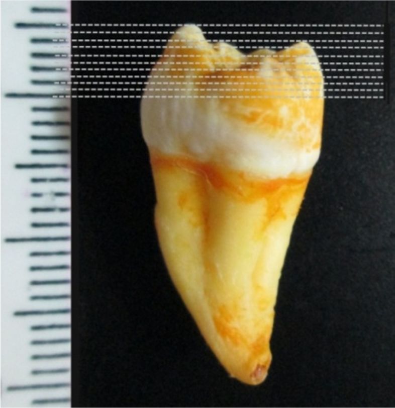
M. Koopaie, et al.

                                                                                                                                               images of the tooth preparation were taken.
                                                                                                                                               The teeth were mounted to examine the
                                                                                                                                               cross-section of the tooth images as shown
                                                                                                                                               in Figure1.
                                                                                                                                                 An example of the cutting lines on the
                                                                                                                                               crown of the maxillary third molar is
                                                                                                                                               shown in Figure 2. Pictures, taken in each
Downloaded from mjiri.iums.ac.ir at 7:29 IRST on Tuesday November 12th 2019

                                                                                                                                               level, were used in the teeth simulation.
                                                                                                                                                 In the first step, CT-Scan images were
                                                                                                                                               used, with a CT-Scan device (GE
                                                                                                                                               HiSPEED NX / I Pro CT) of the space of
                                                                                                                                               50 microns between the sections. Time
                                                                                                                                               measured for each frame was about 20 sec-
                                                                                 Fig. 1. The Teeth Were Mounted to Examine the                 onds. For a tooth measurement, an average
                                                                                 Cross-section of the Tooth Images                             time of an hour was spent. On average, 80
                                                                                                                                               images of each tooth were prepared. The
                                                                                                                                               space between the cross- sectional images
                                                                                                                                               was about 200 to 500 micrometers. In the
                                                                                                                                               next step, the hard tissue margin was de-
                                                                                                                                               tected in each image. Matlab (R2009b) was
                                                                                                                                               used as an image processing software.
                                                                                                                                                 The detection algorithm was based on the
                                                                                                                                               difference of color in the images, and ulti-
                                                                                                                                               mately this software defined the boundaries
                                                                                                                                               of the hard tissue. The boundaries were
                                                                                                                                               specified in the Matlab (R2009b) for each
                                                                                                                                               image. An example of image processing for
                                                                                                                                               the second premolar is shown in Figure 3.
                                                                                                                                                 The images were transferred to Solid-
                                                                                                                                               works 2015 software. Tooth border curve
                                                                                         Fig. 2. Cutting Lines for Tooth Slices                was fitted on B-spline curves, using least
                                                                                                                                               square-curve fitting algorithm with less
                                                                               to the root, with a space of 0.5 millimeters                    than 5%error. An example of the fitted
                                                                               between the cuts. Then the cross-sectional

                                                                                                           Fig. 3. An Example of the Detection of the Tooth Margin in Matlab

                                                                                        Fig. 4. An Example of Marginal Detection in Matlab and Its Adaptation on B-Spline Curve in Solidworks

                                                                              Med J Islam Repub Iran 2016 (24 December). Vol. 30:461.     3                                     http://mjiri.iums.ac.ir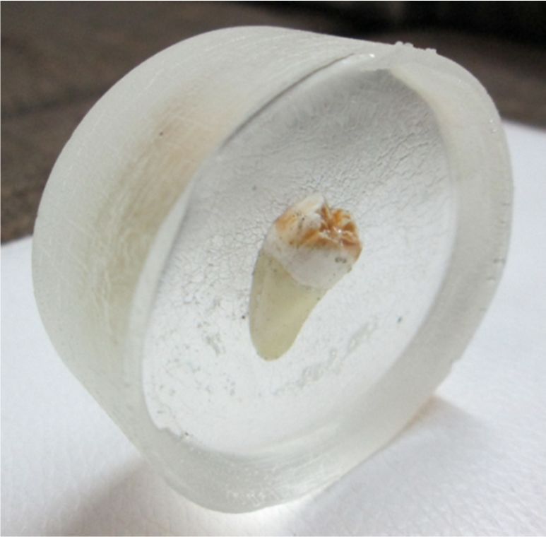
3-D simulation of dental teeth in education
Downloaded from mjiri.iums.ac.ir at 7:29 IRST on Tuesday November 12th 2019

                                                                                            Fig. 5. A Set of B-Spline Curves Used for Molar Teeth Modeling in Solidworks 2015

                                                                              boundary lines is shown in Figure 4 (the                 attributed (22,23). The three-dimensional
                                                                              red line is the example of curve fitting on              modeling algorithm used in the study is
                                                                              the tooth border in Solidworks 2015 soft-                shown in Figure 8.
                                                                              ware).                                                     Rendering is the final step of the simula-
                                                                                To create boundary curves tooth, a cross-              tions that providesthe model with real ef-
                                                                              sectional image of each tooth was prepared               fects. The virtual three-dimensional model,
                                                                              with the space of 0.2mm from each other.                 which was created with real teeth, was used
                                                                              The curves were transferred to Solidworks                for comparison (Fig. 9).
                                                                              2015 in compliance with the order. A series
                                                                              of images produced by this method are
                                                                              shown in Figure 5 (mandibular molars).
                                                                                After transferring all curves to Solid-
                                                                              works for each tooth, the surface was creat-
                                                                              ed based on the surface fitting technique.
                                                                              This surface was meshed in Meshlab-v132
                                                                              software, and the optimization of the sur-
                                                                              face was done based on the remeshing
                                                                              technique. Remeshing technique was based
                                                                              on the downsizing of the model meshes,
                                                                              and the pyramid tetrahedral mesh was used.
                                                                              An example of this method and creation of
                                                                              the model is shown in Figure 6. Initial
                                                                              model is shown in part A. Part D in Figure
                                                                              6 displays the model after the remeshing                     Fig. 6. Steps to Prepare and Improve the Quality of
                                                                              technique. All apparent faults in this model                 the Model Surface (A to D)
                                                                              have been corrected.
                                                                                To assign the mechanical properties, we
                                                                              had to create an intrinsic structure of the
                                                                              tooth other than its surface. Cutting the sec-
                                                                              tion of intrinsic structure of the tooth is
                                                                              shown in Figure 7. Finally, the models
                                                                              were created with the format of "STEP".
                                                                                To perform the finite element analysis on
                                                                              the model, the mechanical properties of the
                                                                              teeth should be allocated. The Young's
                                                                              modulus of tooth enamel (E=88GPa) was                       Fig. 7. Cutting the Section Images of the Tooth in
                                                                                                                                          Different Levels
                                                                              http://mjiri.iums.ac.ir                              4     Med J Islam Repub Iran 2016 (24 December). Vol. 30:461.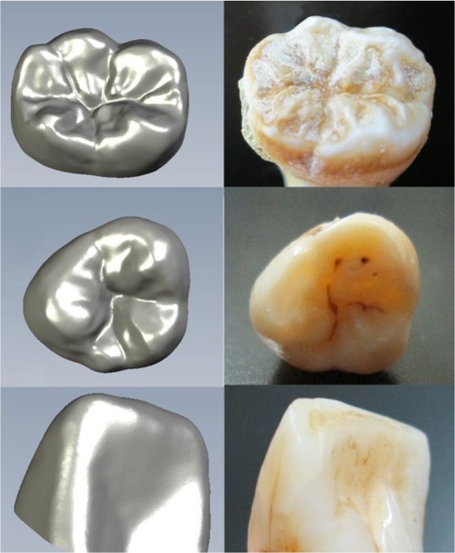
M. Koopaie, et al.
cost and time were reduced compared to
other studies. This modeling can be per-
formed without destroying the structure of
real teeth, and could be used in the finite
element analysis for designing implants,
dental prosthesis. To our knowledge, there
was no reported document on the educa-
Downloaded from mjiri.iums.ac.ir at 7:29 IRST on Tuesday November 12th 2019
tional function of the finite element analy-
sis in the literature. Without precise geo-
metric dimensions of the teeth, the finite
element analysis and E-learning will not be
accurate. This study presented a methodol-
ogy for communication between CT-scan
images and the finite element and training
software through which modeling and sim-
ulation teeth were performed.
Measuring the tooth dimensions demon-
strate that the error was in the range of 5%.
It is possible to obtain detailed models with
high accuracy and less error, but this great-
ly increases the time and cost of modeling.
The method described in this study has
some advantages to the laser scanning
method. One of the problems in laser scan-
ning method is the lack of adequate access
and lack of sufficient precision in the mod-
Fig. 8. The Three-Dimensional Algorithm el.
Modeling of Teeth Simulation
Discussion
The three-dimensional models of the hu-
man body anatomy could improve medical
education, describe a variety of diseases,
and help determine the types of injury. In
this study, by using images obtained from
CT-scanning and the cutting sections of the
teeth, three-dimensional models of teeth
were created, whereas in other studies
cloud points were used to create the three-
dimensional model of teeth (23,24). In this
study, the possibility of creating a virtual
three-dimensional model of hard tissue
(teeth) was provided. This model increases
the quality of specialized training in dentis-
Fig. 9. Comparison of the Virtual Three-
Dimensional Model of the Teeth with Real
try and significantly improves the quality
Teeth and accuracy of dental procedures.
Results Conclusion
In this study, the cross-sectional images The method presented in this study has
were used for modeling. In this model, the the following basic characteristics:
Med J Islam Repub Iran 2016 (24 December). Vol. 30:461. 5 http://mjiri.iums.ac.ir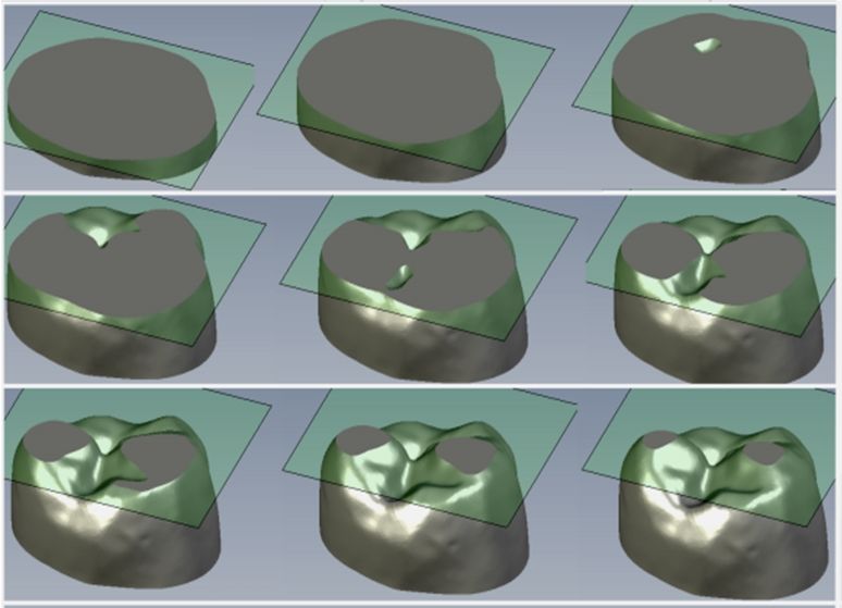
3-D simulation of dental teeth in education
The three-dimensional model was more of dental restorative procedures using micro-CT
useful to the dental students and dentists. data. Dental Materials 2007;23:539-548.
10. Ichim I, Schmidlin PR, Kieser JA, Swain MV.
The method presented in this study, in Mechanical evaluation of cervical glass-ionomer
comparison with the other methods, is more restorations: 3D finite element study. Journal of
accurate and less expensive, and the possi- dentistry 2007;35:28-35.
bility of creating a three-dimensional model 11. Ichim I, Li Q, Loughran J, Swain MV, Kieser
J. Restoration of non-carious cervical lesions Part I.
of the tooth with respect to CT-Scan imag-
Downloaded from mjiri.iums.ac.ir at 7:29 IRST on Tuesday November 12th 2019
Modelling of restorative fracture Dental Materials
es is higher. 2007;23:1553-1561.
Based on the three-dimensional model 12. Li W, Swain MV, Li Q, Steven GP. Towards
proposed, analysis of the design and manu- automated 3D finite element modeling of direct fi-
facturing of implants and dental prosthesis ber reinforced composite dental bridge. Journal of
Biomedical Materials Research Part B: Applied Bi-
are possible. omaterials 2005;74(1):520-528.
Itis possible to create a detailed model- 13. Rahimi A, Keilig L, Bendels G, Klein R,
with maximum error of 50 micrometers by Buzug TM, Abdelgader I, et al. 3D Reconstruction
this model. of dental specimens from 2D histological images
and CT-Scans. Computer Methods in Biomechanics
and Biomedical Engineering 2005;8(3):167-176.
14. Liang X, Lambrichts I, Sun Y, Denis K, Has-
References san B, Li L, et al. A comparative evaluation of
1. Wang D, Zhang Y, Wang Y, Lee YS, Lu P, Cone Beam Computed Tomography (CBCT) and
Wang Y. Cutting on triangle mesh: local model- Multi-Slice CT (MSCT). Part II: On 3D model ac-
based haptic display for dental preparation surgery curacy. European Journal of Radiology 2010;
simulation.IEEE Transactions on Visualization and 75(2):270-274.
Computer Graphics 2005;11(6):671-683. 15. Nagasawa S, Yoshida T, Tamura K,
2. Verdonschot N, Fennis WM, Kuijs RH, Stolk Yamazoe M, Hayano K, Arai Y, et al. Construction
J, Kreulen CM, Creugers NH. Generation of 3D of database for three-dimensional human tooth
finite element models of restored human teeth using models and its ability for education and research -
micro-CT techniques. The International Journal of Carious tooth models. Dental Materials Journal
Prosthodontics 2001;14(4):310–315. 2010;29(2):132-137.
3. Macchiarelli R, Bondioli L, Debenath A, Ma- 16. Hohmann A, Kober C, Young P, Dorow C,
zurier A, Tournepiche JF, Birch W, et al. How Ne- Geiger M, Boryor A, et al. Influence of different
anderthal molar teeth grew. Nature 2006; modeling strategies for the periodontal ligament on
444(7120):748-751. finite element simulation results. American Journal
4. Moritomo H, Goto A, Sato Y, Sugamoto K, of Orthodontics and Dentofacial Orthopedics
Murase T, Yoshikawa H. The triquetrum-hamate 2011;139(6):775-783.
joint: an anatomic and in vivo three-dimensional 17. Pohlenz P, Gröbe A, Petersik A, von Stern-
kinematic study.The Journal of Hand Surgery berg N, Pflesser B, Pommert A, et al. Virtual dental
2003;28(5):797-805. surgery as a new educational tool in dental school.
5. Sun ZJ, Liu B, Zhao YF. Radiopacity in syn- Journal of Cranio-Maxillofacial Surgery 2010;
drome keratocysticodontogenictumour. Den- 38(8):560-564.
tomaxillofacial Radiology 2008;37(3):175-178. 18. de Boer IR, Wesselink PR, Vervoorn JM. The
6. Kato A, Ohno N. Construction of three- creation of virtual teeth with and without tooth pa-
dimensional tooth model by micro-computed to- thology for a virtual learning environment in dental
mography and application for data sharing. Clinical education.European Journal of Dental Education
oral investigations 2009;13(1);43-46. 2013;17(4):191-197.
7. Clement R, Schneider J, Brambs HJ, Wunder- 19. Moschos G, Nikolaidis N, Pitas I, Lyroudia
lich A, Geiger M, Sander FG. Quasi-automatic 3D K. A Virtual Anatomical 3D Head, Oral Cavity and
finite element model generation for individual sin- Teeth Model for Dental and Medical Applications.
glerooted teeth and periodontal ligament.Computer Man-Machine Interactions 2, Advances in Intelli-
Methods and Programs in Biomedicine 2004;73: gent and Soft Computing 2011;103:197-206.
135-144. 20. Canellas JV, Araujo MM, Arce JP. The use of
8. Gomes de Oliveira S, Seraidarian PI, Landre J anatomical models for learning anesthesia tech-
Jr, Oliveira DD, Cavalcanti BN. Tooth displace- niques in oral surgery.Indian Journal of Dental Re-
ment due to occlusal contacts: a three-dimensional search 2013;24(3):326-230.
finite element study. Journal of Oral Rehabilitation 21. Soares PV, de Almeida Milito G, Pereira FA,
2006;33:874-880. Reis BR, Soares CJ, de Sousa Menezes M, et al.
9. Magne P. Efficient 3D finite element analysis Rapid prototyping and 3D-virtual models for opera-
http://mjiri.iums.ac.ir 6 Med J Islam Repub Iran 2016 (24 December). Vol. 30:461.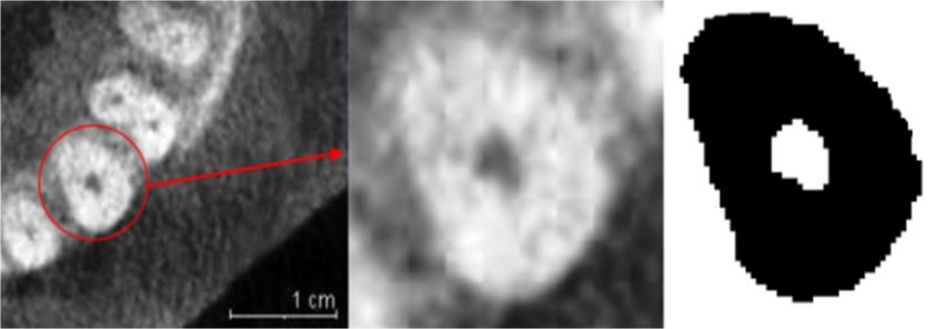
M. Koopaie, et al.

                                                                                tive dentistry education in Brazil. Journal of Dental       nite element modeling from CT images of tooth and
                                                                                Education 2013;77(3):358-363.                               its validation. Dental Materials Journal 2009;
                                                                                  22. He LH, Foster Page L, Purton D. An evalua-            28(2):219-226.
                                                                                tion of dental operative simulation materials. Dental         24. Persson A, Andersson M, Oden A, Sand-
                                                                                Materials Journal 2012;31(4):645-649.                       borgh-Englund G. A three-dimensional evaluation
                                                                                  23. Tajima K, Chen KK, Takahashi N, Noda N,               of a laser scanner and a touch-probe scanner.The
                                                                                Nagamatsu Y, Kakigawa H. Three-dimensional fi-              Journal of Prosthetic Dentistry 2006;95(3):194-200.
Downloaded from mjiri.iums.ac.ir at 7:29 IRST on Tuesday November 12th 2019

                                                                              Med J Islam Repub Iran 2016 (24 December). Vol. 30:461.   7                                 http://mjiri.iums.ac.ir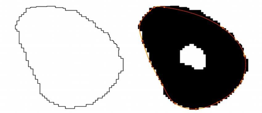
You can also read